Nuclear Medicine Infection Assessment, Protocols, and Interpretation

Earn CME/CE in your profession:


Continuing Education Activity

As the complexity of patients and their comorbidities nowadays increase, the management of infections poses an increasing clinical dilemma. This chapter highlights the application of different nuclear medicine techniques in the evaluation and workup of infectious foci. Modern nuclear medicine practice uses advanced SPECT-CT and PET-CT hybrid imaging techniques to evaluate infections. This activity will review the various indications, techniques, and radiopharmaceuticals used by the interprofessional team.

Objectives:

  • Review different radiopharmaceuticals used clinically to evaluate infections.
  • Outline the impact of SPECT-CT techniques.
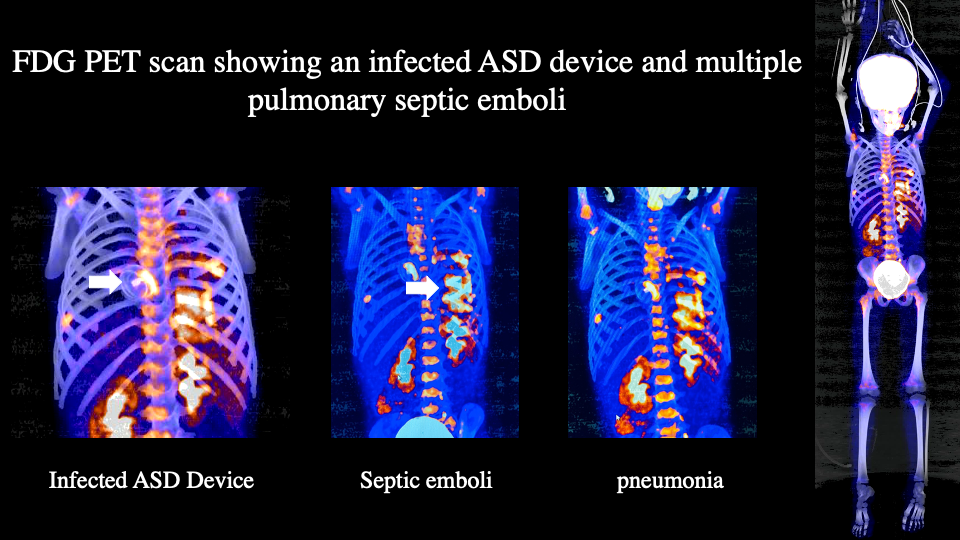
  • Explain the use of FDG PET in cardiovascular device infections.
  • Describe the use of Fluoride PET scans versus FDG PET scans in the evaluation of bony infections.

Introduction

Infectious diseases are a significant cause of morbidity and mortality worldwide. On occasion, nuclear medicine modalities can play a role in helping a clinician evaluate for occult or suspected sites of infection. Nuclear medicine has historically helped evaluate sources of infectious foci through the use of a variety of radiopharmaceuticals when structural imaging modalities such as radiography, sonography, CT, and MRI do not yield sufficient diagnostic information.

While structural imaging modalities can still provide significant diagnostic information, nuclear medicine can play an important role in distinguishing acute from chronic changes, active from healed disease, and information on the abnormal metabolic activity of affected tissues in the human body. More recently, advances in camera technologies with the wider use of hybrid imaging devices such as SPECT-CT, PET-CT, and PET-MR scanners have improved the specificity and sensitivity of different nuclear medicine exams used in evaluation for infection by combining the pathophysiologic information of nuclear medicine modalities with the structural information of cross-sectional imaging.

By itself, CT imaging has a number of limitations, including lack of morphological tissue changes in early infection and the inability to distinguish acute changes in patients with chronic morphological tissue changes. MRI's high sensitivity to water with T2/STIR imaging can play a role in evaluating inflammation and infection; however, it also has a limited role in distinguishing post-surgical and healing changes from active inflammation.

Anatomy and Physiology

18-F-FDG PET

The use of 18F-fluorodeoxyglucose positron emission tomography/computed tomography (18-F-FDG PET/CT) has grown in recent years and has become one of the primary nuclear medicine modalities in evaluating infection. 18-F-FDG PET has a variable normal biodistribution to the liver, brain, kidneys, bladder, heart, and bowel. Glucose transporters, hexokinase, and glycolytic activity are upregulated in inflammatory/infectious conditions allowing for the use of 18-F-FDG PET imaging in the evaluation of inflammatory/infectious conditions. Before an FDG PET scan to evaluate an infectious cardiac focus.[1][2]

67-Ga-Citrate Scan

67-Gallium-Citrate Scans had a historical significance in the evaluation of infection before the introduction of 18-F-FDG PET but are still on occasion used in the assessment of spinal osteomyelitis. Gallium acts as an analog to iron and functions as a helpful radiotracer by being recruited to sites of inflammation via lactoferrin.

White Blood Cell Scans

White blood cells can be most commonly radiolabelled with two isotopes, Technetium-99m (99m-Tc) or Indium-111 (111-In). Depending on the radiopharmaceutical used, white blood cell scans have varying normal biodistributions and different scan accuracy. Therefore the radiotracer used should be tailored to the indication. Radiolabeled human leukocytes, properly prepared and handled ex vivo, behave in vivo in a fashion essentially identical to native leukocytes, including migrating to foci of acute and/or chronic inflammation and infection.  The most common method currently in clinical use involves labeling autologous leukocytes or purified neutrophils with In-111-oxine or 99m-Tc-HMPAO, reinfusion of the labeled leukocytes, and imaging at variable time points.[3] Indium-leukocytes may also be used to evaluate active inflammatory bowel disease sites, such as Crohn's disease and ulcerative colitis. Some of the drawbacks of white blood cell scans include being time-consuming and exposure of technicians to blood.

Bone Scintigraphy

Traditional bone scans using either methylene diphosphonate(MDP) or hydroxydiphosphonate (HDP) radiolabelled with technetium can be of value in assessing bone infections. The radiopharmaceutical targeting mechanism is through chemisorption into the bone matrix via attachment to hydroxyapatite crystals. Bone scans with SPECT or PET are highly sensitive techniques and have a high negative predictive value, and are very good at excluding disease when negative but can lack specificity in certain clinical scenarios especially following bony instrumentation. Tc99m-besilesomab and Tc99m-sulesomab have also been used successfully in certain markets where they are available. The addition of recent hybrid techniques with SPECT-CT is extremely beneficial and more accurate.[4][5][6] These compounds are monoclonal antibodies initially used to evaluate bony infections, with recent reports of benefit in assessing cardiovascular infections.[7][8] 

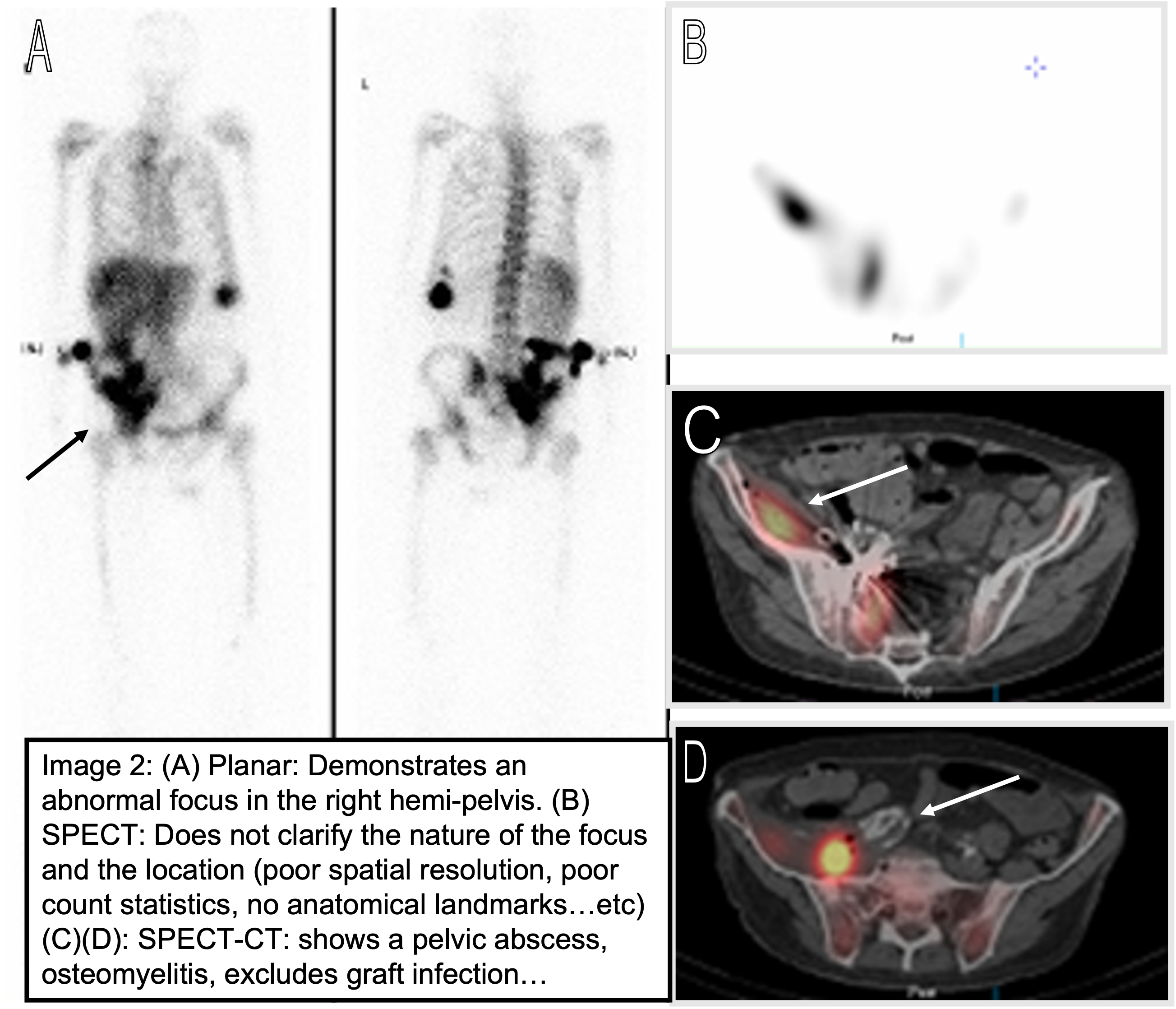
Common practice has been to image patients through 2D planar imaging and occasionally use 3D SPECT data as an adjunct. However, the availability of SPECT-CT cameras has become essential in improving diagnostic accuracy as SPECT lacks much-needed anatomical resolution and correlative information. Recent reviews have found that SPECT-CT is useful in accurately predicting anatomy, increasing reader confidence, and improving scan accuracy compared to SPECT and planar imaging. SPECT-CT of the region of interest should be routinely considered in adults when performing a WBC scan as it has been shown to increase specificity and sensitivity (Figure 1). Beyond correlation, the low dose associated with a CT from the SPECT-CT improves imaging data. CT is used in the reconstruction of SPECT data in creating attenuation correction maps. It also improves spatial, contrast resolution and allows for proper quantification of data.[9][10][11]

Historically radiolabeled leukocytes have been used when suspected infective processes are three weeks or less in duration, whereas 67-Gallium-citrate scans were felt to be more useful when infections are more chronic (>3 weeks). 67-Gallium-citrate scans are more helpful in imaging the chest and in a fever of unknown origin (where the differential diagnosis may include occult malignancy). However, nowadays, this does not have supportive literature data. When available, 18-F-FDG PET is increasingly used over 67-gallium-citrate. When 18-F-FDG PET imaging is not available, 67-Gallium-citrate with the use of a SPECT-CT technique is helpful.[12][13][14]

Indications

Common indications include the evaluation of infections of:

  • Cardiovascular implanted devices
  • Endocarditis
  • Osteomyelitis
  • Soft-tissue infections
  • Muskuloskeletal hardware infection
  • Line infections
  • Charcot joint
  • Arthroplasty infections

Contraindications

Nuclear medicine scans are safe as radiopharmaceuticals contain a trace amount of the biological compound. Contraindications would relate to the appropriateness of the test ordered according to the latest guidelines and appropriateness use criteria. Proper quality control of radiopharmaceuticals injected should be performed. Adverse events are rare but may occur more so with WBC-based radiopharmaceuticals or antibody-based radiopharmaceuticals (though not as frequently used anymore). Additionally, proper workflows need to be in place for safe radiopharmaceutical administration, especially radiolabelled white blood cells.

Equipment

The latest equipment should be used when possible. This not only allows lower doses to be injected but also offers higher sensitivity and spatial resolution. The utilization of hybrid imaging with SPECT-CT and PET-CT allows for higher sensitivity and higher accuracy. Following strict quality control protocols in the radiopharmacy and the scanner is critical to delivering the highest quality and safe patient care. Proper planned maintenance and upkeep of the nuclear medicine equipment is also essential to avoid equipment failures.

Personnel

Fully trained, certified, and licensed nuclear medicine technologists under the supervision of a qualified, licensed, and board-certified nuclear medicine physician must oversee the care of nuclear medicine patients. Close attention should be paid to the training of proper radiolabeling of white blood cells. In addition, nuclear medicine technologists should be well trained in aseptic techniques to prepare radiopharmaceuticals in general and specifically when handling a patient's white blood cells. 

While all radiopharmaceutical misadministrations are considered serious, the inadvertent administration of one patient's leukocytes to another patient may have serious consequences, including anaphylaxis or transmission of hepatitis, HIV, or another inoculant.  Similarly, leukocytes obtained from patients potentially infected with transmittable pathogens must be handled, labeled, and reinfused with the utmost caution to avoid inadvertent contamination. Labeling steps should not be performed in a rush. There should be no shortcuts to the multiple levels of confirmation needed to ensure the correct identity of a given patient's leukocytes.

Completion of the study should require a nuclear medicine physician, technologist, and potentially a radio pharmacist when available.  A nuclear medicine physician should initiate the study by written prescription after receiving a written order from the patient's referring physician.  A technologist will collect the required blood sample and either radiolabel the sample or have a nuclear pharmacist perform the labeling.  Subsequently, the radiolabeled autologous leukocytes will be readministered to the patient, followed by imaging. All personnel involved will be appropriately trained to provide these patient care services.

If any discrepancy is observed regarding the patient-sample identification process, the technologist should immediately return the dosage syringe, injection tray, and the completed radiolabeled blood product administration record to the nuclear pharmacy for disposition.

Preparation

18-F-FDG PET scans have special requirements as follows:[15]

  • Fasting for 4 to 6 hours before the exam
  • No exercise for 24 hours
  • The patient should be well hydrated before the fasting period.
  • A high-fat, low-carb diet should be initiated at least 24 hours optimally 48 to 72 hours before an FDG PET scan for any cardiac site infection.
  • Special instructions should be given to people with diabetes to minimize peaks in blood sugars just before injection to decrease any competition of nonradioactive sugar with the FDG.
  • Peaks in blood sugars in the time frame before injection will cause a peak in endogenous insulin, shifting glucose away from the target in question and into muscles.
  • Patients with a history of diabetes should also have their medication regimen reviewed and adjusted to avoid peaks in exogeneous rapid or ultrarapid insulin. Usually, long-acting insulin should not significantly affect FDG uptake.
  • People with diabetes should also be preferably scheduled in a morning slot.

Regarding labeled leukocytes, it is recommended that the radiolabeled white blood cells be prepared as follows:

  • Collection of heparinized whole blood: The nuclear medicine technologist will obtain a labeled heparinized syringe.  Subsequently, the technologist will locate and appropriately identify the patient according to institutional approved policies.  The technologist will also consult the nursing service or a physician in the identification process and obtain a second signature on the radiolabeled blood product administration record. 50 ml (30 to 80 ml) of whole blood will be collected following proper patient identification. A smaller amount (e.g., 30 to 40 cc) may be drawn in neonates with anemia after consultation with the referring pediatrician. The technologist will immediately identify the syringe with the patient's name, registration number, and date before leaving the patient or per institutional policy. Two staff members witnessing the process and signing the radiolabeled blood product administration record are recommended. The blood sample label must correspond with the identifying information on the prescription and the radiolabeled blood product administration record. Blood samples should not be accepted if there is any doubt regarding proper patient-sample identification. Any discrepancy in the patient-sample identification information should necessitate the provision of a freshly drawn, properly labeled blood sample. Upon completion, the technologist should immediately deliver the patient's blood sample and the radiolabeled blood product administration record to the nuclear pharmacy.
  • Leukocyte isolation and radiolabeling: The nuclear pharmacist/technologist will then isolate and radiolabel the leukocytes by standard procedures utilizing a laminar flow hood, aseptic technique, and sterile containers/reagents.  All patient samples should be labeled appropriately at all stages of preparation regarding content, patient name, and hospital registration number.

For 67-gallium-citrate scans, it is recommended to:

  • Avoid blood transfusions or gadolinium-enhanced MRI scanning within 24 hours.
  • Avoid recent iron transfusions.

Technique or Treatment

WBC Cell Labelling and Imaging

  1. The technologist draws 50 ml venous blood into a syringe containing 300 units of heparin. A 19 gauge needle is preferred. Gloves should be worn during blood drawing, labeling, and re-injection. To avoid inadvertent needle sticks, needles should not be recapped while holding the needle cover. The syringe must be labeled with the patient's name and registration number, date, and initials of the individual drawing the blood sample.
  2. Sample should be delivered to the nuclear pharmacy as early in the morning as practical (preferably by 8 am).
  3. Cells are labeled within the nuclear radiopharmacy, which takes approximately 3 hours.
  4. When labeling is complete, the cells must be injected intravenously within 10 to 15 minutes if possible to preserve cell viability and function, but no later than one hour after calibration. The labeled cells must be resuspended just before injection by gently inverting the syringe several times.
  5. Because blood products are being injected, it is important to carefully match the patient's name and registration number with the dispensing label before injection. 
  6. The activity injected is based on SNMMI/EANM guidelines. 
  7. Following cell labeling and injection, patients have been routinely imaged 24 hours postinjection. Nowadays, with SPECT-CT, early imaging at 4-6 hours may be sufficient to yield a diagnosis. However, except in very unusual circumstances, imaging later than 24 hours postinjection is usually not helpful.

FDG PET

18-F-FDG is injected based on SNMMI/EANM guidelines. Following an uptake phase of 45 to 90 minutes, images are acquired either as a whole-body acquisition or a limited acquisition for certain indications. One should pay close attention to the patient preparation as it has a high likelihood of impacting results and decreasing the sensitivity of the test. 

Gallium 67

Gallium 67 is less used nowadays and has been replaced by FDG PET for similar indications due to better accuracy and better radiation exposure profiles. However, in cases where 18-F-FDG PET imaging is not available, Gallium 67 may be used. Gallium 67 biodistribution can be affected by blood transfusions or gadolinium-enhanced MRI scanning when performed within 24 hrs. Recent iron transfusions can also interfere with uptake. Image acquisition should include three main photopeak windows (93, 185, and 300 keV). Imaging usually occurs at 48 hours and 72 hours after injection. Further imaging can be performed for up to 5 days.[16][17][18] Hybrid imaging with SPECT-CT can be useful. Hybrid imaging has renewed the use of gallium 67 and improved its accuracy, but FDG PET is still favored when available.

Tc99m-besilesomab or Tc99m-sulesomab

Following injection of the radiopharmaceutical as per guidelines, imaging occurs at 4 and 24 hours with planar and preferably SPECT-CT.

Bone Scintigraphy

Following injection of the radiopharmaceutical as per guidelines, imaging occurs at 3-4 hours with planar and preferably SPECT-CT.

Complications

Severe complications can occur from misadministration of the radiotracer or misinterpretation of the study, and caution and knowledge should be obtained regarding false positives and false negatives.

False Negative

False-negative examinations may occur when the chemotactic function of the leukocytes has been altered, e.g., improper cell labeling. Chronicity of infection (>2 weeks duration) has been found to reduce the sensitivity of the WBC scan, presumably due to reduced numbers of leukocytes infiltrating the more chronic lesions. Whether antibiotic therapy decreases the sensitivity of any of the scans described above is controversial. Several reports have suggested this concept not to be true, and data suggests a high sensitivity and NPV even in cases of concurrent antibiotic usage.[19]

False Positive

Virtually any process resulting in a white cell response (inflammation) can cause uptake of labeled cells, uptake of FDG, gallium-67, or monoclonal antibodies even in the absence of infection. Some reported cases of false-positive scans are:

  1. Vascular grafts
  2. Gastrointestinal bleeding
  3. Infected sputum, particularly with a tracheostomy.
  4. Accessory spleen(s)
  5. Non-infected fractures
  6. Drainage tubes, tracheostomy sites
  7. Infarction - cerebral, myocardial, and bowel 
  8. Recent hematomas
  9. Rarely, tumors and bony metastases
  10. Diffuse pulmonary activity may be seen on the 4 to 6-hour image but usually disappears in 24 hours on a WBC scan.
  11. Infectious and non-infectious processes (e.g., ARDS) may cause inflammatory changes in the lungs, and differentiation may be difficult. In addition, a focal pattern of uptake is more likely to be associated with infection.
  12. Charcot (neuropathic) joint. Anecdotal evidence suggests that comparison with early (2 to 4 hours) and 24 imaging may be helpful to differentiate neuropathic osseous changes from osteomyelitis when using a WBC scan. The former will be more diffuse and more pronounced on the early images compared to the delayed images. In addition, comparison with supplemental bone marrow imaging may be useful since there is evidence of a mildly inflammatory component in neuropathic joints. Nowadays, hybrid imaging with SPECT-CT and PET-CT can frequently resolve this issue. Bone marrow imaging is performed when hybrid imaging is unavailable or when equivocal findings persist. 
  13. Quality assurance - pulmonary clumps or prolonged pulmonary residence may indicate poor labeling technique of leucocytes.
  14. Allergic reactions and the development of human antimouse antibodies have been reported with Tc99m besilesomab.[20][21]

Clinical Significance

Nuclear medicine plays a major role in assessing infectious processes. Newer advanced imaging techniques using hybrid PET-CT, SPECT-CT, and less commonly available PET-MR scanning are essential to provide added value and the highest sensitivity, specificity, and accuracy of diagnosing infection.[22] WBC scans with technetium offer a higher spatial resolution and higher count rate than with In-111 and are preferred for musculoskeletal indications. In-111 WBC scans are preferred when evaluating intrabdominal and vascular foci.[19] 

Intraabdominal foci can also be evaluated with Tc99m WBC scans, but early imaging before 4 hours is recommended. When available, monoclonal antibodies using hybrid imaging techniques can offer a great value beyond standard morphological imaging. Sulesomab may offer a better safety profile than besilesomab when available, although severe reactions have been seen even with sulesomab.[23][24] Other SPECT agents such as gallium 67, diphosphonate-based bone scans, and less commonly bone marrow scans are of use, but accuracy is significantly impacted by using hybrid imaging techniques. Regarding PET agents used, FDG and Fluoride PET are highly diagnostic accuracy depending on the indication.

  1. Fever of unknown origin: A variety of etiologies can be explored using 18-F-FDG PET scans ranging from systemic inflammatory conditions, rheumatologic diseases/vasculidities, infectious processes, and oncology pathology.[25][26][27][28][29] Additionally, 18-F-FDG PET scans are useful in evaluating conditions with increased inflammatory markers even in the absence of fever. 18-F-FDG PET scans can be used to evaluate response to therapies as well.[30][31][32]
  2. Evaluation of device infections: 18-F-FDG PET scans are used to evaluate cardiovascular devices and implanted musculoskeletal hardware.[33][34][35] They have demonstrated very high sensitivity and specificity.[36] It has also been shown to be prognostic and correlates well with patient outcomes.[37][38] In contrast, white blood cell scans can also be used in the absence of FDG PET availability and, when used with hybrid imaging, SPECT-CT techniques offer high sensitivity, high specificity, and high reader confidence.[39][19] Monoclonal antibodies using Tc99m-besilesomab or Tc99m-sulesomab have been traditionally useful in evaluating musculoskeletal and post arthroplasty infections. Still, recent reports have also hinted at their accurate use in assessing cardiovascular infections with the aid of SPECT-CT techniques.[40][41][23][7][42][8] Gallium 67 has been reported recently to show value when using a SPECT-CT technique to assess cardiovascular and musculoskeletal device infections.[43][14][12]
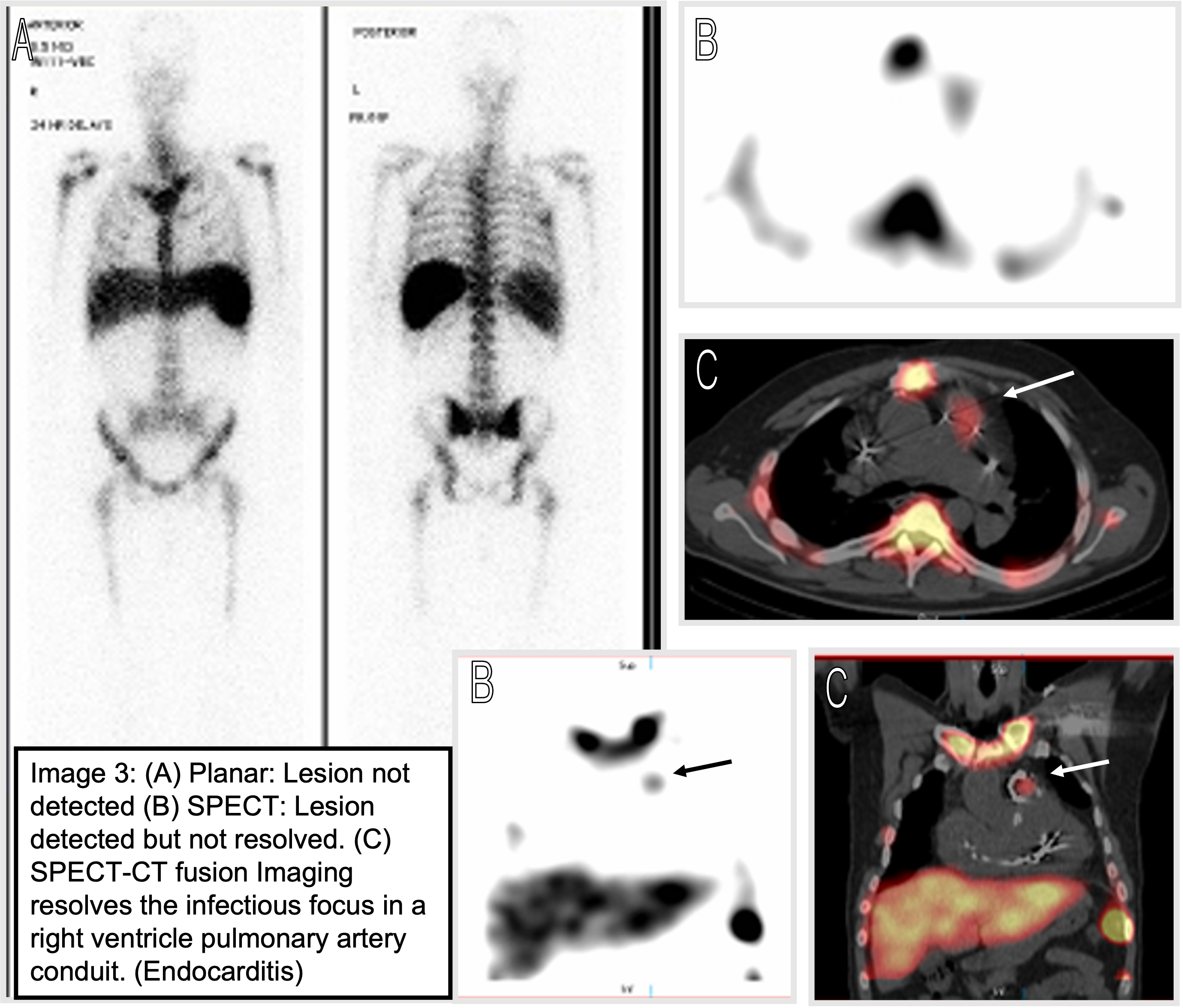
  3. Endocarditis: FDG PET scans, labeled WBC scans, gallium 67, and monoclonal antibody scans have also been used effectively in assessing native valve and prosthetic valve endocarditis with high accuracy.[42][44] They have proven to exhibit higher sensitivity and show earlier changes compared to CT. It is essential to use a SPECT-CT technique to increase the accuracy of non-PET radiopharmaceuticals. FDG PET scans are of added value to the modified Duke criteria when confirming suspected endocarditis.[45]
  4. Osteomyelitis and soft tissue infections: FDG PET scans have been added recently successfully to the armamentarium of musculoskeletal imaging infections.[46][47][22] Attention should be placed on the potential for false positives close to surgery. Bone scans with SPECT and PET are highly sensitive techniques and have high negative predictive values. This specificity is of the highest benefit in native bone with no instrumentation and no implants. A negative bone PET or SPECT study completely excludes osteomyelitis. However, in the presence of an implant or status post osseous instrumentation, abnormal bony remodeling changes can persist for up to 12 months, creating a false positive pattern. The use of SPECT-CT techniques can help localize the infection in case of hardware to a specific screw or an element of the hardware that can be targetted instead of replacing the entire hardware apparatus. A three-phase bone SPECT or PET study can be performed to evaluate the soft tissue component and bone elements.[48][49] Dynamic imaging has also been explored with FDG PET in assessing post-surgical animal models of osteomyelitis and diagnosing chronic osteomyelitis following trauma.[50][51] Dual time point and dual/combined radiotracer PET scans with FDG and Fluoride have shown high accuracy in guiding the management of orthopedic surgical sites and chronic osteomyelitis in defining the culprit sequestrum for resection.[52][53] Labeled WBC scans, gallium 67, and monoclonal antibody scans have shown value in assessing bony infections.[54][43][14][12][40] This can aid in distinguishing between bone marrow changes and cortical bone foci and also reduce the need for correlative bone marrow scans. 
  5. Diabetic foot and Charcot joint: Diagnosis can be challenging in patients with Charcot arthropathy. Diabetes is a frequent cause of Charcot's foot. However, even in the absence of Charcot arthropathy, diagnosing an infection in a diabetic foot can be challenging. The diabetic foot can have open sores and changes that can be chronic and either get acutely infected superficially or extend deep into the bone. Accurate clinical and imaging evaluation is paramount to guide management properly. Labeled WBC scans have shown similar accuracy to MRI in diagnosing foot osteomyelitis when using SPECT-CT techniques in the diabetic foot.[54] A recent study evaluating different nuclear medicine modalities found similar sensitivity; however, specificity was best obtained with Tc-HMPAO-labeled WBC and 18-F-FDG PET scans.[55] Gallium-67, on the other hand, shows a low sensitivity at 44% and a specificity of 77% in diabetic foot osteomyelitis diagnosis.[56] Charcot joint infections pose a serious clinical diagnostic dilemma as they frequently have chronic changes on morphological and functional imaging. 18-F-FDG PET scans have been shown to have a high sensitivity and accuracy of 100 and 93.8% compared to MRI 76.9 and 75%, respectively.[57][46][58]

Although morphological/anatomical imaging is usually the first study ordered in evaluating infectious foci, it frequently fails to address the clinical question. A variety of nuclear medicine techniques can significantly impact clinical management when used in the appropriate setting, in the appropriate timing (acute, subacute, or acute on chronic), as well as with the appropriate technique and choice of the radiopharmaceutical.

Enhancing Healthcare Team Outcomes

Nowadays, the number of patients with chronic conditions, multiple comorbidities, implanted cardiovascular devices, and musculoskeletal hardware steadily increases. With the increased complexity of the modern patient, accurate diagnosis of infections becomes challenging. To optimize outcomes, decrease complications, guide surgical and medical management, it is imperative to use the most appropriate and up-to-date nuclear medicine modalities available.

Optimization of outcomes and medical practice requires interprofessional coordination and seeking expert opinion. Nuclear medicine functional imaging techniques play a major role. Hybrid imaging techniques offer the best diagnostic accuracy. The interprofessional team must include expertise in the most advanced imaging techniques, and an expert team with a nuclear medicine physician, nuclear medicine technologist, infectious disease specialist, and other allied health practitioners is necessary. Levels [2,3,4,5]


(Click Image to Enlarge)
Endocarditis
Endocarditis
Contributed by Mehdi Djekidel, MD

(Click Image to Enlarge)
Endocarditis
Endocarditis
Contributed by Mehdi Djekidel, MD

(Click Image to Enlarge)
white blood cell scan with SPECT-CT
Contributed by Mehdi Djekidel, MD
Details

Editor:

Dawood Tafti

Updated:

5/22/2023 5:31:55 PM

References


[1]

Dietz M,Paulmier B,Berthier F,Civaia F,Mocquot F,Serrano B,Nataf V,Hugonnet F,Faraggi M, An Intravenous 100-mL Lipid Emulsion Infusion Dramatically Improves Myocardial Glucose Metabolism Extinction in Cardiac FDG PET Clinical Practice. Clinical nuclear medicine. 2021 Jun 1     [PubMed PMID: 33630808]


[2]

Gazzilli M,Albano D,Durmo R,Cerudelli E,Mesquita Tinoco C,Bertagna F,Giubbini R, Improvement of diagnostic accuracy of 18fluorine-fluorodeoxyglucose PET/computed tomography in detection of infective endocarditis using a 72-h low carbs protocol. Nuclear medicine communications. 2020 Aug     [PubMed PMID: 32404648]


[3]

Signore A, Jamar F, Israel O, Buscombe J, Martin-Comin J, Lazzeri E. Clinical indications, image acquisition and data interpretation for white blood cells and anti-granulocyte monoclonal antibody scintigraphy: an EANM procedural guideline. European journal of nuclear medicine and molecular imaging. 2018 Sep:45(10):1816-1831. doi: 10.1007/s00259-018-4052-x. Epub 2018 May 31     [PubMed PMID: 29850929]


[4]

Tagiling N,Mohd-Rohani MF,Wan-Sohaimi WF,Faisham WI,Nawi NM, Combined Techniques of Non-invasive {sup}99m{/sup}Tc-Besilesomab/{sup}99m{/sup}Tc-Sulfur Colloid with Hybrid SPECT/CT Imaging in Characterising Cellulitis from Symptomatic Perimegaprosthetic Infection: A Case Report. Malaysian orthopaedic journal. 2020 Nov;     [PubMed PMID: 33403085]

Level 3 (low-level) evidence

[5]

Frölich A,Knoll G,Bähre M,Neumann C, Diagnosis of chronic osteomyelitis by combined {sup}99m{/sup}Tc-bone and {sup}99m{/sup}Tc-anti-granulocyte scintigraphy. Nuklearmedizin. Nuclear medicine. 2016 Dec 6;     [PubMed PMID: 27922152]


[6]

Frölich A,Schwarze I,Neumann C, Polymyalgia rheumatica detected by SPECT/CT using {sup}99m{/sup}Tc-labeled monoclonal antibody Fab'-fragments. Nuklearmedizin. Nuclear medicine. 2017 Feb 14;     [PubMed PMID: 28004845]


[7]

Bouter C,Meller B,Sahlmann CO,Meller J, {sup}99m{/sup}Tc-Besilesomab-SPECT/CT in Infectious Endocarditis: Upgrade of a Forgotten Method? Frontiers in medicine. 2019;     [PubMed PMID: 30873410]


[8]

Schiavo R,Ricci A,Pontillo D,Bernardini G,Melacrinis FF,Maccafeo S, Implantable cardioverter-defibrillator lead infection detected by 99mTc-sulesomab single-photon emission computed tomography/computed tomography 'fusion' imaging. Journal of cardiovascular medicine (Hagerstown, Md.). 2009 Nov;     [PubMed PMID: 19680133]


[9]

Boudjelal A,Elmoataz A,Attallah B,Messali Z, A Novel Iterative MLEM Image Reconstruction Algorithm Based on Beltrami Filter: Application to ECT Images. Tomography (Ann Arbor, Mich.). 2021 Jul 28     [PubMed PMID: 34449726]


[10]

Nakamura Y,Kangai Y,Abe T,Nakahara Y, [Improvement of Standardized Uptake Value Accuracy in the {sup}99m{/sup}Tc Body SPECT and SPECT/CT: Optimization of the Phantom for Calculating Becquerel Calibration Factor and Correction Method]. Nihon Hoshasen Gijutsu Gakkai zasshi. 2021;     [PubMed PMID: 34544916]


[11]

Zhang R,Wang M,Zhou Y,Wang S,Shen Y,Li N,Wang P,Tan J,Meng Z,Jia Q, Impacts of acquisition and reconstruction parameters on the absolute technetium quantification of the cadmium-zinc-telluride-based SPECT/CT system: a phantom study. EJNMMI physics. 2021 Sep 26     [PubMed PMID: 34568990]


[12]

Kwok HM,Luk WH,Cheng LF,Pan NY,Chan HF,Ma JK, Gallium-67 Scan with Single Photon Emission Computed Tomography for the Evaluation and Monitoring of Infected Abdominal Aortic Aneurysms: A 10-Year Case Series. Vascular specialist international. 2021 Jun 29;     [PubMed PMID: 34183473]

Level 2 (mid-level) evidence

[13]

Kubota K,Tanaka N,Miyata Y,Ohtsu H,Nakahara T,Sakamoto S,Kudo T,Nishiyama Y,Tateishi U,Murakami K,Nakamoto Y,Taki Y,Kaneta T,Kawabe J,Nagamachi S,Kawano T,Hatazawa J,Mizutani Y,Baba S,Kirii K,Yokoyama K,Okamura T,Kameyama M,Minamimoto R,Kunimatsu J,Kato O,Yamashita H,Kaneko H,Kutsuna S,Ohmagari N,Hagiwara A,Kikuchi Y,Kobayakawa M, Comparison of {sup}18{/sup}F-FDG PET/CT and {sup}67{/sup}Ga-SPECT for the diagnosis of fever of unknown origin: a multicenter prospective study in Japan. Annals of nuclear medicine. 2021 Jan     [PubMed PMID: 33037581]


[14]

Kimura Y,Seguchi O,Mochizuki H,Iwasaki K,Toda K,Kumai Y,Kuroda K,Nakajima S,Tateishi E,Watanabe T,Matsumoto Y,Fukushima S,Kiso K,Yanase M,Fujita T,Kobayashi J,Fukushima N, Role of Gallium-SPECT-CT in the Management of Patients With Ventricular Assist Device-Specific Percutaneous Driveline Infection. Journal of cardiac failure. 2019 Oct;     [PubMed PMID: 31454687]


[15]

Jamar F,Buscombe J,Chiti A,Christian PE,Delbeke D,Donohoe KJ,Israel O,Martin-Comin J,Signore A, EANM/SNMMI guideline for 18F-FDG use in inflammation and infection. Journal of nuclear medicine : official publication, Society of Nuclear Medicine. 2013 Apr     [PubMed PMID: 23359660]


[16]

Bombardieri E,Aktolun C,Baum RP,Bishof-Delaloye A,Buscombe J,Chatal JF,Maffioli L,Moncayo R,Mortelmans L,Reske SN, 67Ga scintigraphy: procedure guidelines for tumour imaging. European journal of nuclear medicine and molecular imaging. 2003 Dec;     [PubMed PMID: 14989225]


[17]

Seabold JE,Palestro CJ,Brown ML,Datz FL,Forstrom LA,Greenspan BS,McAfee JG,Schauwecker DS,Royal HD, Procedure guideline for gallium scintigraphy in inflammation. Society of Nuclear Medicine. Journal of nuclear medicine : official publication, Society of Nuclear Medicine. 1997 Jun;     [PubMed PMID: 9189159]


[18]

Al-Suqri B,Al-Bulushi N, Gallium-67 Scintigraphy in the Era of Positron Emission Tomography and Computed Tomography: Tertiary centre experience. Sultan Qaboos University medical journal. 2015 Aug;     [PubMed PMID: 26357553]


[19]

Djekidel M,Brown RK,Piert M, Benefits of hybrid SPECT/CT for (111)In-oxine- and Tc-99m-hexamethylpropylene amine oxime-labeled leukocyte imaging. Clinical nuclear medicine. 2011 Jul     [PubMed PMID: 21637042]


[20]

Richter WS,Ivancevic V,Meller J,Lang O,Le Guludec D,Szilvazi I,Amthauer H,Chossat F,Dahmane A,Schwenke C,Signore A, 99mTc-besilesomab (Scintimun) in peripheral osteomyelitis: comparison with 99mTc-labelled white blood cells. European journal of nuclear medicine and molecular imaging. 2011 May;     [PubMed PMID: 21321791]


[21]

Losman MJ,DeJager RL,Monestier M,Sharkey RM,Goldenberg DM, Human immune response to anti-carcinoembryonic antigen murine monoclonal antibodies. Cancer research. 1990 Feb 1;     [PubMed PMID: 2297721]


[22]

Gemmel F,Van den Broeck B,Vanelstraete S,Van Innis B,Huysse W, Hybrid imaging of complicating osteomyelitis in the peripheral skeleton. Nuclear medicine communications. 2021 Sep 1;     [PubMed PMID: 33852533]


[23]

Gratz S,Reize P,Pfestroff A,Höffken H, Intact versus fragmented 99mTc-monoclonal antibody imaging of infection in patients with septically loosened total knee arthroplasty. The Journal of international medical research. 2012;     [PubMed PMID: 22971485]


[24]

Jacobs J,Coorevits L,Verscuren R,Frans G,Schrijvers R,Breynaert C, Severe Anaphylaxis on Murine Antibodies in Sulesomab. Journal of investigational allergology     [PubMed PMID: 33541849]


[25]

Li Q, Tian R, Sun X. More Evidence Is Warranted to Establish the Role of 18FDG-PET/CT in Fever of Unknown Origin (FUO) Investigations Among Children. Clinical infectious diseases : an official publication of the Infectious Diseases Society of America. 2021 Nov 2:73(9):e2842-e2844. doi: 10.1093/cid/ciaa1574. Epub     [PubMed PMID: 33064795]


[26]

Pijl JP, Kwee TC, Legger GE, Peters HJH, Armbrust W, Schölvinck EH, Glaudemans AWJM. Role of FDG-PET/CT in children with fever of unknown origin. European journal of nuclear medicine and molecular imaging. 2020 Jun:47(6):1596-1604. doi: 10.1007/s00259-020-04707-z. Epub 2020 Feb 7     [PubMed PMID: 32030452]


[27]

Shimizu M, Ikawa Y, Mizuta M, Takakura M, Inoue N, Nishimura R, Yachie A. FDG-PET in macrophage activation syndrome associated with systemic juvenile idiopathic arthritis. Pediatrics international : official journal of the Japan Pediatric Society. 2017 Apr:59(4):509-511. doi: 10.1111/ped.13238. Epub     [PubMed PMID: 28401744]


[28]

Shimizu M,Saikawa Y,Yachie A, Role of 18-fluoro-2-deoxyglucose positron emission tomography in detecting acute inflammatory lesions of non-bacterial osteitis in patients with a fever of unknown origin: A comparative study of 18-fluoro-2-deoxyglucose positron emission tomography, bone scan, and magnetic resonance imaging. Modern rheumatology. 2018 Nov;     [PubMed PMID: 27321232]

Level 2 (mid-level) evidence

[29]

Garg G, DaSilva R, Bhalakia A, Milstein DM. Utility of fluorine-18-fluorodeoxyglucose positron emission tomography/computed tomography in a child with chronic granulomatous disease. Indian journal of nuclear medicine : IJNM : the official journal of the Society of Nuclear Medicine, India. 2016 Jan-Mar:31(1):62-4. doi: 10.4103/0972-3919.172366. Epub     [PubMed PMID: 26917900]


[30]

Chang L, Cheng MF, Jou ST, Ko CL, Huang JY, Tzen KY, Yen RF. Search of Unknown Fever Focus Using PET in Critically Ill Children With Complicated Underlying Diseases. Pediatric critical care medicine : a journal of the Society of Critical Care Medicine and the World Federation of Pediatric Intensive and Critical Care Societies. 2016 Feb:17(2):e58-65. doi: 10.1097/PCC.0000000000000601. Epub     [PubMed PMID: 26649939]


[31]

del Rosal T,Goycochea WA,Méndez-Echevarría A,García-Fernández de Villalta M,Baquero-Artigao F,Coronado M,Marín MD,Albajara L, ¹⁸F-FDG PET/CT in the diagnosis of occult bacterial infections in children. European journal of pediatrics. 2013 Aug;     [PubMed PMID: 23479196]


[32]

Jasper N,Däbritz J,Frosch M,Loeffler M,Weckesser M,Foell D, Diagnostic value of [(18)F]-FDG PET/CT in children with fever of unknown origin or unexplained signs of inflammation. European journal of nuclear medicine and molecular imaging. 2010 Jan     [PubMed PMID: 19526234]


[33]

Kawamura J,Ueno K,Taimura E,Matsuba T,Imoto Y,Jinguji M,Kawano Y, Case Report: {sup}18{/sup}F-FDG PET-CT for Diagnosing Prosthetic Device-Related Infection in an Infant With CHD. Frontiers in pediatrics. 2021     [PubMed PMID: 33763393]

Level 3 (low-level) evidence

[34]

Aslanidis IP, Pursanova DM, Mukhortova OV, Shurupova IV, Ekaeva IV, Arakelyan VS, Golukhova EZ, Mironenko VA, Garmanov SV, Popov DA. [18F-fluorodeoxyglucose PET/CT in the diagnosis of vascular graft infection]. Khirurgiia. 2021:(2):58-66. doi: 10.17116/hirurgia202102158. Epub     [PubMed PMID: 33570356]


[35]

Jiménez D, Soto ME, Martínez-Martínez LA, Hernández-López D, Lerma C, Barragán-Garfias JA, Pérez-Torres I, Hernández-Lemus E, Guarner-Lans V. Assessment of inflammatory activity in Takayasu's arteritis: performance of clinical scores and common biomarkers versus 18F-FDG PET/CT. Clinical and experimental rheumatology. 2021 Sep-Oct:39(5):1011-1020. doi: 10.55563/clinexprheumatol/a71bzh. Epub 2020 Oct 29     [PubMed PMID: 33124558]


[36]

Treglia G. Diagnostic Performance of (18)F-FDG PET/CT in Infectious and Inflammatory Diseases according to Published Meta-Analyses. Contrast media & molecular imaging. 2019:2019():3018349. doi: 10.1155/2019/3018349. Epub 2019 Jul 25     [PubMed PMID: 31427907]


[37]

Chen W, Sajadi MM, Dilsizian V. Merits of FDG PET/CT and Functional Molecular Imaging Over Anatomic Imaging With Echocardiography and CT Angiography for the Diagnosis of Cardiac Device Infections. JACC. Cardiovascular imaging. 2018 Nov:11(11):1679-1691. doi: 10.1016/j.jcmg.2018.08.026. Epub     [PubMed PMID: 30409329]


[38]

Diemberger I, Bonfiglioli R, Martignani C, Graziosi M, Biffi M, Lorenzetti S, Ziacchi M, Nanni C, Fanti S, Boriani G. Contribution of PET imaging to mortality risk stratification in candidates to lead extraction for pacemaker or defibrillator infection: a prospective single center study. European journal of nuclear medicine and molecular imaging. 2019 Jan:46(1):194-205. doi: 10.1007/s00259-018-4142-9. Epub 2018 Sep 8     [PubMed PMID: 30196365]


[39]

Blanc P,Bonnet E,Giordano G,Monteil J,Salabert AS,Payoux P, The use of labelled leucocyte scintigraphy to evaluate chronic periprosthetic joint infections: a retrospective multicentre study on 168 patients. European journal of clinical microbiology     [PubMed PMID: 31218592]

Level 2 (mid-level) evidence

[40]

Loessel C,Mai A,Starke M,Vogt D,Stichling M,Willy C, Value of antigranulocyte scintigraphy with Tc-99m-sulesomab in diagnosing combat-related infections of the musculoskeletal system. BMJ military health. 2021 Feb;     [PubMed PMID: 30787111]


[41]

Albano D,Bosio G,Paghera B,Bertagna F, Comparison Between 99mTc-Sulesomab and 18F-FDG PET/CT in a Patient With Suspected Prosthetic Joint Infection. Clinical nuclear medicine. 2016 Jun;     [PubMed PMID: 27055130]


[42]

Lawal IO,Kleyhans J,Mokoala KMG,Vorster M,Ebenhan T,Zeevaart JR,Sathekge MM, Single Photon Emission Tomography in the Diagnostic Assessment of Cardiac and Vascular Infectious Diseases. Current radiopharmaceuticals. 2020 Jun 21;     [PubMed PMID: 32564768]


[43]

Puius YA,Parkar F,Tlamsa AP,Nistico JA,Muggia VA,Minamoto GY,Jorde UP,Goldstein DJ,Moadel RM, Gallium-67 Single-Photon Emission Computed Tomography Affects Management of Infections of Left Ventricular Assist Devices. ASAIO journal (American Society for Artificial Internal Organs : 1992). 2021 Jul 1;     [PubMed PMID: 33196482]


[44]

McWilliams ET,Yavari A,Raman V, Aortic root abscess: multimodality imaging with computed tomography and gallium-67 citrate single-photon emission computed tomography/computed tomography hybrid imaging. Journal of cardiovascular computed tomography. 2011 Mar-Apr;     [PubMed PMID: 21130063]


[45]

Pretet V,Blondet C,Ruch Y,Martinez M,El Ghannudi S,Morel O,Hansmann Y,Schindler TH,Imperiale A, Advantages of 18F-FDG PET/CT Imaging over Modified Duke Criteria and Clinical Presumption in Patients with Challenging Suspicion of Infective Endocarditis. Diagnostics (Basel, Switzerland). 2021 Apr 18;     [PubMed PMID: 33919643]


[46]

Diez AIG,Fuster D,Morata L,Torres F,Garcia R,Poggio D,Sotes S,Del Amo M,Isern-Kebschull J,Pomes J,Soriano A,Brugnara L,Tomas X, Comparison of the diagnostic accuracy of diffusion-weighted and dynamic contrast-enhanced MRI with {sup}18{/sup}F-FDG PET/CT to differentiate osteomyelitis from Charcot neuro-osteoarthropathy in diabetic foot. European journal of radiology. 2020 Nov;     [PubMed PMID: 33032207]


[47]

Casali M,Lauri C,Altini C,Bertagna F,Cassarino G,Cistaro A,Erba AP,Ferrari C,Mainolfi CG,Palucci A,Prandini N,Baldari S,Bartoli F,Bartolomei M,D'Antonio A,Dondi F,Gandolfo P,Giordano A,Laudicella R,Massollo M,Nieri A,Piccardo A,Vendramin L,Muratore F,Lavelli V,Albano D,Burroni L,Cuocolo A,Evangelista L,Lazzeri E,Quartuccio N,Rossi B,Rubini G,Sollini M,Versari A,Signore A, State of the art of {sup}18{/sup}F-FDG PET/CT application in inflammation and infection: a guide for image acquisition and interpretation. Clinical and translational imaging. 2021 Jul 10;     [PubMed PMID: 34277510]


[48]

Lee SJ,Won KS,Choi HJ,Choi YY, Early-Phase SPECT/CT for Diagnosing Osteomyelitis: A Retrospective Pilot Study. Korean journal of radiology. 2021 Apr;     [PubMed PMID: 33289359]

Level 2 (mid-level) evidence

[49]

Freesmeyer M,Stecker FF,Schierz JH,Hofmann GO,Winkens T, First experience with early dynamic (18)F-NaF-PET/CT in patients with chronic osteomyelitis. Annals of nuclear medicine. 2014 May;     [PubMed PMID: 24474597]


[50]

Brown TL,Spencer HJ,Beenken KE,Alpe TL,Bartel TB,Bellamy W,Gruenwald JM,Skinner RA,McLaren SG,Smeltzer MS, Evaluation of dynamic [18F]-FDG-PET imaging for the detection of acute post-surgical bone infection. PloS one. 2012;     [PubMed PMID: 22860021]


[51]

Stecker FF,Schierz JH,Opfermann T,Driesch D,Hofmann GO,Winkens T,Freesmeyer M, Early dynamic 18F-FDG PET/CT to diagnose chronic osteomyelitis following lower extremity fractures. A pilot study. Nuklearmedizin. Nuclear medicine. 2014;     [PubMed PMID: 23780221]

Level 3 (low-level) evidence

[52]

Lee JW,Yu SN,Yoo ID,Jeon MH,Hong CH,Shim JJ,Chang SH,Lee SM, Clinical application of dual-phase F-18 sodium-fluoride bone PET/CT for diagnosing surgical site infection following orthopedic surgery. Medicine. 2019 Mar;     [PubMed PMID: 30882648]


[53]

Christersson A,Larsson S,Sörensen J, Presurgical localization of infected avascular bone segments in chronic complicated posttraumatic osteomyelitis in the lower extremity using dual-tracer PET/CT. EJNMMI research. 2018 Jul 21;     [PubMed PMID: 30032355]


[54]

La Fontaine J,Bhavan K,Lam K,Van Asten S,Erdman W,Lavery LA,Öz OK, Comparison Between Tc-99m WBC SPECT/CT and MRI for the Diagnosis of Biopsy-proven Diabetic Foot Osteomyelitis. Wounds : a compendium of clinical research and practice. 2016 Aug;     [PubMed PMID: 27560470]


[55]

Lauri C,Tamminga M,Glaudemans AWJM,Juárez Orozco LE,Erba PA,Jutte PC,Lipsky BA,IJzerman MJ,Signore A,Slart RHJA, Detection of Osteomyelitis in the Diabetic Foot by Imaging Techniques: A Systematic Review and Meta-analysis Comparing MRI, White Blood Cell Scintigraphy, and FDG-PET. Diabetes care. 2017 Aug;     [PubMed PMID: 28733376]

Level 1 (high-level) evidence

[56]

Delcourt A,Huglo D,Prangere T,Benticha H,Devemy F,Tsirtsikoulou D,Lepeut M,Fontaine P,Steinling M, Comparison between Leukoscan (Sulesomab) and Gallium-67 for the diagnosis of osteomyelitis in the diabetic foot. Diabetes     [PubMed PMID: 15959418]


[57]

Heidari N,Oh I,Li Y,Vris A,Kwok I,Charalambous A,Rogero R, What Is the Best Method to Differentiate Acute Charcot Foot From Acute Infection? Foot     [PubMed PMID: 31322932]


[58]

Basu S, Chryssikos T, Houseni M, Scot Malay D, Shah J, Zhuang H, Alavi A. Potential role of FDG PET in the setting of diabetic neuro-osteoarthropathy: can it differentiate uncomplicated Charcot's neuroarthropathy from osteomyelitis and soft-tissue infection? Nuclear medicine communications. 2007 Jun:28(6):465-72     [PubMed PMID: 17460537]