Learning Outcome
- List the causes of heart block
- Describe the presentation of heart block
- Recall the types of heart block
- Summarize the treatment of heart block
Atrioventricular (AV) conduction is evaluated by assessing the relationship between the P waves and QRS complexes. Normally, there is a P wave that precedes each QRS complex by a fixed PR interval of 120 to 200 milliseconds. AV block represents a delayed electrical impulse from the atria to the ventricles. This can be due to an anatomical or functional impairment in the heart’s conduction system. This disruption in normal electrical activity can be transient or permanent. In general, there are three degrees of AV nodal blocks: first degree, second degree (Mobitz type 1 or 2), and third-degree.[1][2][3]
At this time, there is no well-characterized large study about the relationship between different types of AV block with age, race, or gender. AV block is sometimes seen in athletes and in patients with congenital heart disorders.
Higher degrees of AV block often suggest some underlying pathology. This is known as a pathophysiologic AV block. About half of such cases are a result of chronic idiopathic fibrosis and sclerosis of the conduction system.
Another common source is ischemic heart disease which is responsible for around 40 percent of cases of AV block [4].
AV block is also associated with cardiomyopathies, including hypertrophic obstructive cardiomyopathy and infiltrative conditions such as sarcoidosis and amyloidosis. Infectious causes such as Lyme disease, rheumatic fever, endocarditis, viruses as well as autoimmune disease such as systemic lupus erythematosus should also be explored [5][6][7][8].
Other potential triggers include cardiac surgery, medications, and inherited conditions [9].
First-degree AV block can originate from various locations within the conduction system. The levels of conduction delay include the atrium, AV node (most common in first-degree heart block), Bundle of His, bundle branches, fascicles, Purkinje system. Mobitz type I second-degree AV block usually occurs within the AV node, while Mobitz type II second-degree AV block mainly originates from conduction system disease below the level of the AV node (in the bundle of His and in the bundle branches). In third-degree AV block, no atrial impulses reach the ventricle- it can occur in the AV node or in the infranodal specialized conduction system. [10]
The following medications can affect different levels of conduction delay:
History taking for patients with concerns for AV block should include:
The following symptoms should raise concerns:
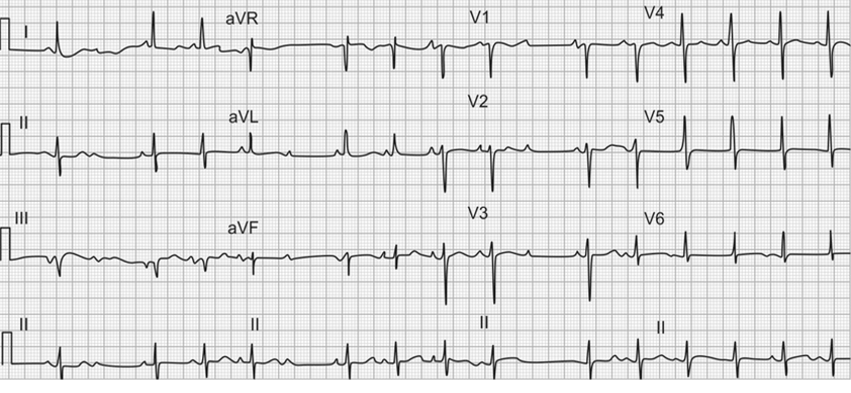
First degree. In first-degree AV block, the P waves always precede the QRS complexes, but there is a prolongation of the PR interval. The PR interval will be greater than 200 milliseconds in duration without any dropped beats. There is a delay, without interruption, in conduction from the atrium to the ventricle. All atrial activation is eventually transmitted to the ventricles. The delay is typically due to a minor AV conduction defect occurring at or below the AV node.
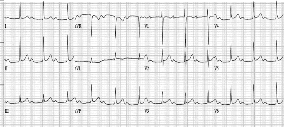
Second degree, Mobitz type 1 (Wenckebach). In second-degree Mobitz type 1 AV block, there is a progressive prolongation of the PR interval, which eventually culminates in a non-conducting P wave. The PR interval continues to prolong with each beat of the cycle, and the subsequent PR lengthening is progressively shorter. The PR interval before the dropped beat is the longest of the cycle, and the PR interval after the dropped beat is the shortest as the cycle starts over.
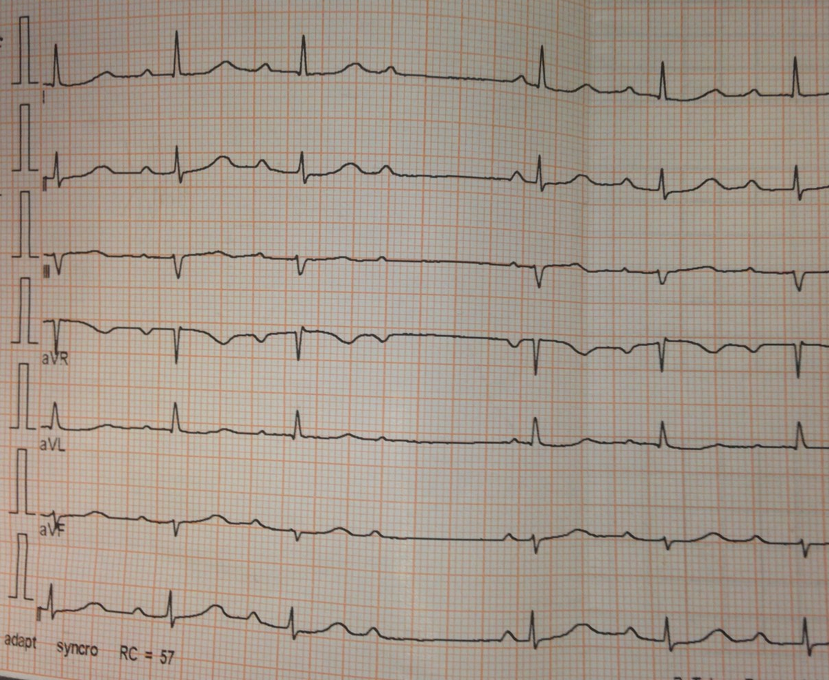
Second degree, Mobitz type 2. In second-degree Mobitz type 2 AV block, there are intermittent non-conducted P waves without warning. Unlike Mobitz type 1 (Wenckebach), there is no progressive prolongation of the PR interval; instead, the PR interval remains constant, and the P waves occur at a constant rate with unchanged P-P intervals. Because the P waves continue to occur at normal intervals, the R-R interval surrounding the dropped beat is simply a multiple of the preceding R-R interval and remains unchanged.
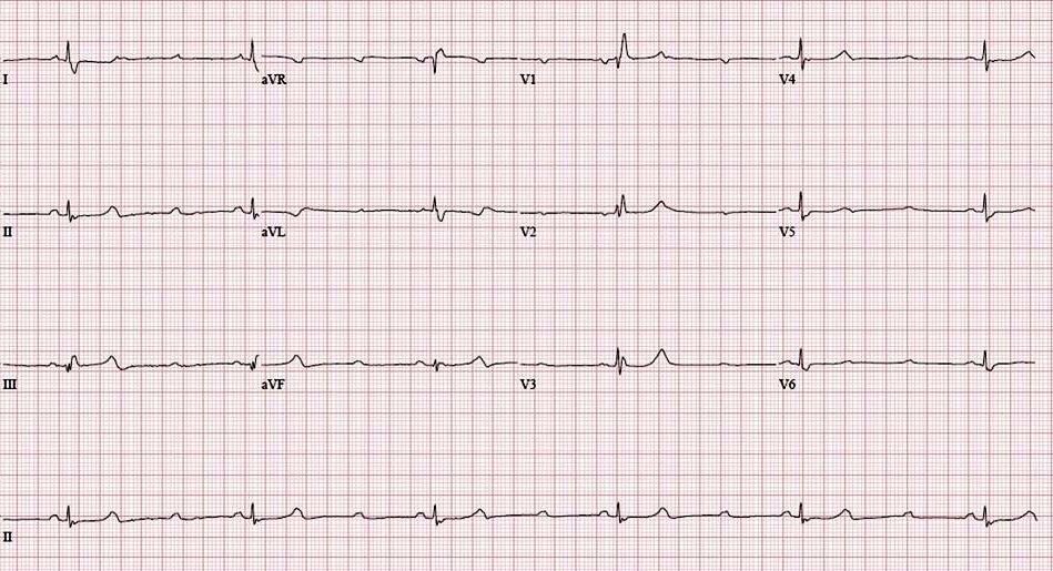
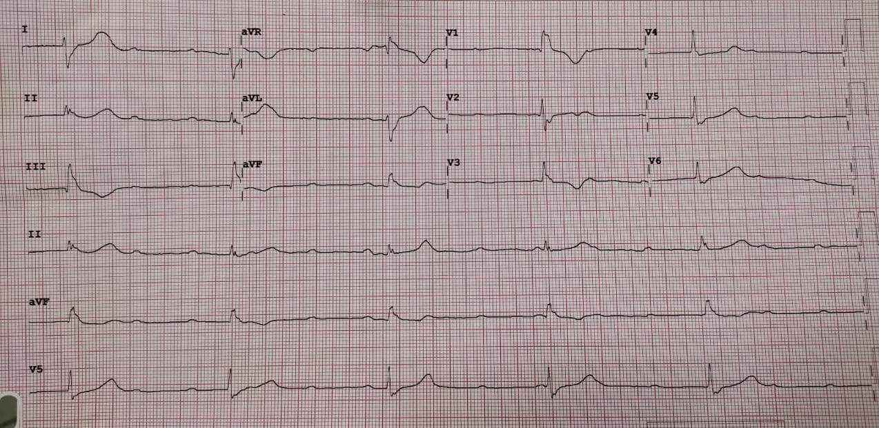
Third-degree (complete). In third-degree, or complete, heart block there is an absence of AV nodal conduction, and the P waves are never related to the QRS complexes. If ventricular conduction occurs, it is maintained by a junctional or ventricular escape rhythm. There is a complete dissociation between the atria and ventricles and they conduct independently of each other. The P waves (atrial activity) are said to “march through” the QRS complexes at their regular, faster rate. The QRS complexes (ventricular activity) also occur at a regular, but slower rate. There are two independent rhythms occurring simultaneously.
In general, patients that present with first-degree or second-degree Mobitz type 1 AV block do not require treatment. Any provoking medications can be removed, and patients can be monitored on an outpatient basis. However, patients with higher degrees of AV block (Mobitz type 2 AV block, 3rd degree) tend to have severe damage to the conduction system. They are at a much greater risk of progressing into asystole, ventricular tachycardia, or sudden cardiac death. Hence, they require urgent admission for cardiac monitoring, backup temporary cardiac pacing, and insertion of a permanent pacemaker.[11][12][13][14]
Prognosis depends on the various factors that include age and other chronic medical conditions such as diabetes mellitus, chronic kidney disease, underlying heart disease, and underlying types of AV block.
The management of heart block is best done with an interprofessional team because if the diagnosis is missed (esp higher degrees of heart block), the condition can have significant morbidity and mortality.
Except for a first-degree heart block, the rest of the patients should be referred to a cardiologist for a more definitive workup. Some of these patients may require a pacemaker which can be life-saving. Following treatment, the cardiology nurse should follow up on the patients to ensure that the heart rate has normalized and the patients have no symptoms.[15]
Anytime patients with a pacemaker undergo surgery, the cardiologist should be consulted first.
Patients with first-degree and asymptomatic Mobitz type 1 AV block usually can continue their usual activities but should be advised to avoid medications that can prolong the PR interval. Patients with Mobitz type 2 and third-degree AV block should discuss with their cardiologists about the need for pacemakers. All patients should be educated on alarming symptoms of hypoperfusion such as fatigue, lightheadedness, syncope, presyncope, or angina and seek timely medical treatment
Żuchowski B, Błaszyk K, Piskorski J, Wykrętowicz A, Guzik P. Dependence of the Atrioventricular Conduction Time on the Conduction through the Atrioventricular Node and His-Purkinje System. Journal of clinical medicine. 2023 Feb 7:12(4):. doi: 10.3390/jcm12041330. Epub 2023 Feb 7 [PubMed PMID: 36835864]
Wu J, Zipes DP. Mechanisms underlying atrioventricular nodal conduction and the reentrant circuit of atrioventricular nodal reentrant tachycardia using optical mapping. Journal of cardiovascular electrophysiology. 2002 Aug:13(8):831-4 [PubMed PMID: 12212708]
Prystowsky EN, Jackman WM, Rinkenberger RL, Heger JJ, Zipes DP. Effect of autonomic blockade on ventricular refractoriness and atrioventricular nodal conduction in humans. Evidence supporting a direct cholinergic action on ventricular muscle refractoriness. Circulation research. 1981 Aug:49(2):511-8 [PubMed PMID: 7249285]
Kwok CS, Rashid M, Beynon R, Barker D, Patwala A, Morley-Davies A, Satchithananda D, Nolan J, Myint PK, Buchan I, Loke YK, Mamas MA. Prolonged PR interval, first-degree heart block and adverse cardiovascular outcomes: a systematic review and meta-analysis. Heart (British Cardiac Society). 2016 May:102(9):672-80. doi: 10.1136/heartjnl-2015-308956. Epub 2016 Feb 15 [PubMed PMID: 26879241]
Epstein AE, DiMarco JP, Ellenbogen KA, Estes NA 3rd, Freedman RA, Gettes LS, Gillinov AM, Gregoratos G, Hammill SC, Hayes DL, Hlatky MA, Newby LK, Page RL, Schoenfeld MH, Silka MJ, Stevenson LW, Sweeney MO, Smith SC Jr, Jacobs AK, Adams CD, Anderson JL, Buller CE, Creager MA, Ettinger SM, Faxon DP, Halperin JL, Hiratzka LF, Hunt SA, Krumholz HM, Kushner FG, Lytle BW, Nishimura RA, Ornato JP, Page RL, Riegel B, Tarkington LG, Yancy CW, American College of Cardiology/American Heart Association Task Force on Practice Guidelines (Writing Committee to Revise the ACC/AHA/NASPE 2002 Guideline Update for Implantation of Cardiac Pacemakers and Antiarrhythmia Devices), American Association for Thoracic Surgery, Society of Thoracic Surgeons. ACC/AHA/HRS 2008 Guidelines for Device-Based Therapy of Cardiac Rhythm Abnormalities: a report of the American College of Cardiology/American Heart Association Task Force on Practice Guidelines (Writing Committee to Revise the ACC/AHA/NASPE 2002 Guideline Update for Implantation of Cardiac Pacemakers and Antiarrhythmia Devices) developed in collaboration with the American Association for Thoracic Surgery and Society of Thoracic Surgeons. Journal of the American College of Cardiology. 2008 May 27:51(21):e1-62. doi: 10.1016/j.jacc.2008.02.032. Epub [PubMed PMID: 18498951]
Sheldon RS, Lei LY, Solbiati M, Chew DS, Raj SR, Costantino G, Morillo C, Sandhu RK. Electrophysiology studies for predicting atrioventricular block in patients with syncope: A systematic review and meta-analysis. Heart rhythm. 2021 Aug:18(8):1310-1317. doi: 10.1016/j.hrthm.2021.04.010. Epub 2021 Apr 20 [PubMed PMID: 33887450]
Dhingra RC, Amat-y-Leon F, Pouget JM, Rosen KM. Infranodal block: diagnosis, clinical significance, and management. The Medical clinics of North America. 1976 Jan:60(1):175-92 [PubMed PMID: 1107691]
Warnes CA. Transposition of the great arteries. Circulation. 2006 Dec 12:114(24):2699-709 [PubMed PMID: 17159076]
Karpawich PP, Gillette PC, Garson A Jr, Hesslein PS, Porter CB, McNamara DG. Congenital complete atrioventricular block: clinical and electrophysiologic predictors of need for pacemaker insertion. The American journal of cardiology. 1981 Dec:48(6):1098-102 [PubMed PMID: 7304459]
Ambrosi A, Wahren-Herlenius M. Congenital heart block: evidence for a pathogenic role of maternal autoantibodies. Arthritis research & therapy. 2012 Apr 26:14(2):208. doi: 10.1186/ar3787. Epub 2012 Apr 26 [PubMed PMID: 22546326]
Buyon JP, Hiebert R, Copel J, Craft J, Friedman D, Katholi M, Lee LA, Provost TT, Reichlin M, Rider L, Rupel A, Saleeb S, Weston WL, Skovron ML. Autoimmune-associated congenital heart block: demographics, mortality, morbidity and recurrence rates obtained from a national neonatal lupus registry. Journal of the American College of Cardiology. 1998 Jun:31(7):1658-66 [PubMed PMID: 9626848]
Ye Z, Wang L, Yang T, Chen L, Wang T, Chen L, Zhao L, Zhang S, Zheng Z, Luo L, Qin J. Maternal Viral Infection and Risk of Fetal Congenital Heart Diseases: A Meta-Analysis of Observational Studies. Journal of the American Heart Association. 2019 May 7:8(9):e011264. doi: 10.1161/JAHA.118.011264. Epub [PubMed PMID: 30995883]
Barra SN, Providência R, Paiva L, Nascimento J, Marques AL. A review on advanced atrioventricular block in young or middle-aged adults. Pacing and clinical electrophysiology : PACE. 2012 Nov:35(11):1395-405. doi: 10.1111/j.1540-8159.2012.03489.x. Epub 2012 Aug 16 [PubMed PMID: 22897386]
LENEGRE J. ETIOLOGY AND PATHOLOGY OF BILATERAL BUNDLE BRANCH BLOCK IN RELATION TO COMPLETE HEART BLOCK. Progress in cardiovascular diseases. 1964 Mar:6():409-44 [PubMed PMID: 14153648]
LEV M. ANATOMIC BASIS FOR ATRIOVENTRICULAR BLOCK. The American journal of medicine. 1964 Nov:37():742-8 [PubMed PMID: 14237429]
Alboni P, Holz A, Brignole M. Vagally mediated atrioventricular block: pathophysiology and diagnosis. Heart (British Cardiac Society). 2013 Jul:99(13):904-8. doi: 10.1136/heartjnl-2012-303220. Epub 2013 Jan 2 [PubMed PMID: 23286970]
Kottkamp H. Fibrotic atrial cardiomyopathy: a specific disease/syndrome supplying substrates for atrial fibrillation, atrial tachycardia, sinus node disease, AV node disease, and thromboembolic complications. Journal of cardiovascular electrophysiology. 2012 Jul:23(7):797-9. doi: 10.1111/j.1540-8167.2012.02341.x. Epub 2012 May 3 [PubMed PMID: 22554187]
Rajdev A, Groh WJ. Arrhythmias in the muscular dystrophies. Cardiac electrophysiology clinics. 2015 Jun:7(2):303-8. doi: 10.1016/j.ccep.2015.03.011. Epub 2015 Mar 29 [PubMed PMID: 26002394]
Ferrari AD, Süssenbach CP, Guaragna JC, Piccoli Jda C, Gazzoni GF, Ferreira DK, Albuquerque LC, Goldani MA. Atrioventricular block in the postoperative period of heart valve surgery: incidence, risk factors and hospital evolution. Revista brasileira de cirurgia cardiovascular : orgao oficial da Sociedade Brasileira de Cirurgia Cardiovascular. 2011 Jul-Sep:26(3):364-72 [PubMed PMID: 22086572]
Siontis GC, Jüni P, Pilgrim T, Stortecky S, Büllesfeld L, Meier B, Wenaweser P, Windecker S. Predictors of permanent pacemaker implantation in patients with severe aortic stenosis undergoing TAVR: a meta-analysis. Journal of the American College of Cardiology. 2014 Jul 15:64(2):129-40. doi: 10.1016/j.jacc.2014.04.033. Epub [PubMed PMID: 25011716]
Chandler SF, Fynn-Thompson F, Mah DY. Role of cardiac pacing in congenital complete heart block. Expert review of cardiovascular therapy. 2017 Nov:15(11):853-861. doi: 10.1080/14779072.2017.1376655. Epub 2017 Sep 13 [PubMed PMID: 28875729]
Upshaw CB Jr. Comparison of the prevalence of first-degree atrioventricular block in African-American and in Caucasian patients: an electrocardiographic study III. Journal of the National Medical Association. 2004 Jun:96(6):756-60 [PubMed PMID: 15233485]
Moak JP, Barron KS, Hougen TJ, Wiles HB, Balaji S, Sreeram N, Cohen MH, Nordenberg A, Van Hare GF, Friedman RA, Perez M, Cecchin F, Schneider DS, Nehgme RA, Buyon JP. Congenital heart block: development of late-onset cardiomyopathy, a previously underappreciated sequela. Journal of the American College of Cardiology. 2001 Jan:37(1):238-42 [PubMed PMID: 11153745]
Bourke JP. Atrioventricular block and problems with atrioventricular conduction. Clinics in geriatric medicine. 2002 May:18(2):229-51 [PubMed PMID: 12180245]
Abu Rmilah AA, Al-Zu'bi H, Haq IU, Yagmour AH, Jaber SA, Alkurashi AK, Qaisi I, Kowlgi GN, Cha YM, Mulpuru S, DeSimone CV, Deshmukh AJ. Predicting permanent pacemaker implantation following transcatheter aortic valve replacement: A contemporary meta-analysis of 981,168 patients. Heart rhythm O2. 2022 Aug:3(4):385-392. doi: 10.1016/j.hroo.2022.05.001. Epub 2022 May 12 [PubMed PMID: 36097458]
Writing Committee Members, Kusumoto FM, Schoenfeld MH, Barrett C, Edgerton JR, Ellenbogen KA, Gold MR, Goldschlager NF, Hamilton RM, Joglar JA, Kim RJ, Lee R, Marine JE, McLeod CJ, Oken KR, Patton KK, Pellegrini CN, Selzman KA, Thompson A, Varosy PD. 2018 ACC/AHA/HRS guideline on the evaluation and management of patients with bradycardia and cardiac conduction delay: A Report of the American College of Cardiology/American Heart Association Task Force on Clinical Practice Guidelines and the Heart Rhythm Society. Heart rhythm. 2019 Sep:16(9):e128-e226. doi: 10.1016/j.hrthm.2018.10.037. Epub 2018 Nov 6 [PubMed PMID: 30412778]
Zeltser D, Justo D, Halkin A, Rosso R, Ish-Shalom M, Hochenberg M, Viskin S. Drug-induced atrioventricular block: prognosis after discontinuation of the culprit drug. Journal of the American College of Cardiology. 2004 Jul 7:44(1):105-8 [PubMed PMID: 15234417]
Glikson M, Nielsen JC, Kronborg MB, Michowitz Y, Auricchio A, Barbash IM, Barrabés JA, Boriani G, Braunschweig F, Brignole M, Burri H, Coats AJS, Deharo JC, Delgado V, Diller GP, Israel CW, Keren A, Knops RE, Kotecha D, Leclercq C, Merkely B, Starck C, Thylén I, Tolosana JM. Corrigendum to: 2021 ESC Guidelines on cardiac pacing and cardiac resynchronization therapy: Developed by the Task Force on cardiac pacing and cardiac resynchronization therapy of the European Society of Cardiology (ESC): With the special contribution of the European Heart Rhythm Association (EHRA). Europace : European pacing, arrhythmias, and cardiac electrophysiology : journal of the working groups on cardiac pacing, arrhythmias, and cardiac cellular electrophysiology of the European Society of Cardiology. 2022 Apr 5:24(4):699. doi: 10.1093/europace/euac023. Epub [PubMed PMID: 35253863]
Knabben V, Chhabra L, Slane M. Third-Degree Atrioventricular Block. StatPearls. 2025 Jan:(): [PubMed PMID: 31424783]
STACK MF, RADER B, SOBOL BJ, FARBER SJ, EICHNA LW. Cardiovascular hemodynamic functions in complete heart block and the effect of isopropylnorepinephrine. Circulation. 1958 Apr:17(4, Part 1):526-36 [PubMed PMID: 13523763]
Morin DP, Bernard ML. Extreme "cannon A waves" and pulsatile skin color in complete heart block. HeartRhythm case reports. 2017 Oct:3(10):493. doi: 10.1016/j.hrcr.2017.06.012. Epub 2017 Aug 7 [PubMed PMID: 29062706]
Oldroyd SH, Quintanilla Rodriguez BS, Makaryus AN. First-Degree Heart Block. StatPearls. 2024 Jan:(): [PubMed PMID: 28846254]
Lewalter T, Pürerfellner H, Ungar A, Rieger G, Mangoni L, Duru F, INSIGHT XT study investigators. "First-degree AV block-a benign entity?" Insertable cardiac monitor in patients with 1st-degree AV block reveals presence or progression to higher grade block or bradycardia requiring pacemaker implant. Journal of interventional cardiac electrophysiology : an international journal of arrhythmias and pacing. 2018 Aug:52(3):303-306. doi: 10.1007/s10840-018-0439-7. Epub 2018 Aug 13 [PubMed PMID: 30105427]
Burri H, Zimmermann M. Infranodal Wenckebach conduction block and illustration of the gap phenomenon. HeartRhythm case reports. 2021 Feb:7(2):63-64. doi: 10.1016/j.hrcr.2020.11.005. Epub 2020 Nov 13 [PubMed PMID: 33665102]
Surawicz B, Childers R, Deal BJ, Gettes LS, Bailey JJ, Gorgels A, Hancock EW, Josephson M, Kligfield P, Kors JA, Macfarlane P, Mason JW, Mirvis DM, Okin P, Pahlm O, Rautaharju PM, van Herpen G, Wagner GS, Wellens H, American Heart Association Electrocardiography and Arrhythmias Committee, Council on Clinical Cardiology, American College of Cardiology Foundation, Heart Rhythm Society. AHA/ACCF/HRS recommendations for the standardization and interpretation of the electrocardiogram: part III: intraventricular conduction disturbances: a scientific statement from the American Heart Association Electrocardiography and Arrhythmias Committee, Council on Clinical Cardiology; the American College of Cardiology Foundation; and the Heart Rhythm Society. Endorsed by the International Society for Computerized Electrocardiology. Journal of the American College of Cardiology. 2009 Mar 17:53(11):976-81. doi: 10.1016/j.jacc.2008.12.013. Epub [PubMed PMID: 19281930]
Barold SS. 2:1 Atrioventricular block: order from chaos. The American journal of emergency medicine. 2001 May:19(3):214-7 [PubMed PMID: 11326349]
Linzer M, Yang EH, Estes NA 3rd, Wang P, Vorperian VR, Kapoor WN. Diagnosing syncope. Part 1: Value of history, physical examination, and electrocardiography. Clinical Efficacy Assessment Project of the American College of Physicians. Annals of internal medicine. 1997 Jun 15:126(12):989-96 [PubMed PMID: 9182479]
Linzer M, Yang EH, Estes NA 3rd, Wang P, Vorperian VR, Kapoor WN. Diagnosing syncope. Part 2: Unexplained syncope. Clinical Efficacy Assessment Project of the American College of Physicians. Annals of internal medicine. 1997 Jul 1:127(1):76-86 [PubMed PMID: 9214258]
Coplan NL, Morales MC, Romanello P, Wilentz JR, Moses JW. Exercise-related atrioventricular block. Influence of myocardial ischemia. Chest. 1991 Dec:100(6):1728-30 [PubMed PMID: 1959424]
Fontana M, Pica S, Reant P, Abdel-Gadir A, Treibel TA, Banypersad SM, Maestrini V, Barcella W, Rosmini S, Bulluck H, Sayed RH, Patel K, Mamhood S, Bucciarelli-Ducci C, Whelan CJ, Herrey AS, Lachmann HJ, Wechalekar AD, Manisty CH, Schelbert EB, Kellman P, Gillmore JD, Hawkins PN, Moon JC. Prognostic Value of Late Gadolinium Enhancement Cardiovascular Magnetic Resonance in Cardiac Amyloidosis. Circulation. 2015 Oct 20:132(16):1570-9. doi: 10.1161/CIRCULATIONAHA.115.016567. Epub 2015 Sep 11 [PubMed PMID: 26362631]
Fisher JD. Role of electrophysiologic testing in the diagnosis and treatment of patients with known and suspected bradycardias and tachycardias. Progress in cardiovascular diseases. 1981 Jul-Aug:24(1):25-90 [PubMed PMID: 7019962]
Podoleanu C, DaCosta A, Defaye P, Taieb J, Galley D, Bru P, Maury P, Mabo P, Boveda S, Cellarier G, Anselme F, Kouakam C, Delarche N, Deharo JC, FRESH investigators. Early use of an implantable loop recorder in syncope evaluation: a randomized study in the context of the French healthcare system (FRESH study). Archives of cardiovascular diseases. 2014 Oct:107(10):546-52. doi: 10.1016/j.acvd.2014.05.009. Epub 2014 Sep 16 [PubMed PMID: 25241220]
Ackerman MJ, Priori SG, Willems S, Berul C, Brugada R, Calkins H, Camm AJ, Ellinor PT, Gollob M, Hamilton R, Hershberger RE, Judge DP, Le Marec H, McKenna WJ, Schulze-Bahr E, Semsarian C, Towbin JA, Watkins H, Wilde A, Wolpert C, Zipes DP. HRS/EHRA expert consensus statement on the state of genetic testing for the channelopathies and cardiomyopathies this document was developed as a partnership between the Heart Rhythm Society (HRS) and the European Heart Rhythm Association (EHRA). Heart rhythm. 2011 Aug:8(8):1308-39. doi: 10.1016/j.hrthm.2011.05.020. Epub [PubMed PMID: 21787999]
Glikson M, Nielsen JC, Kronborg MB, Michowitz Y, Auricchio A, Barbash IM, Barrabés JA, Boriani G, Braunschweig F, Brignole M, Burri H, Coats AJS, Deharo JC, Delgado V, Diller GP, Israel CW, Keren A, Knops RE, Kotecha D, Leclercq C, Merkely B, Starck C, Thylén I, Tolosana JM, ESC Scientific Document Group. [2021 ESC Guidelines on cardiac pacing and cardiac resynchronization therapy Developed by the Task Force on cardiac pacing and cardiac resynchronization therapy of the European Society of Cardiology (ESC) With the special contribution of the European Heart Rhythm Association (EHRA)]. Giornale italiano di cardiologia (2006). 2022 Jul:23(7 Suppl 1):e1-e94. doi: 10.1714/3824.38087. Epub [PubMed PMID: 35771031]
Brady WJ, Swart G, DeBehnke DJ, Ma OJ, Aufderheide TP. The efficacy of atropine in the treatment of hemodynamically unstable bradycardia and atrioventricular block: prehospital and emergency department considerations. Resuscitation. 1999 Jun:41(1):47-55 [PubMed PMID: 10459592]
Hickey AR, Wenger TL, Carpenter VP, Tilson HH, Hlatky MA, Furberg CD, Kirkpatrick CH, Strauss HC, Smith TW. Digoxin Immune Fab therapy in the management of digitalis intoxication: safety and efficacy results of an observational surveillance study. Journal of the American College of Cardiology. 1991 Mar 1:17(3):590-8 [PubMed PMID: 1993775]
Kennebäck G, Tabrizi F, Lindell P, Nordlander R. High-degree atrioventricular block during anti-arrhythmic drug treatment: use of a pacemaker with a bradycardia-detection algorithm to study the time course after drug withdrawal. Europace : European pacing, arrhythmias, and cardiac electrophysiology : journal of the working groups on cardiac pacing, arrhythmias, and cardiac cellular electrophysiology of the European Society of Cardiology. 2007 Mar:9(3):186-91 [PubMed PMID: 17255148]
Bjørnstad CC, Gjertsen E, Thorup F, Gundersen T, Tobiasson K, Otterstad JE. Temporary cardiac pacemaker treatment in five Norwegian regional hospitals. Scandinavian cardiovascular journal : SCJ. 2012 Jun:46(3):137-43. doi: 10.3109/14017431.2012.672763. Epub 2012 Mar 27 [PubMed PMID: 22390277]
Strasberg B, Amat-Y-Leon F, Dhingra RC, Palileo E, Swiryn S, Bauernfeind R, Wyndham C, Rosen KM. Natural history of chronic second-degree atrioventricular nodal block. Circulation. 1981 May:63(5):1043-9 [PubMed PMID: 7471363]
Barold SS. Indications for permanent cardiac pacing in first-degree AV block: class I, II, or III? Pacing and clinical electrophysiology : PACE. 1996 May:19(5):747-51 [PubMed PMID: 8734740]
Simon AB, Zloto AE. Atrioventricular block: natural history after permanent ventricular pacing. The American journal of cardiology. 1978 Mar:41(3):500-7 [PubMed PMID: 626128]
Bhakta D, Shen C, Kron J, Epstein AE, Pascuzzi RM, Groh WJ. Pacemaker and implantable cardioverter-defibrillator use in a US myotonic dystrophy type 1 population. Journal of cardiovascular electrophysiology. 2011 Dec:22(12):1369-75. doi: 10.1111/j.1540-8167.2011.02200.x. Epub 2011 Oct 28 [PubMed PMID: 22035077]
Connolly SJ, Kerr CR, Gent M, Roberts RS, Yusuf S, Gillis AM, Sami MH, Talajic M, Tang AS, Klein GJ, Lau C, Newman DM. Effects of physiologic pacing versus ventricular pacing on the risk of stroke and death due to cardiovascular causes. Canadian Trial of Physiologic Pacing Investigators. The New England journal of medicine. 2000 May 11:342(19):1385-91 [PubMed PMID: 10805823]
Sweeney MO, Hellkamp AS, Ellenbogen KA, Greenspon AJ, Freedman RA, Lee KL, Lamas GA, MOde Selection Trial Investigators. Adverse effect of ventricular pacing on heart failure and atrial fibrillation among patients with normal baseline QRS duration in a clinical trial of pacemaker therapy for sinus node dysfunction. Circulation. 2003 Jun 17:107(23):2932-7 [PubMed PMID: 12782566]
SANGHVI LM. Isorhythmic dissociation. The American journal of cardiology. 1961 Aug:8():293-6 [PubMed PMID: 13746248]
Edhag O, Swahn A. Prognosis of patients with complete heart block or arrhythmic syncope who were not treated with artificial pacemakers. A long-term follow-up study of 101 patients. Acta medica Scandinavica. 1976:200(6):457-63 [PubMed PMID: 1015354]
Johansson BW. Complete heart block. A clinical, hemodynamic and pharmacological study in patients with and without an artificial pacemaker. Acta medica Scandinavica. Supplementum. 1966:451():1-127 [PubMed PMID: 5223645]
Lopez-Jimenez F, Goldman L, Orav EJ, Ellenbogen K, Stambler B, Marinchak R, Wilkoff BL, Mangione CM, Yoon C, Vitale K, Lamas GA. Health values before and after pacemaker implantation. American heart journal. 2002 Oct:144(4):687-92 [PubMed PMID: 12360166]
Edhag O. Long-term cardiac pacing. Experience of fixed-rate pacing with an endocardial electrode in 260 patients. Acta medica Scandinavica. Supplementum. 1969:502():9-110 [PubMed PMID: 5265649]
Jeon JH, Her SH, Chin JY, Park KH, Yoon HJ, Lee JM, Jin SW. Complete atrioventricular block-induced Torsade de pointes, manifested by epilepsy. The Korean journal of internal medicine. 2011 Mar:26(1):99-102. doi: 10.3904/kjim.2011.26.1.99. Epub 2011 Mar 2 [PubMed PMID: 21437170]
Irwin ME. Cardiac pacing device therapy for atrial dysrhythmias: how does it work? AACN clinical issues. 2004 Jul-Sep:15(3):377-90 [PubMed PMID: 15475812]