Continuing Education Activity
Diphtheria is a toxin-mediated infection caused by Corynebacterium species, primarily Corynebacterium diphtheria. It causes skin and mucosal infection and can rarely cause focal infection after bacteremia. The manifestations of C. diphtheria infection are influenced by the anatomic site of infection, the immune status of the host, and the production and systemic distribution of toxin. This activity outlines the epidemiology, pathogenesis, clinical features, diagnosis, and management of this highly contagious disease and highlights the role of the interprofessional team in evaluating and managing patients with this condition.
Objectives:
- Outline the pathogenesis of diphtheria.
- Describe the clinical findings and laboratory investigations required to diagnose diphtheria.
- Summarize the treatment options available for managing a case of diphtheria.
- Review the importance of improving care coordination among interprofessional team members including infectious disease specialists, pediatricians, providers, and anesthesiologists to improve outcomes of patients affected by diphtheria.
Introduction
Corynebacterium diphtheria is the causative agent of diphtheria. Corynebacterium diphtheria is a nonencapsulated, nonmotile, gram-positive bacillus that appears club-shaped. The predisposing factor for this disease is the failure to immunize during childhood. It mainly affects the respiratory system, integumentary system, or be present in an asymptomatic carrier state. Humans are the only hosts of the organism and are present in the upper respiratory tract. The organisms are transmitted via airborne droplets.
Exotoxins production is the key to the pathogenesis of the organism. The disease occurs mostly in the tropics but is prevalent worldwide with cases rarely seen in the United States. Patients present with the thick, gray, adherent pseudomembrane over the tonsils and throat. Diagnosis mainly involves isolating the organism, culturing the organism, and slowing toxin production. Management involves isolating the patient and treating with the antitoxin and antibiotics. Diphtheria vaccination is present in the regular vaccination schedule with diphtheria toxoid, which is given as a combination of diphtheria & tetanus toxoid, and acellular pertussis vaccine (DTaP).
Etiology
C. diphtheria is a nonencapsulated, nonmotile, gram-positive bacillus that appears club-shaped and is arranged in palisades or V- or L- shaped formation. These are non-spore-forming gram-positive rods. Besides C. diphtheria, Corynebacterium ulcerans causes cutaneous diphtheria and, in rare cases, is the cause of respiratory diphtheria.[1]
Epidemiology
After the introduction of the vaccine, the incidence of the disease has rapidly decreased. Before 1920, in the United States, there were approximately 200,000 cases of the disease annually. But after the immunization program, the number has rapidly declined, and now about 1000 cases are being reported annually. The majority of the cases are seen in those with low socioeconomic status, living in overcrowded conditions, lack of immunizations, travel history from endemic regions, and comorbid conditions. These numbers, however, are higher in certain parts of the world like South-east Asia and Africa.
In terms of mortality in the United States, the number has decreased from 100 to 200 cases to 0.001 cases per 100,000 population after the introduction of vaccinations. There is no racial and sexual predilection of the disease. Diphtheria is considered to be the disease of childhood, mainly affecting the children of age group below 12; however, people around 40 and those with comorbid conditions are at risk of getting infected. As the immunity against the bacteria gradually wanes off, the likelihood of being infected increases if the individual is not up to date with immunizations with the booster doses.[2]
Pathophysiology
Exotoxin production is the primary mechanism by which the organism shows its typical clinical characteristics. Exotoxin is a single polypeptide consisting of two subunits; A and B. B subunit mediates the binding of the toxin to the receptor on the cell membrane. A subunit has an enzymatic property that cleaves nicotinamide from nicotinamide adenine dinucleotide (NAD), thus inhibiting protein synthesis by ADP-ribosylation of elongation factor 2 (EF-2). The host response to the bacteria results in the local inflammation in the throat and pharynx, forming a tough, gray pseudomembrane, which is the characteristic physical finding of the diseases.
History and Physical
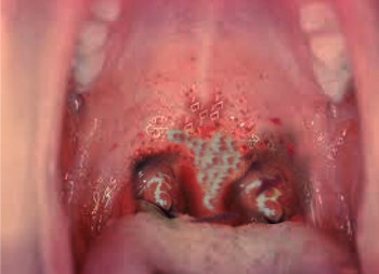
Diphtheria initially presents with nonspecific flu-like symptoms like fever, sore throat, and cervical lymphadenopathy. Generally, the average incubation period is 2 to 5 days (ranges between 1 to 10 days). In most of the cases, the history of the patients points toward travel from an endemic region with no vaccination records. In such cases, the provider should be aware of the most prominent features of the diseases, i.e., the thick, gray, adherent pseudomembrane over the tonsils and throat. Respiratory tract involvement is a common feature of the disease. It begins with mild erythema, which later changes into coalescing pseudomembrane.
Pseudomembrane consists of red blood cells, white blood cells, dead cell debris, and organisms. This pseudomembrane often bleeds if attempts are made to scrape it off from the adhering surface. Typical symptoms and signs of diphtheria include low-grade fever, sore throat, malaise, cervical lymphadenopathy, headache, and dysphagia. In a study by Pancharoen C et al., the most common manifestations of diphtheria included patch followed by fever and upper respiratory tract infection.[3] Further, systemic involvement occurs once the toxin leaks into the lymphatic and hematologic system after local tissue destruction.
In the case of cutaneous diphtheria, ulcerating skin lesions are present, which are covered with a gray membrane. These lesions do not spread or invade the surrounding tissues. The site for the cutaneous lesions are previously injured tissues from trauma or previous dermatologic lesions.[4][5]
Evaluation
Laboratory Diagnosis: The laboratory testing must be correlated with physical examination findings for the prompt and definitive diagnosis.
Bacteriologic Testing: A smear of the throat sample is stained with Gram stain and methylene blue. Although the Gram staining does not confirm the diagnosis, it is the initial test that is done in suspected cases. The Gram stain shows club-shaped, nonencapsulated, nonmotile bacilli found in clusters. The methylene blue stain reveals the typical metachromatic granules.
Culture: Culture from the throat swab is done either on Loffler medium or Tindale media, a telluride plate, and blood agar. A black colony with halos on Tindale media, metachromatic granules seen on a Loffler medium, the typical gray-black color of tellurium shows the presence of the organism in these media.
Toxin Testing: Toxin detection in the case of C. diphtheria helps to differentiate toxigenic strain from non-toxigenic variant. This can be achieved via the Elek test, PCR testing, and enzyme immunoassay (EIA) test.
Other Laboratory Studies:
- Complete blood count: It may show moderate leukocytosis.
- Troponin I: This helps in finding out the extent of the myocardial injury.
Imaging Studies: Chest and neck x-ray may reveal swelling of the soft tissue structure in and around the pharynx, epiglottis, and chest.
Treatment / Management
The two most important treatment modalities for diphtheria are antitoxins and antibiotics. Besides these two, the patient should be assessed for any respiratory and cardiovascular instabilities. When the patient is suspected of having diphtheria, antitoxin should be given immediately on a clinical basis without waiting for laboratory confirmation. The suspected cases must be kept in the isolation unit, and proper droplet precautions should be initiated. Further, the patient should be assessed for respiratory distress, and definite airway must be secured if needed. Cardiac monitoring is also an essential component of early management.
Diphtheria Antitoxin (DAT)
Diphtheria antitoxin is an antiserum derived from the horse.[6] Antitoxin functions by neutralizing the unbound diphtheria toxin in the blood. Once the toxin gets bound to the cell membrane, antitoxin has no role in neutralizing the antitoxin. The dosing of the antitoxin depends upon the clinical state and severity of the condition. It can be administered by either the intramuscular or intravenous route. Before administering the antitoxin, the patient must be tested for hypersensitivity, and emergency medication for anaphylaxis must be available at the bedside.
Antibiotic Treatment
The choice of antibiotics for diphtheria are erythromycin or penicillin G. Antibiotic must be initiated as soon as possible for the eradication of the organism. This helps to limit the toxin released into the system, quickens the recovery phase in the patient, and prevents the spread of the infection to the close contacts. Further, in the case of antibiotic resistance, linezolid or vancomycin can be used.
Differential Diagnosis
Diphtheria must be differentiated from other infections of the upper respiratory tract with similar presentations. Other differentials that must be kept in mind while diagnosing diphtheria are as follows:
- Epiglottitis: It is an acute inflammation involving the supraglottic region of the oropharynx with inflammation of the epiglottis and surrounding structures.[7][8]
- Retropharyngeal Abscess: It manifests with high spiking fevers and requires urgent drainage.[9]
- Angioedema: It manifests as generalized swelling due to the involvement of lower dermis and subcutaneous/submucosal tissues.[10]
- Infectious Mononucleosis: It manifests with fatigue, malaise, sore throat, fever, nausea, anorexia, cough. The classic triad is children who present with fever, pharyngitis, and lymphadenopathy.[11][12]
- Pharyngitis: It presents with a sore throat that is usually sudden in onset, odynophagia, fever, and cough.[13]
- Oral Candidiasis: Grayish pseudomembrane, in the case of diphtheria, must be differentiated from oral candidiasis.[14][15]
Prognosis
The prognosis depends on multiple factors:
- Age of Onset: High mortality rates are seen in individuals younger than five years and those older than 40 years.
- Duration of Onset of Symptoms: High mortality is seen in cases with onset of duration greater than four days.
- Cardiac Involvement: It is associated with a very poor prognosis, particularly AV and left bundle-branch blocks.
- Systemic Disease: High mortality rate is seen in cases of systemic involvement.
Complications
The most frequent complications are myocarditis and neuritis.[16][17] Death occurs in 5%-10% of cases. Severe complication includes pseudomembrane formation in the upper respiratory tract leading to respiratory obstruction and needing immediate mechanical ventilation and intubation.[18]
Cardiac Complications
It presents with myocarditis accompanied by cardiac arrhythmias with either first, second, or third-degree heart block[19] and circulatory collapse. ECG changes noted on these patients are prolonged P-R interval and ST/T wave changes.
Neurologic Complications
Neurological complications in diphtheria include nerve weakness or paralysis, especially involving the cranial nerves and also affecting the nerves in the extremity leading to weakness of the muscle of the extremity. The involvement of the muscles of the pharynx and the soft palate results in the regurgitation of the foods and fluids through the nose. In rare instances, encephalitis due to complications of diphtheria is seen in children.[20]
Consultations
The following consultations may be necessary for the management of diphtheria:
- Centers for Disease Control and Prevention (CDC): Antitoxins are not available commercially. CDC must be contacted and informed about the case before obtaining the antitoxins.
- Infectious Disease Center: Infectious disease centers must be informed as soon as the case is suspected.
- Cardiology: In the case of cardiac complications, cardiology consultation is necessary to assess the extent of the disease and to carry out measures required in case of cardiac dysrhythmia and heart block.
- Critical Care Service: Patients with severe diseases and septicemia must be admitted to the intensive unit and managed accordingly.
- ENT/ Anesthesia: ENT and anesthesia need to be consulted to find out the extent of the disease spread and in the case where intubations are required for respiratory distress.
- Pulmonology: Helps to assess the disease extent in the respiratory tract.
Deterrence and Patient Education
Vaccination
Vaccines for diphtheria are in the form of toxoids. Toxoid is the denatured protein (a bacterial toxin) with an intact receptor binding site and has the ability for antibody production. Generally, vaccination against the diphtheria is given in combination with other vaccines against tetanus and pertussis. Some of the forms of the combined diphtheria vaccine are as follows:
- DTaP: Consists of vaccination against diphtheria, tetanus, and pertussis.
- Tdap: Consists of vaccination against tetanus, diphtheria, and pertussis.
- DT: It consists of vaccination against diphtheria and tetanus.
- td: It consists of vaccination against tetanus and diphtheria.
In the United States, the DTaP vaccination schedule is followed for the neonate. For the infants, the vaccination schedule for the diphtheria vaccination includes 2, 4, 6 months. The 4th dose of the vaccination is given in between 15 to 18 months, and the 5th dose is given between the age of 4 to 6 years of age. The booster dose is given at the age of 11 to 12 years. During this time, the Tdap form of the vaccine is used. After that, the booster dose of Tdap or td is given every ten years for the remainder of the life. In the case of pregnancy, previously fully immunized females should receive a Tdap vaccine between 27 to 36 weeks of gestation.
Vaccination is the most crucial step in preventing the disease. Parents should be advised about the benefits of routine immunization to prevent the disease. In case of a missed vaccination schedule or lost vaccination records, the parents should immediately contact the primary care provider and report the case. As the immunity against diphtheria wanes off with time, the booster dose of vaccination is of utmost importance in the general population as well. People in the community must be made aware of the benefits of immunizations. In case of contact with the suspected case, the individual must immediately contact the health care professional to seek proper consultation. Similarly, a patient diagnosed with diphtheria must be made aware of the importance of isolation and limiting contact with the general population until cleared to do so by the healthcare professional.
In the case of close contacts who had recent exposure to diphtheria, management includes close surveillance for any respiratory or cutaneous symptoms. Patients should be kept under isolation, swabs should be taken for culture, and erythromycin utilized for 7 to 10 days. If the immunization status of the individual is unknown, the booster dose of the diphtheria toxoid should be given as well.
Enhancing Healthcare Team Outcomes
Diphtheria initially can present with vague upper respiratory tract symptoms like fever, sore throat, dysphagia, and headache. These nonspecific signs and symptoms tend to have a long list of differentials and frequently pose a diagnostic dilemma. Thus, during history taking, travel history, and vaccination status of the patient can be of importance which will help the health care providers to narrow down the differential diagnosis. The appearance of a pseudomembrane is a strong pointer to diphtheria. While the pediatrician or the provider is almost always involved in the care of patients with diphtheria, it is essential to consult with an interprofessional team of specialists that include an otorhinolaryngologist and a dermatologist.
Involving infectious disease experts in the case is equally important. The nurses are also vital members of the interprofessional group, as they will monitor the patient's vital signs. The radiologist and pathologist also play a crucial role in helping the provider arrive at a correct diagnosis. Preventive medicine specialists can significantly minimize the outbreaks of diphtheria by spreading awareness through vaccination programs. Cardiologists and neurologist's involvement early in the case can help to manage the complication if seen in the disease's course. Public health experts and international organizations must be notified and made aware of any suspected outbreaks.
The outcome of diphtheria depends on early diagnosis and treatment. A well-formed health care team can help to achieve this goal and prevent any morbidity and mortality associated with the diseases. However, to improve outcomes, prompt consultation with an interprofessional group of specialists is recommended.[21]