Oral Surgery, Instruments

Earn CME/CE in your profession:


Continuing Education Activity

Oral surgical procedures involve the use of numerous instruments. Each has its specific use, and multiple instruments are often required during a single procedure; hence it is crucial to understand the role of each to achieve optimal results with minimal complications. These instruments can have diagnostic as well as therapeutic purposes. This article reviews those most commonly utilized instruments and explains their function and application in treating patients that require oral surgical procedures.

Objectives:

  • Outline the local anesthetic delivery systems used in oral surgery.
  • Identify instruments used in non-surgical and surgical dental extractions and oral surgical procedures.
  • Explain the difference between luxators and elevators.
  • Summarise the importance of having a full range of instruments available.

Introduction

During oral surgical procedures, numerous instruments are utilized, each having a specific role. Dental practitioners should be able select appropriate instruments in given situations, preventing iatrogenic damage and traumatic procedures, thus increasing patient safety. This article introduces several commonly used instruments during various oral surgical procedures but does not provide an exhaustive list.

Function

Examination Kit

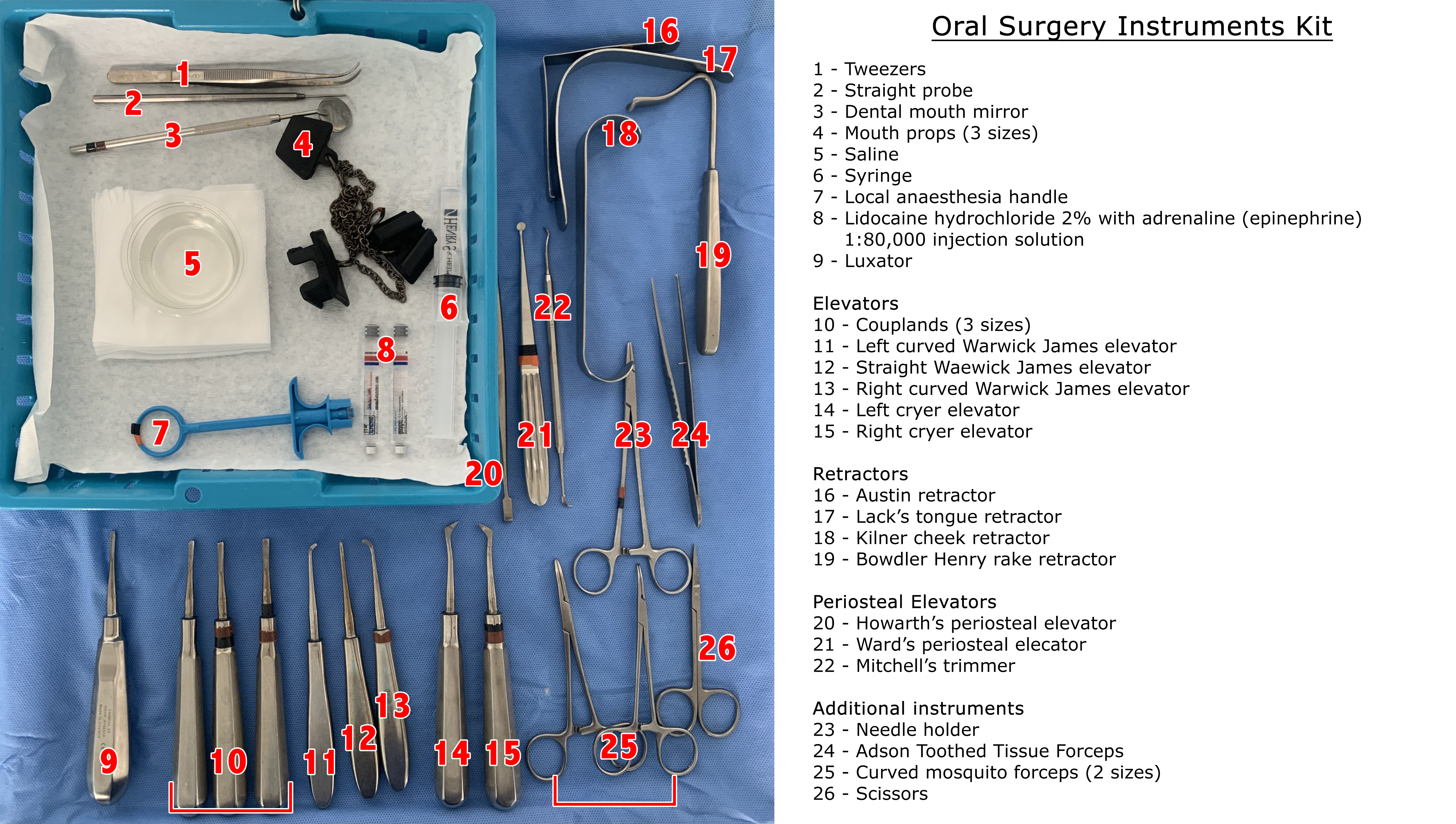
A clinical examination must be performed before performing any oral surgical procedure. A standard kit consists of a dental mouth mirror, probe, and tweezers. These instruments can be combined within separate disposable kits or included within a standard oral surgery procedure kit.[1] 

After administering local anesthesia, its efficacy can be tested using objective signs, which can put the patient at ease before initiating the surgical procedure. This includes applying firm pressure with a probe adjacent to the tooth to be extracted and down the periodontal ligament to ensure the absence of pain.[2]

Local Anesthetic Delivery Systems 

Local anesthesia is essential in dentistry to eliminate pain and anxiety during dental procedures. Conventional syringes and needles are commonly used with a handle to administer local anesthesia.

In the 1990s, computer-assisted systems were developed, which aim to provide predictable pulpal anesthesia, lowering the overall dose delivered and, in turn, reducing the risk of collateral anesthesia.[3] Additionally, to minimize the risk of administering anesthesia directly into a blood vessel, self-aspirating syringes are frequently used.[4] 

Needles and sharp instruments can pose a risk of needle-stick injury to the operator. Modern safety syringes minimize this risk through the application of a sheath that, when activated, locks over the needle, preventing potential accidental injury.[5]

Periotomes and Luxators 

These fine instruments are used to shear the periodontal ligament and act as a wedge to expand the socket.[6][7] These are inserted between the tooth and socket and moved around the tooth to create space for the use of elevators and forceps while applying gentle pressure apically.[8][9] By careful widening of a socket and making space for appropriate instruments, a more 'atraumatic technique' can be achieved, facilitating a good surface for implant placement through bone preservation.[10] 

Where the root apex fractures within the socket, a root pick elevator is useful in luxating the apical tip. Where the surgeon struggles in finding a definitive application point, bone removal at the level of the apical root tip may be required to improve access. These fine instruments have a posterior curve of the working end, allowing for successful application and luxation.[9]

Elevators 

A variety of dental elevators exist to 'lever' and mobilize teeth. They are used to apply forces to the tooth, which in turn sever the periodontal ligament and expand the socket. Care is necessary not to apply so much force that damage is caused to surrounding structures, ultimately leading to iatrogenic injury, like alveolar fractures.[9]

The most commonly used elevators are Coupland, Warwick-James, and Cryer elevators.[8][11] These instruments can be placed between the crestal bone and the side of the tooth or root using the principles of a lever, wedge, and wheel to elevate the tooth.[7][12] 

Cryer elevators have a sharp point and impart rotational forces. When engaged in the interseptal bone, it is particularly useful to remove either a mesial or distal root of a mandibular molar.[8] 

Warwick James elevators are a set of three; straight, curved left, and curved right.[11] 

Coupland elevators commonly come in a set of three sizes, and as more space is created, a larger size can be used to elevate the tooth further.[9]

Dental Extraction Forceps 

Dental extraction forceps are used to remove teeth from their sockets and grip onto either the crown or the root surface. They usually act as levers; however, some will also create a wedge effect.[7] They are positioned as apically as possible, and rotational, buccolingual, or a combination of these movements are applied to remove the teeth.

A variety of forceps is available to use depending on whether the tooth is in the maxillary or mandibular arch, a molar, premolar, or anterior tooth. Maxillary root forceps are suitable for use with any maxillary root, and similarly, mandibular root forceps can be applied to any mandibular root. Cowhorn forceps can be used for either the maxillary molar or mandibular molar teeth and are designed so that their beak is seated into the furcation of molar teeth. Application of pressure then luxates the tooth coronally, which subsequently allows for simple delivery. They can sometimes also be used to section molar teeth, which facilitates individual root delivery. Care should be taken to not fracture the buccal plate due to excessive force.[8]

Mouth Props

Mouth props or bite blocks are placed in the mouth to hold the teeth apart. Certain oral surgical procedures require patients to keep their mouths open for extended periods. The use of mouth props allows patients to bite on these, so they do not have to hold their mouths open for the duration of the procedure.[12] 

Gutters are present within the top and bottom of the prop, in which the teeth or alveolar ridge sit. They are usually made of rubber and have an attached chain that sits outside the mouth to retrieve the prop easily.[13] In patients with a hyperactive gag reflex, the insertion of a mouth prop can trigger their response, and they may not be able to tolerate it during the procedure.[14] Mouth props come in varying sizes, so clinical judgment should be applied regarding the most appropriate option.

Retractors 

These instruments retract soft tissue structures, such as raised flaps.[8] Retraction is essential for handling soft tissues to perform the surgical procedure optimally. Retractors commonly used include Lack, Laster, Bowdley Henry rake, Minnesota, Austin, and Kilner retractors, which can be used in various clinical situations.[15] 

The Lack's tongue retractor helps to stabilize the tongue, improving visualization.[16] The Laster's retractor can be useful when extracting upper third molars to both retract the soft tissues and direct light posteriorly towards the maxillary tuberosity region.[15] Bowdler Henry rake, Minnesota, or Austin retractors are often employed during surgical extractions to retract the buccal mucoperiosteal flap, aiding visibility and access while preventing damage to the flap.[17] The Kilner cheek retractor is routinely used to retract the cheeks or the lips, improving access to the surgical field within the oral cavity.[18]

Scalpel Handle and Blade 

A scalpel is required for certain oral surgical procedures, such as raising a mucoperiosteal flap; commonly, a 15-blade is used in a Bard Parker Handle.[8][12] A scalpel can also be used to carry out incisional and excisional intra-oral biopsies. Usually, the inferior incision is made first so as not to obscure vision due to the hemorrhage. Tissue forceps are then used to lift the sample and transfer it into a suitable medium.[19] A scalpel is the instrument of choice when carrying out incision and drainage of intra-oral and extra-oral abscesses.[20][21]

Periosteal Elevators 

When raising a full-thickness mucoperiosteal flap, the mucosa and periosteum must be cleanly reflected together completely down to the bone to limit trauma to the underlying tissues and improve the healing process when the flap is repositioned. This is usually carried out with a periosteal elevator, such as Howarth's periosteal elevator, in a pushing motion. Sometimes periosteal elevators are also used to retract the flap for purposes such as lingual nerve protection.[12][22]

The Ward's periosteal elevator has one spoon-shaped end, which is useful in scooping up and reflecting mucoperiosteal flaps.[23] Similarly, Mitchell's trimmer can be used as an elevator because it has one spoon-shaped end. The curvature of these two instruments is beneficial during cyst enucleation within bony structures, enabling the surgeon to carefully separate the cyst lining from the underlying bone and prevent cyst rupture, which can then be sent for histopathological assessment completely intact.[24]

Surgical Straight Handpiece 

A surgical straight handpiece that does not expel air into the surgical site can be used to section teeth and remove bone when necessary. It is prudent to use a handpiece that does not expel air to avoid surgical emphysema, and it must be cooled with sterile saline.[8][12][25] 

Most commonly, round burs are used to remove the alveolar bone. Surgeons may use a fissure bur for crown and root sectioning during tooth removal.[26][27] Fissure burs are also used for coronectomy procedures, which involve removing the crown of the tooth and reducing the remaining roots 3mm below the crest of the buccal and lingual plates.[28] 

Bone rongeurs are instruments that are also used to aid bone removal. They can remove bony prominences or irregular ridges, which can then be smoothed using a bone file, allowing for improved healing.[8]

Suction

Good suction is necessary to facilitate a clear field for good visualization of the surgical site and prevent ingestion or aspiration by the patient.[29][30] Many suction tips exist, with Frazier and Yankaeur most commonly used in oral surgical procedures.

Yankauer suction is usually plastic tubing, while Frazier suction is usually a metal suction tip.[29] Frazier suction is particularly useful during surgical procedures such as surgical tooth extractions. It has a bend in the shaft, making it easier to access deeper structures, including the apical area of extraction sockets. The hole where it is held can be occluded with a finger to provide stronger suction when needed; lighter suction is usually required when on soft tissues compared to hard tissues.[30]

Sutures 

Various suture materials can be utilized depending on the procedure and clinician preference. They can be grouped into resorbable or non-resorbable. Resorbable sutures include those made from polygalactic, polydiossanone, and polyglycolic acid. Non-resorbable sutures include those made from silk, polyamides, and polyesters.[31] Resorbable sutures are most commonly used for oral surgical procedures.[12]

Suture needles are available in different shapes, sizes, and alloys depending on the size and depth of the tissue being closed.[32] The suture needle is held in a suture holder, which aids in passing the needle through soft tissues; they are often locking suture holders to keep the needle secure.[32] The needle should be grasped by the suture holder approximately one-third from the swage to the tip to prevent needle fracture, bending, and twisting.[32]

Scissors 

A variety of scissors are available in different shapes and sizes to cut soft tissues, sutures, and materials such as oxidized resorbable cellulose and also to dissect tissues.[12] Different scissors have different uses; for example, curved McIndoe scissors are desirable for dissection and cutting of tissue.[33] Mayo scissors can have curved or straight blades and are often used for cutting tissues and sutures.[34]

Clinical Significance

The instruments reviewed are fundamental to many oral surgical procedures. As such, it is necessary to be aware of their existence, appreciate the clinical situations they are used in and understand how they can be applied. This knowledge will facilitate their safe use, as improper instrumentation in combination with uncontrolled excessive force can have detrimental effects such as iatrogenic fracture of the mandible.[35] 

This reading should be supplemented with practical experience as one method for a particular procedure does not always work in another clinician's hands. Often, several different instruments can be used in the same clinical scenario, and clinicians develop a preference.

Enhancing Healthcare Team Outcomes

The World Health Organisation launched the Surgical Safety Checklist in 2009, allowing different treatment stages to be checked before commencing a procedure. This can be modified for oral surgical procedures to minimize the risk of adverse outcomes.[36] The whole dental team involved in the surgery must engage in the pre-operative checks, and patient involvement is encouraged.

Pauses in the form of verbal checks may be utilized before an invasive process, such as local anesthesia administration, or proceeding to use an oral surgical instrument such as an elevator or forceps.[37] It is necessary for all team members involved in oral surgical procedures to have an awareness of the available instruments, prepare the surgical kits appropriately, and request and use specific instruments.

Nursing, Allied Health, and Interprofessional Team Interventions

Dental nurses often undertake decontamination tasks when there is no dedicated member of staff on duty for cleaning and sterilizing instruments. All dental care professionals must understand and be trained in cross-infection control, which is essential in dentistry due to exposure-prone procedures.[38] Following thorough decontamination protocols allows for instruments that are safe for patient use.

During oral surgical procedures, sharp instruments are utilized by the dental practitioner, such as the scalpel and local anesthetic needle. It is, therefore, essential that at the end of the surgery, the dentist takes the responsibility to dispose of these sharp instruments carefully. Proper sharps management reduces the risk of sharps injuries and contact with potentially infectious material to the person who clears the instrument tray.[39]

Nursing, Allied Health, and Interprofessional Team Monitoring

Four-handed dentistry can be utilized to maximize efficiency during oral surgical procedures. The dental nurse recognizes the instrument required next or is instructed by the dental practitioner and then provides this to them.[40]

A sound understanding of oral surgical instruments is imperative as there are usually many instruments on the tray, and delays in treatment can result if the assistant cannot identify each instrument and pass it to the operator in good time. Additionally, the wider team needs to be aware of various instruments to identify faulty pieces and report and replace them.


(Click Image to Enlarge)
An Oral Surgery Instruments Kit
Contributed by Dr Glenda Aruede
Details

Editor:

Glenda Aruede

Updated:

9/20/2022 10:29:54 AM

References


[1]

Kumar S,Kumar S,Hassan N,Anjan R,Shaikh S,Bhowmick D, A Comparative Assessment of the Effect of Professional Oral Hygiene Measures on the Periodontal Health of Patients Undergoing Fixed Orthodontic Appliance Therapy. Journal of pharmacy     [PubMed PMID: 35017981]

Level 2 (mid-level) evidence

[2]

M M A,Khatri A,Kalra N,Tyagi R,Khandelwal D, Pain perception and efficacy of local analgesia using 2% lignocaine, buffered lignocaine, and 4% articaine in pediatric dental procedures. Journal of dental anesthesia and pain medicine. 2019 Apr;     [PubMed PMID: 31065592]


[3]

Sivaramakrishnan G,Sridharan K, Local Anaesthetic Drug Administration in Dentistry Using Computer Assisted Anaesthetic Delivery System: A Systematic Review. The open dentistry journal. 2016;     [PubMed PMID: 27733872]

Level 1 (high-level) evidence

[4]

Henderson S, Drug interactions with local anaesthetic preparations used by primary care dental practitioners. Dental update. 2010 May;     [PubMed PMID: 20527498]


[5]

Saxena P,Gupta SK,Newaskar V,Chandra A, Advances in dental local anesthesia techniques and devices: An update. National journal of maxillofacial surgery. 2013 Jan;     [PubMed PMID: 24163548]

Level 3 (low-level) evidence

[6]

Sambrook PJ,Goss AN, Contemporary exodontia. Australian dental journal. 2018 Mar;     [PubMed PMID: 29574811]


[7]

McKenzie WS, Principles of Exodontia. Oral and maxillofacial surgery clinics of North America. 2020 Nov;     [PubMed PMID: 32839095]


[8]

Dym H,Weiss A, Exodontia: tips and techniques for better outcomes. Dental clinics of North America. 2012 Jan;     [PubMed PMID: 22117954]


[9]

Mamoun J, Use of elevator instruments when luxating and extracting teeth in dentistry: clinical techniques. Journal of the Korean Association of Oral and Maxillofacial Surgeons. 2017 Jun;     [PubMed PMID: 28770164]


[10]

Sharma SD,Vidya B,Alexander M,Deshmukh S, Periotome as an Aid to Atraumatic Extraction: A Comparative Double Blind Randomized Controlled Trial. Journal of maxillofacial and oral surgery. 2015 Sep;     [PubMed PMID: 26225052]

Level 2 (mid-level) evidence

[11]

Gelbier S, William Warwick James {i}OBE{/i} FRCS MCh FDS FLS (1874 to 1965). Journal of medical biography. 2021 Dec 14;     [PubMed PMID: 34904471]


[12]

DeBowes LJ, Simple and surgical exodontia. The Veterinary clinics of North America. Small animal practice. 2005 Jul;     [PubMed PMID: 15979521]

Level 3 (low-level) evidence

[13]

Rapaport BHJ,Smith MT,Brown JS, An audit of routine practice: a prospective study on the use of mouth props in oral surgery. British dental journal. 2020 Jun     [PubMed PMID: 32591709]


[14]

Kil WJ,Kulasekere C,Derrwaldt R,Bugno J,Hatch C, Decreased radiation doses to tongue with     [PubMed PMID: 27447973]


[15]

Graham RM,Bussell MA,Laster Z, Laster's upper third molar and cheek retractor. The British journal of oral     [PubMed PMID: 19692154]


[16]

Davari P,Fazel N, Practical pearls for oral procedures. Clinics in dermatology. 2016 Jul-Aug;     [PubMed PMID: 27343958]


[17]

Malden NJ,Maidment YG, Lingual nerve injury subsequent to wisdom teeth removal--a 5-year retrospective audit from a high street dental practice. British dental journal. 2002 Aug 24;     [PubMed PMID: 12222906]

Level 2 (mid-level) evidence

[18]

Mubeen S,Mubeen S,Falconer D, Use of the Alexis ring retractor in oral and maxillofacial surgery. The British journal of oral     [PubMed PMID: 24685476]


[19]

Avon SL,Klieb HB, Oral soft-tissue biopsy: an overview. Journal (Canadian Dental Association). 2012     [PubMed PMID: 22889502]

Level 3 (low-level) evidence

[20]

Stephens MB, Wiedemer JP, Kushner GM. Dental Problems in Primary Care. American family physician. 2018 Dec 1:98(11):654-660     [PubMed PMID: 30485039]


[21]

Priyamvada S,Motwani G, A Study on Deep Neck Space Infections. Indian journal of otolaryngology and head and neck surgery : official publication of the Association of Otolaryngologists of India. 2019 Oct;     [PubMed PMID: 31742093]


[22]

Bailey E,Kashbour W,Shah N,Worthington HV,Renton TF,Coulthard P, Surgical techniques for the removal of mandibular wisdom teeth. The Cochrane database of systematic reviews. 2020 Jul 26;     [PubMed PMID: 32712962]

Level 1 (high-level) evidence

[23]

Pogrel MA,Goldman KE, Lingual flap retraction for third molar removal. Journal of oral and maxillofacial surgery : official journal of the American Association of Oral and Maxillofacial Surgeons. 2004 Sep;     [PubMed PMID: 15346365]


[24]

Hill CM,Renton T, Oral surgery II: Part 3. Cysts of the mouth and jaws and their management. British dental journal. 2017 Oct 27;     [PubMed PMID: 29074909]


[25]

Chong BS,Rhodes JS, Endodontic surgery. British dental journal. 2014 Mar;     [PubMed PMID: 24651333]


[26]

Stajčić Z,Stojčev Stajčić LJ,Kalanović M,Đinić A,Divekar N,Rodić M, Removal of dental implants: review of five different techniques. International journal of oral and maxillofacial surgery. 2016 May;     [PubMed PMID: 26688293]


[27]

Bhati B,Kukreja P,Kumar S,Rathi VC,Singh K,Bansal S, Piezosurgery versus Rotatory Osteotomy in Mandibular Impacted Third Molar Extraction. Annals of maxillofacial surgery. 2017 Jan-Jun;     [PubMed PMID: 28713729]


[28]

Martin A,Perinetti G,Costantinides F,Maglione M, Coronectomy as a surgical approach to impacted mandibular third molars: a systematic review. Head     [PubMed PMID: 25890111]

Level 1 (high-level) evidence

[29]

Nakao S,Nishida T,Sonoda KH, Removal of MIRAgel scleral buckle implants using Yankauer suction catheter with adjusted diameter. American journal of ophthalmology case reports. 2022 Jun;     [PubMed PMID: 35282604]

Level 3 (low-level) evidence

[30]

Yadav RP,Jaisani MR,Acharya P,Mahat AK, Frazier Metallic Suction Tube: An Adjunct in Irrigation of Deep Fascial Spaces. Journal of maxillofacial and oral surgery. 2017 Sep     [PubMed PMID: 28717302]


[31]

Minozzi F,Bollero P,Unfer V,Dolci A,Galli M, The sutures in dentistry. European review for medical and pharmacological sciences. 2009 May-Jun;     [PubMed PMID: 19673173]


[32]

Byrne M,Aly A, The Surgical Needle. Aesthetic surgery journal. 2019 Mar 14     [PubMed PMID: 30869752]


[33]

Renton T,Durham J,Hill CM, Oral surgery II: Part 2. The maxillary sinus (antrum) and oral surgery. British dental journal. 2017 Oct 13     [PubMed PMID: 29026224]


[34]

El-Sedfy A,Chamberlain RS, Surgeons and their tools: a history of surgical instruments and their innovators--part I: place the scissors on the Mayo stand. The American surgeon. 2014 Nov;     [PubMed PMID: 25347498]


[35]

Woldenberg Y,Gatot I,Bodner L, Iatrogenic mandibular fracture associated with third molar removal. Can it be prevented? Medicina oral, patologia oral y cirugia bucal. 2007 Jan 1;     [PubMed PMID: 17195834]


[36]

Moore R,Miller R,Henderson S, Risk management in oral surgery. British dental journal. 2019 Dec     [PubMed PMID: 31873258]


[37]

Tagar H,Devine M,Obisesan O, How to create local safety standards for invasive procedures (LocSSIPs) by engaging the team in patient safety. British dental journal. 2019 Jan 18     [PubMed PMID: 30655621]


[38]

Smith A,Creanor S,Hurrell D,Bagg J,McCowan M, Management of infection control in dental practice. The Journal of hospital infection. 2009 Apr     [PubMed PMID: 19162375]


[39]

Miller CH, Infection control. Dental clinics of North America. 1996 Apr;     [PubMed PMID: 8641531]


[40]

Mamoun J, Basic principles of maximizing dental office productivity. General dentistry. 2012 Mar-Apr;     [PubMed PMID: 22414506]