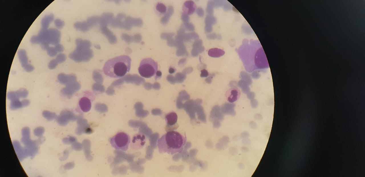
Plasmacytoma

Earn CME/CE in your profession:


Continuing Education Activity

Plasmacytoma is a tumor of plasma cells of bony or soft tissue and can occur anywhere in the body without evidence of systemic disease. It may present as a solitary or multiple mass anywhere in the body. It can progress to multiple myeloma if not evaluated and appropriately managed. This activity reviews the disease process, evaluation, and management of Plasmacytoma and highlights the role of the interprofessional team in improving care for patients with this condition.

Objectives:

  • Review the presentation of plasmacytoma and its types.
  • Identify the epidemiology of the disease.
  • Review the evaluation of plasmacytoma.
  • Outline the treatment options for patients with plasmacytoma.

Introduction

Plasmacytoma is a rare plasma cell dyscrasia that grows within the axial skeleton or soft tissue structures as solitary or multiple masses without evidence of systemic disease. It is an early plasma cell malignancy and is considered an intermediate phase between monoclonal gammopathy of undetermined significance(MGUS) and multiple myeloma (MM).[1][2] Plasmacytoma can be divided into the following types according to the International Myeloma Working Group:[3]

Solitary Plasmacytoma

It is further divided into:

  • Solitary plasmacytoma of bone (SPB), which arises from plasma cells of the bone marrow.
  • Extramedullary plasmacytoma (EMP), which arises from plasma cells of the mucosal surfaces.

Multiple Solitary Plasmacytomas (MSP)

Solitary plasmacytoma is the most common form of plasmacytoma. Most cases of plasmacytomas cause paraproteinemia. Metastatic spread of plasmacytoma occurs to soft tissues frequently and occasionally to bones. Plasmacytoma is treated with surgery, radiotherapy, and chemotherapy as required. Therapy for local disease gives prolonged survival, and disseminated disease treatment gives longer remission.

According to the World Health Organization (WHO) update, SPB is further composed of two subtypes:[2][4]

  1. SPB with minimal bone marrow involvement:SPB type that has less than 10% clonal marrow cells apart from the plasmacytoma itself. The rate of progression of this type of SPB to MM is 20-60% in 3 years.
  2. SPB with no bone marrow involvement: SPB type that lacks clonal marrow cells apart from the plasmacytoma. The rate of progression of this type to MM is 10% in 3 years.

MSP consists of monoclonal cell infiltrates in one or more lytic bone lesions or extramedullary tissue. Only a few cases of MSP have been reported. Imaging modalities used for solitary plasmacytoma may reveal the presence of multiple lesions leading to MSP diagnosis. MSP is diagnosed in up to 5% of cases of solitary plasmacytoma.[3] Long term prognosis of MSP is not clear. Surgery, radiotherapy, chemotherapy, and stem cell transplantation are indicated for the treatment of MSP.[5][3][6]

Etiology

The etiology of plasmacytoma is unknown.[7][8] However, it is suggested that EMP may be caused by inhalation of chemicals, chronic stimulation, an overdose of irradiation, viral infection, and genetic disorders in the reticuloendothelial system.[9]

Epidemiology

Plasmacytoma is a rare disease, with SPB being the most common type. Solitary plasmacytoma has an annual incidence of fewer than 450 cases. The incidence of SPB is 40% more than EMP, with SPB constituting 2% to 5% of all plasma cell malignancies and EMP constituting 4% of all plasma cell malignancies.[1] Plasmacytoma is prevalent in older individuals, African Americans, and males.[10] It presents in the middle to old aged people with the mean age of presentation of 55 to 60 years.[11] Plasmacytoma is more prevalent in males than females, with a male to female ratio of 2 to 1 in SPB and 3:1 in EMP.

Pathophysiology

Plasmacytomas share many biological features of other plasma cell disorders and can arise in any part of the body. SPB arises from plasma cells of the bone marrow, and EMP arises from the plasma cells of mucosal surfaces. According to some authors, EMP and marginal zone B-cell lymphomas share many histological and clinical similarities, with EMP having an extensive degree of plasmacytic differentiation. Interleukin-6 (IL-6) is considered the principal growth factor in plasma cell disorders.[12] Cytogenetic studies show that losses of chromosome 13 arm 1p and 14q and gains in arm 19,1q and 9q play an essential part in tumorigenesis of plasmacytoma.[13] High histological grade and angiogenesis in plasmacytoma are associated with increased progression to multiple myeloma, and plasmacytoma is considered an intermediate phase from the conversion of MGUS.[12][14]  

The occurrence of plasmacytoma at the surgical wound site may be due to direct wound contamination, host immune response, or tumor manipulation and aerosolization, among other factors. A dominant theory explaining the pathophysiology of plasmacytoma explains the increased expression of stromal cell-derived factor 1 (SDF-1) in areas of inflammation and trauma, which is a highly potent immune cell attractant. After chemotherapy, myeloma cells expressing CXCR-4 chemokine receptors attach to SDF-1.[15] The release of inflammatory cytokines causes proliferation of these plasma cells leading to tumor formation.[15]

Histopathology

Routine hematoxylin and eosin stain is used for plasmacytoma. The histologic picture resembles multiple myeloma and contains dense infiltrate of plasmablastic or anaplastic plasma cells that may be mature or immature.[16] Amyloid deposits may be seen in extraosseous tumor mass as pink deposits containing multinucleated giant cells.

History and Physical

SPB presents with pain in the bones containing red bone marrow, e.g., ribs, vertebrae(thoracic more than lumber and cervical), femur, and pelvis.[3][17] An uncommon presentation is in the skull with headaches, diplopia, strabismus, exophytic mass, otalgia, and dizziness.[18][19][20] Compression fractures of bones may occur with infiltrating bony lesions along with signs of local soft tissue damage. Significant vertebral involvement may lead to the involvement of the spinal cord or nerve root.

EMP can occur anywhere in the body with 80% to 90% in the head and neck region and aerodigestive tract, mainly in the oral cavity, tonsillar fossa, nasal cavity, and paranasal sinuses, which may present with headache, nasal discharge, dysphagia, sore throat, epistaxis, and nasal obstruction.[17][21][22] 

Uncommon presentations include involvement of the larynx causing hoarseness, dysphonia, dyspnea, airway obstruction, and wheezing.[23]

Gastrointestinal involvement is rare and presents with epigastric pain, hemoptysis, loss of appetite, bleeding, and abdominal discomfort. Involvement of mesentery is also seen, with only 10 cases reported.[24][25][26]

Plasmacytoma may mimic a pulmonary tumor and present as a nodular parenchymal or perihilar opacity, associated with mediastinal lymphadenopathy, rarely alveolar interstitial or endobronchial damage.[27] It may also be rarely present in the thyroid, causing thyromegaly.[28]

Evaluation

Laboratory Studies

Monoclonal proteins in serum or urine are detected. According to the diagnostic criteria, no M-protein should be detected on immunofixation of the serum and urine. Still, approximately 50% of patients show a small M-protein in the serum or urine.[3]  

Uninvolved immunoglobulins and other blood tests are in average values.

Histology

Lesional biopsy or fine-needle aspiration techniques are used to diagnose plasmacytoma. In the case of the spine, fluoroscopic guided biopsy or CT guided lesional biopsy is used. 

Tissue biopsy in SPB shows infiltration of monoclonal cells, and EMP shows submucosal growth and may require excision or tissue biopsy according to the location. A biopsy is done to check the phenotype of plasma cells. Additionally, bone marrow aspiration or trephine biopsy is done to rule out multiple myeloma. Furthermore, an immunohistological analysis is performed to check the presence of CD138 and CD38 markers.

Imaging Studies

Plain film radiography, MRI and PET CT are used to assess the lesion. Whole-body CT/MRI is used to detect other primary lesions elsewhere in the body.[17][21]

MRI due to better soft-tissue contrast and multiplanar images is a better modality for evaluating radiation volumes in plasmacytomas and the staging of the disease. It is also helpful in the verification of tumor reduction after therapy.[29]

Features suggesting plasmacytoma include bulky soft tissue mass and features of high cellularity exhibited by relatively isointense T2 weighted MRI images.[22] 

Other imaging techniques include flow cytometry or kappa lambda labeling to check the degree of clonal plasma cell infiltration. Moreover, endoscopy is used to detect any associated lesions, resectability, mass effect, and destruction of surrounding tissue in EP. In addition to CT, which is used to detect underlying bony abnormalities, 18 fluorodeoxyglucose (18F-FDG) PET/CT is used in patients when MRI cannot be done.[30]

Patients with plasmacytoma should be evaluated to exclude systemic disease by performing skeletal surveys, bone marrow biopsy, and blood tests. MM should be ruled out by performing the recommended tests, including spine MRI or FDG-PET, multicolor flow cytometry (MFC), immunoglobulins levels, and free light chain levels. Evaluation and diagnosis of systemic disease in this way may reduce the percentage of patients with plasmacytoma evolving into MM.[11]

According to the international myeloma working group, the diagnostic criteria for plasmacytoma is as follows:[3]

SPB

One bone lesion containing clonal plasma cells, absence of M-protein in serum and urine (may be present in some cases), bone marrow histology not consistent with MM, normal skeletal survey, no end-organ damage.

EMP 

Extramedullary tumor of clonal plasma cells, absence of M-protein in serum and urine (may be present in some cases), normal bone marrow, normal skeletal survey, no end-organ damage.

MSP

More than one bone lesion or extramedullary tumor of plasma cells that may be recurrent, absence of M-protein in serum and urine (may be present in some cases), normal bone marrow, normal skeletal survey, no end-organ damage.

Treatment / Management

Plasmacytomas are treated with radiation therapy (RT), surgery, and chemotherapy as required, given the disease condition.

Radiotherapy

Plasmacytoma is radiosensitive, and radiotherapy is the treatment of choice in SPB and EMP, which helps achieve an 80% control rate.[17][11][31][21] 

Fractionated radiotherapy dose of 40-50 Gy over a duration of 4 weeks is given at the rate of 1.8 to 2.0 Gy per fraction.[17]  

Conformal RT with parallel fields is commonly used to cover planning target volume (PTV). However, intensely modulated radiotherapy (IMRT) may be used to spare important structures like eyes and glands.

The treatment field includes the tumor along with a 1.5 to 2.0 cm tumor-free margin. Furthermore, some authors recommend including the whole bone-in RT margins due to fear of recurrence, while others recommend the partially involved bone to be included in radiation therapy.[29] In cases of vertebral involvement, RT fields include 1 or 2 vertebrae above and below the affected vertebral level. Lymph node involvement in the radiation field is recommended if lymph nodes are involved in the disease process. The addition of regional lymph nodes in the radiation margin when not involved in the disease process decreases the recurrence rate. Still, it is associated with increased morbidity and hence not preferred.

Surgery

Some patients get partial or complete removal of tumors for diagnostic purposes. For therapeutic purposes, surgery combined with RT provides better progression-free survival (PFS). Radical surgery of head and neck plasmacytoma should be avoided as these tumors are highly radiosensitive, and radical surgery is mutilating. In other areas, surgery should be done where feasible. Moreover, surgery is also indicated for vertebral instability, fractures, or neurologic involvement. RT can also be delayed after surgery. However, surgery without RT shows increased rates of recurrence.[17][29] 

Chemotherapy

In most studies, chemotherapy shows no beneficial effect on disease control or prevention of complications. Chemotherapy does not decrease the incidence of progression of plasmacytoma to multiple myeloma (MM), but it does increase the duration of progression to MM. Adjuvant chemotherapy may be considered for tumors greater than 5cm and in tumors unresponsive to RT.[17][29] 

Autologous Stem Cell transplantation (ASCT)

ASCT is indicated in patients with high risk, recurrent and large numbers of solitary plasmacytoma. Trials have been conducted in patients with recurrent SPB in the radiation zone, distal recurrence, and high-risk plasmacytomas, showing promising results.[32][33]

Differential Diagnosis

Plasmacytoma shows a few similarities with other diseases and hence needs to be differentiated from them. Differential diagnosis of plasmacytoma includes:

Multiple Myeloma (MM)

Multiple myeloma is a tumor of plasma cells. Plasmacytoma may progress to multiple myeloma over 2-3 years. Plasmacytoma is a localized bone disease and is further differentiated from multiple myeloma by the presence of CRABS (hypercalcemia, renal failure, anemia, bone disease), multiple lytic bone lesions, end-organ damage, and serum or urinary monoclonal proteins.[5][34][29][34]

Non-Hodgkin Lymphoma (NHL)

NHL, in some cases, shows plasmacytic differentiation, which makes it difficult to differentiate them from plasmacytoma. In these cases, immunophenotyping can be used to differentiate these entities—plasma cells in lymphoma express CD19 and CD45. The phenotype of SPB lesion is similar to that of myeloma, but that of EMP lesion is similar to lymphomas suggesting that they may represent NHL with plasmacytic differentiation.[35]  

Reactive Plasmacytosis

It consists of follicular hyperplasia. There is no light chain restriction in reactive plasmacytosis as compared to plasmacytoma.

Plasmablastic Lymphoma

The tumor has plasmablastic morphology, often occurs in HIV-positive or immunosuppressed individuals. It occurs in the oral cavity or the mucosal surfaces of the head. It is associated with the Epstein-Barr virus.

Staging

Soft tissue plasmacytoma staging is as follows:[36] 

  1. Limited to extramedullary site
  2. Involvement of the regional lymph nodes
  3. Multiple metastases 

Extramedullary plasmacytoma staging is also done using a staging system for multiple myeloma because of the lack of a unique staging system of EMP to check survival and subsequently design treatment.[37] Two prognostic factors for its staging include lymph node metastasis and tumor size more than 5 cm.

  • Stage 1: Absence of both factors
  • Stage 2: The presence of one factor
  • Stage 3: The presence of both factors 

Prognosis

The median time of progression to MM is 2 to 3 years. SBP has a poor prognosis and an increased progression to MM as compared to EMP.[38] SPB progresses to MM in 50% of cases, and EMP progresses to MM in 15% of cases.[3]

The causes of poor prognosis include local recurrence, incomplete resection with functional damage, local lymph node metastasis, progression to MM, or new bone lesions formation with MM. Anaplastic plasmacytomas with high histologic grade and increased vascularity are a poor prognostic factor. For SPB, the poor prognostic factors include:

  1.   Age more than 40 years
  2.   Tumour size of 5cm or more
  3.   Spine lesions
  4.   Neurologic symptoms associated with SPB 
  5.   RT dose[39]
  6.   Existence of light chains 
  7.   High serum M protein levels[39]
  8.   Persistence of M proteins after treatment
  9.   Bone marrow infiltration by clonal plasma cells
  10.   Osteopenia[12] 

The prognostic factors for EP are unclear due to fewer cases and changing biological behavior.[40] Age, size and grade of tumor, origin (extramedullary versus bone), regional lymph node metastasis, serum M-protein, light chains, and RT dose are associated factors.[40][29][41]

Complications

Plasmacytoma can occur anywhere in the body and leads to different complications.

SPB causes lytic bone lesions that lead to bone pain and fractures of bones, and associated nerve and soft tissue damages.[42]

POEMS (polyneuropathy, organomegaly, endocrinopathy, multiple myeloma, and skin changes) syndrome is a paraneoplastic disorder associated with plasmacytoma in the bone.[43]

Plasma cell disorders produce immunoglobulins that lead to relative immunodeficiency reducing antigen immune response.[42]

Renal impairment is primarily related to light chains with decreased molecular weight eliminated as the Bence–Jones protein in the urine as described in a case of plasmacytoma in the chest wall of a 44 years old male.[44] Kidney failure may result from reduced immunoglobulin elimination in the urine as immunoglobulins produced by plasmacytoma may be insoluble at cold temperatures. Cryoglobulinemia has been reported in cases where chronic Hepatitis C is associated with plasmacytoma. Amyloidosis may also be a consequence of light chain production by plasmacytoma.[42] A recent case report published in 2019 reported the association of acquired von Willebrand syndrome with intracranial plasmacytoma.[45]

Plasmacytoma is also associated with gastrointestinal bleeding in some cases of MM.[46]

A rare case was reported in which an extramedullary plasmacytoma was ulcerating through the gastric mucosa and presenting as a massive upper GI bleed. Such cases can be complicated by obstruction and perforation in the gastrointestinal system.[47]

Moreover, drug toxicity should be considered if the patient is treated with melphalan, as certain complications like acute leukemia and sepsis are well-known complications of chemotherapy-induced neutropenia.[48] Additionally, thalidomide-induced thrombosis has also been reported.[49]

Postoperative and Rehabilitation Care

Patients treated for plasmacytoma need lifelong follow-up. Evaluation is done at a six-week interval for the first six months, and clinical visits are prolonged after that.[29] Evaluation includes complete blood count, complete metabolic panel, C reactive protein, erythrocyte sedimentation rate, M protein, and skeletal bone survey.

NCCN recommends yearly imaging for five years by the same techniques used at diagnosis. Patients with SPB and EMP are also recommended for follow-up evaluation with orthopedics and ear, nose, throat department, respectively.

Deterrence and Patient Education

Patient education is essential in every disease as it can help them understand their condition and get the appropriate treatment. In the case of plasmacytoma, discussion topics with patients include:

  • Understanding the disease
  • Disease workup 
  • Complications of disease 
  • Possible treatment options and outcomes
  • Possibility of disease recurrence and progression
  • Counseling about long term followup

Enhancing Healthcare Team Outcomes

Management of plasmacytoma requires close cooperation by different departments. Patients getting referred from the primary care physician for further management should be evaluated and treated by an interprofessional approach, including a team of radiotherapists, hematologists, surgeons, and oncologists. Collaboration with nurses, pharmacists, and psychologists (if required) is essential for overall patient care. After treatment, patients need to continue to follow up. Patients need to be checked for recurrence or progression of the disease and may be referred to orthopedics or the ear, nose, throat department. 

In case of occurrence of new bone pain, additional workup and imaging should be done.[29]

Care should be taken to check for recurrence and progression of the disease to multiple myeloma. Early diagnosis, treatment, and careful monitoring may help provide better healthcare to the patients and improve their quality of life affected by the disease.


(Click Image to Enlarge)
Plasmacytoma
Plasmacytoma
Contributed by Shaikh Zayed Hospital
Details

Updated:

7/31/2023 8:38:34 PM

References


[1]

Meyer HJ, Ullrich S, Hamerla G, Surov A. [Extramedullary Plasmacytoma]. RoFo : Fortschritte auf dem Gebiete der Rontgenstrahlen und der Nuklearmedizin. 2018 Nov:190(11):1006-1009. doi: 10.1055/a-0604-2831. Epub 2018 Oct 8     [PubMed PMID: 30296807]


[2]

Rajkumar SV. Updated Diagnostic Criteria and Staging System for Multiple Myeloma. American Society of Clinical Oncology educational book. American Society of Clinical Oncology. Annual Meeting. 2016:35():e418-23     [PubMed PMID: 27249749]


[3]

International Myeloma Working Group. Criteria for the classification of monoclonal gammopathies, multiple myeloma and related disorders: a report of the International Myeloma Working Group. British journal of haematology. 2003 Jun:121(5):749-57     [PubMed PMID: 12780789]


[4]

Ohana N, Rouvio O, Nalbandyan K, Sheinis D, Benharroch D. Classification of Solitary Plasmacytoma, Is it more Intricate than Presently Suggested? A Commentary. Journal of Cancer. 2018:9(21):3894-3897. doi: 10.7150/jca.26854. Epub 2018 Oct 10     [PubMed PMID: 30410592]

Level 3 (low-level) evidence

[5]

Dattolo P, Allinovi M, Michelassi S, Pizzarelli F. Multiple solitary plasmacytoma with multifocal bone involvement. First clinical case report in a uraemic patient. BMJ case reports. 2013 May 23:2013():. doi: 10.1136/bcr-2013-009157. Epub 2013 May 23     [PubMed PMID: 23709144]

Level 3 (low-level) evidence

[6]

Ooi GC, Chim JC, Au WY, Khong PL. Radiologic manifestations of primary solitary extramedullary and multiple solitary plasmacytomas. AJR. American journal of roentgenology. 2006 Mar:186(3):821-7     [PubMed PMID: 16498114]


[7]

Lombardo EM, Maito FLDM, Heitz C. Solitary plasmacytoma of the jaws: therapeutical considerations and prognosis based on a case reports systematic survey. Brazilian journal of otorhinolaryngology. 2018 Nov-Dec:84(6):790-798. doi: 10.1016/j.bjorl.2018.05.002. Epub 2018 Jun 11     [PubMed PMID: 29941386]

Level 3 (low-level) evidence

[8]

Lalla F, Vinciguerra A, Lissoni A, Arrigoni G, Lira Luce F, Abati S. Solitary Extramedullary Plasmacytoma Presenting as Asymptomatic Palatal Erythroplakia: Report of a Case. International journal of environmental research and public health. 2021 Apr 4:18(7):. doi: 10.3390/ijerph18073762. Epub 2021 Apr 4     [PubMed PMID: 33916539]

Level 3 (low-level) evidence

[9]

Gholizadeh N, Mehdipour M, Rohani B, Esmaeili V. Extramedullary Plasmacytoma of the Oral Cavity in a Young Man: a Case Report. Journal of dentistry (Shiraz, Iran). 2016 Jun:17(2):155-8     [PubMed PMID: 27284562]

Level 3 (low-level) evidence

[10]

Dores GM, Landgren O, McGlynn KA, Curtis RE, Linet MS, Devesa SS. Plasmacytoma of bone, extramedullary plasmacytoma, and multiple myeloma: incidence and survival in the United States, 1992-2004. British journal of haematology. 2009 Jan:144(1):86-94. doi: 10.1111/j.1365-2141.2008.07421.x. Epub 2008 Nov 11     [PubMed PMID: 19016727]


[11]

Grammatico S, Scalzulli E, Petrucci MT. Solitary Plasmacytoma. Mediterranean journal of hematology and infectious diseases. 2017:9(1):e2017052. doi: 10.4084/MJHID.2017.052. Epub 2017 Aug 23     [PubMed PMID: 28894561]


[12]

Dimopoulos MA, Moulopoulos LA, Maniatis A, Alexanian R. Solitary plasmacytoma of bone and asymptomatic multiple myeloma. Blood. 2000 Sep 15:96(6):2037-44     [PubMed PMID: 10979944]


[13]

Aalto Y, Nordling S, Kivioja AH, Karaharju E, Elomaa I, Knuutila S. Among numerous DNA copy number changes, losses of chromosome 13 are highly recurrent in plasmacytoma. Genes, chromosomes & cancer. 1999 Jun:25(2):104-7     [PubMed PMID: 10337993]


[14]

Low SF, Mohd Tap NH, Kew TY, Ngiu CS, Sridharan R. Non Secretory Multiple Myeloma With Extensive Extramedullary Plasmacytoma: A Diagnostic Dilemma. Iranian journal of radiology : a quarterly journal published by the Iranian Radiological Society. 2015 Jul:12(3):e11760. doi: 10.5812/iranjradiol.11760v2. Epub 2015 Jul 22     [PubMed PMID: 26528383]


[15]

Gaba RC, Kenny JP, Gundavaram P, Katz JR, Escuadro LR, Gaitonde S. Subcutaneous plasmacytoma metastasis precipitated by tunneled central venous catheter insertion. Case reports in oncology. 2011 May:4(2):315-22. doi: 10.1159/000330044. Epub 2011 Jun 30     [PubMed PMID: 21738502]

Level 3 (low-level) evidence

[16]

Torne R, Su WP, Winkelmann RK, Smolle J, Kerl H. Clinicopathologic study of cutaneous plasmacytoma. International journal of dermatology. 1990 Oct:29(8):562-6     [PubMed PMID: 2242944]


[17]

Caers J, Paiva B, Zamagni E, Leleu X, Bladé J, Kristinsson SY, Touzeau C, Abildgaard N, Terpos E, Heusschen R, Ocio E, Delforge M, Sezer O, Beksac M, Ludwig H, Merlini G, Moreau P, Zweegman S, Engelhardt M, Rosiñol L. Diagnosis, treatment, and response assessment in solitary plasmacytoma: updated recommendations from a European Expert Panel. Journal of hematology & oncology. 2018 Jan 16:11(1):10. doi: 10.1186/s13045-017-0549-1. Epub 2018 Jan 16     [PubMed PMID: 29338789]


[18]

Sweeney AD, Hunter JB, Rajkumar SV, Lane JI, Jevremovic D, Carlson ML. Plasmacytoma of the Temporal Bone, a Great Imitator: Report of Seven Cases and Comprehensive Review of the Literature. Otology & neurotology : official publication of the American Otological Society, American Neurotology Society [and] European Academy of Otology and Neurotology. 2017 Mar:38(3):400-407. doi: 10.1097/MAO.0000000000001317. Epub     [PubMed PMID: 28192381]

Level 3 (low-level) evidence

[19]

Wang L, Ren NJ, Cai H, Cheng HF, Zhang HL, Peng XB, He ZW. Solitary plasmacytoma of the occipital bone: a case report. The Journal of international medical research. 2020 Aug:48(8):300060520914817. doi: 10.1177/0300060520914817. Epub     [PubMed PMID: 32780654]

Level 3 (low-level) evidence

[20]

Siyag A, Soni TP, Gupta AK, Sharma LM, Jakhotia N, Sharma S. Plasmacytoma of the Skull-base: A Rare Tumor. Cureus. 2018 Jan 15:10(1):e2073. doi: 10.7759/cureus.2073. Epub 2018 Jan 15     [PubMed PMID: 29552435]


[21]

Tanrivermis Sayit A, Elmali M, Gün S. Evaluation of Extramedullary Plasmacytoma of the Larynx with Radiologic and Histopathological Findings. Radiologia. 2020 Oct 22:():. pii: S0033-8338(20)30121-1. doi: 10.1016/j.rx.2020.07.006. Epub 2020 Oct 22     [PubMed PMID: 33268135]


[22]

Agarwal A. Neuroimaging of plasmacytoma. A pictorial review. The neuroradiology journal. 2014 Sep:27(4):431-7. doi: 10.15274/NRJ-2014-10078. Epub 2014 Aug 29     [PubMed PMID: 25196616]


[23]

Jizzini MN, Shah M, Yeung SJ. Extramedullary Plasmacytoma Involving the Trachea: A Case Report and Literature Review. The Journal of emergency medicine. 2019 Sep:57(3):e65-e67. doi: 10.1016/j.jemermed.2019.05.032. Epub 2019 Jun 29     [PubMed PMID: 31266689]

Level 3 (low-level) evidence

[24]

Thambi SM, Nair SG, Benson R. Plasmacytoma of the mesentery. Journal of postgraduate medicine. 2018 Oct-Dec:64(4):255-257. doi: 10.4103/jpgm.JPGM_296_18. Epub     [PubMed PMID: 30207325]


[25]

Galhotra R, Saggar K, Gupta K, Singh P. Primary isolated extramedullary plasmacytoma of mesentry: a rare case report. The Gulf journal of oncology. 2012 Jul:(12):81-4     [PubMed PMID: 22773223]

Level 3 (low-level) evidence

[26]

Mitropoulou G, Zizi-Sermpetzoglou A, Moschouris H, Kountourogiannis A, Myoteri D, Dellaportas D. Solitary Plasmacytoma of the Mesentery: A Systematic Clinician's Diagnosis. Case reports in oncological medicine. 2017:2017():5901503. doi: 10.1155/2017/5901503. Epub 2017 May 11     [PubMed PMID: 28584670]

Level 3 (low-level) evidence

[27]

Elmorabit B, Derhem N, Khouchani M. [Solitary plasmacytoma of the lung treated with radiotherapy: case study and literature review]. The Pan African medical journal. 2019:34():92. doi: 10.11604/pamj.2019.34.92.20089. Epub 2019 Apr 23     [PubMed PMID: 31934235]

Level 3 (low-level) evidence

[28]

Kandil EH, Abdel Khalek MS, Alabbas HH, Thethi T, Crawford BE, Jaffe BM. Plasmacytoma in the thyroid. The Journal of the Louisiana State Medical Society : official organ of the Louisiana State Medical Society. 2010 Nov-Dec:162(6):338-40, 342     [PubMed PMID: 21294490]


[29]

Kilciksiz S, Karakoyun-Celik O, Agaoglu FY, Haydaroglu A. A review for solitary plasmacytoma of bone and extramedullary plasmacytoma. TheScientificWorldJournal. 2012:2012():895765. doi: 10.1100/2012/895765. Epub 2012 May 2     [PubMed PMID: 22654647]


[30]

Cavo M, Terpos E, Nanni C, Moreau P, Lentzsch S, Zweegman S, Hillengass J, Engelhardt M, Usmani SZ, Vesole DH, San-Miguel J, Kumar SK, Richardson PG, Mikhael JR, da Costa FL, Dimopoulos MA, Zingaretti C, Abildgaard N, Goldschmidt H, Orlowski RZ, Chng WJ, Einsele H, Lonial S, Barlogie B, Anderson KC, Rajkumar SV, Durie BGM, Zamagni E. Role of (18)F-FDG PET/CT in the diagnosis and management of multiple myeloma and other plasma cell disorders: a consensus statement by the International Myeloma Working Group. The Lancet. Oncology. 2017 Apr:18(4):e206-e217. doi: 10.1016/S1470-2045(17)30189-4. Epub     [PubMed PMID: 28368259]

Level 3 (low-level) evidence

[31]

Tsang RW, Campbell BA, Goda JS, Kelsey CR, Kirova YM, Parikh RR, Ng AK, Ricardi U, Suh CO, Mauch PM, Specht L, Yahalom J. Radiation Therapy for Solitary Plasmacytoma and Multiple Myeloma: Guidelines From the International Lymphoma Radiation Oncology Group. International journal of radiation oncology, biology, physics. 2018 Jul 15:101(4):794-808. doi: 10.1016/j.ijrobp.2018.05.009. Epub 2018 Jun 20     [PubMed PMID: 29976492]


[32]

Jantunen E, Koivunen E, Putkonen M, Siitonen T, Juvonen E, Nousiainen T. Autologous stem cell transplantation in patients with high-risk plasmacytoma. European journal of haematology. 2005 May:74(5):402-6     [PubMed PMID: 15813914]


[33]

Dimopoulos MA, Papadimitriou C, Anagnostopoulos A, Mitsibounas D, Fermand JP. High dose therapy with autologous stem cell transplantation for solitary bone plasmacytoma complicated by local relapse or isolated distant recurrence. Leukemia & lymphoma. 2003 Jan:44(1):153-5     [PubMed PMID: 12691157]


[34]

Di Micco P, Di Micco B. Up-date on solitary plasmacytoma and its main differences with multiple myeloma. Experimental oncology. 2005 Mar:27(1):7-12     [PubMed PMID: 15812350]


[35]

Seegmiller AC, Xu Y, McKenna RW, Karandikar NJ. Immunophenotypic differentiation between neoplastic plasma cells in mature B-cell lymphoma vs plasma cell myeloma. American journal of clinical pathology. 2007 Feb:127(2):176-81     [PubMed PMID: 17210522]


[36]

Nair SK, Faizuddin M, D J, Malleshi SN, Venkatesh R. Extramedullary plasmacytoma of gingiva and soft tissue in neck. Journal of clinical and diagnostic research : JCDR. 2014 Nov:8(11):ZD16-8. doi: 10.7860/JCDR/2014/9305.5176. Epub 2014 Nov 20     [PubMed PMID: 25584334]


[37]

Zhu Q, Zou X, You R, Jiang R, Zhang MX, Liu YP, Qian CN, Mai HQ, Hong MH, Guo L, Chen MY. Establishment of an innovative staging system for extramedullary plasmacytoma. BMC cancer. 2016 Oct 8:16(1):777     [PubMed PMID: 27717354]

Level 2 (mid-level) evidence

[38]

Dimopoulos MA, Hamilos G. Solitary bone plasmacytoma and extramedullary plasmacytoma. Current treatment options in oncology. 2002 Jun:3(3):255-9     [PubMed PMID: 12057071]


[39]

Weber DM. Solitary bone and extramedullary plasmacytoma. Hematology. American Society of Hematology. Education Program. 2005:():373-6     [PubMed PMID: 16304406]


[40]

Liu HY, Luo XM, Zhou SH, Zheng ZJ. Prognosis and expression of lambda light chains in solitary extramedullary plasmacytoma of the head and neck: two case reports and a literature review. The Journal of international medical research. 2010 Jan-Feb:38(1):282-8     [PubMed PMID: 20233540]

Level 3 (low-level) evidence

[41]

Zhu X, Wang L, Zhu Y, Diao W, Li W, Gao Z, Chen X. Extramedullary Plasmacytoma: Long-Term Clinical Outcomes in a Single-Center in China and Literature Review. Ear, nose, & throat journal. 2021 May:100(4):227-232. doi: 10.1177/0145561320950587. Epub 2020 Sep 17     [PubMed PMID: 32941076]

Level 2 (mid-level) evidence

[42]

Marotta S, Di Micco P. Solitary plasmacytoma of the jaw. Journal of blood medicine. 2010:1():33-6. doi: 10.2147/JBM.S8385. Epub 2010 Mar 30     [PubMed PMID: 22282681]


[43]

Singh D, Wadhwa J, Kumar L, Raina V, Agarwal A, Kochupillai V. POEMS syndrome: experience with fourteen cases. Leukemia & lymphoma. 2003 Oct:44(10):1749-52     [PubMed PMID: 14692529]

Level 3 (low-level) evidence

[44]

Kadokura M, Tanio N, Nonaka M, Yamamoto S, Kataoka D, Kushima M, Kimura S, Nakamaki T, Sato I, Takaba T. A surgical case of solitary plasmacytoma of rib origin with biclonal gammopathy. Japanese journal of clinical oncology. 2000 Apr:30(4):191-5     [PubMed PMID: 10830989]

Level 3 (low-level) evidence

[45]

Auge H, Yguel C, Schmitt E, Frotscher B, Busby-Venner H, Morizot R, Moulin C, Feugier P, Perrot A, Filliatre-Clement L. Two Rare Complications in One Patient: Acquired von Willebrand Syndrome Associated with Intracranial Plasmacytoma. Case reports in hematology. 2019:2019():7609308. doi: 10.1155/2019/7609308. Epub 2019 Aug 27     [PubMed PMID: 31534805]

Level 3 (low-level) evidence

[46]

Suvannasankha A, Abonour R, Cummings OW, Liangpunsakul S. Gastrointestinal plasmacytoma presenting as gastrointestinal bleeding. Clinical lymphoma & myeloma. 2008 Oct:8(5):309-11. doi: 10.3816/CLM.2008.n.044. Epub     [PubMed PMID: 18854287]


[47]

Comba IY, Torres Luna NE, Cooper C, W Crespo M, Carilli A. A Rare Case of Extramedullary Plasmacytoma Presenting as Massive Upper Gastrointestinal Bleeding. Cureus. 2019 Jan 31:11(1):e3993. doi: 10.7759/cureus.3993. Epub 2019 Jan 31     [PubMed PMID: 30972272]

Level 3 (low-level) evidence

[48]

Piccinini L, Artusi T, Bonacorsi G, Arigliano V. Acute leukemia of plasmablastic type as terminal phase of multiple myeloma. Haematologica. 2002 Feb:87(2):EIM04     [PubMed PMID: 11836178]


[49]

Rodeghiero F, Elice F. Thalidomide and thrombosis. Pathophysiology of haemostasis and thrombosis. 2003:33 Suppl 1():15-8     [PubMed PMID: 12954993]