Childhood Myopia and Ocular Development

Earn CME/CE in your profession:


Continuing Education Activity

Myopia is the fastest-growing refractive error, and it is expected that by 2030 almost half of the world's population will be suffering from myopia. Childhood myopia is a major challenge faced by ophthalmologists. A better understanding of the pathogenesis of myopia will aid in finding new ways to prevent its progression. Various theories have been proposed for myopia progression. A disproportionate increase in the axial length of the eyeball during the phase of ocular development is one of the most accepted theories of myopia. Patients with pathological myopia risk developing various retinal complications, including retinal detachment, staphyloma formation, and chorioretinal degeneration. This activity describes the pathogenesis of childhood myopia and its relation to ocular development. The article highlights the interprofessional team's role in managing childhood myopia patients.

Objectives:

  • Identify the etiopathogenesis and epidemiology of childhood myopia and its relation to ocular development.
  • Describe the clinical evaluation process for childhood myopia.
  • Explain the pharmaceutical management and other treatment options available for childhood myopia.
  • Review the prognosis and complications of childhood myopia and the role of interprofessional collaboration in preventing and managing childhood myopia.

Introduction

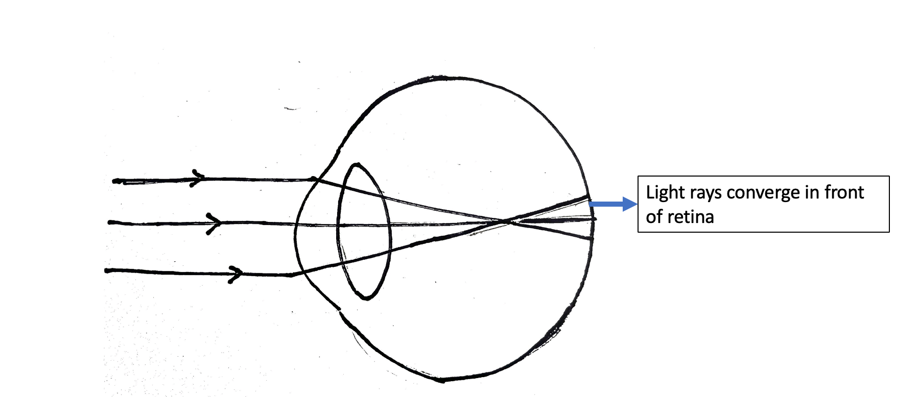
Myopia is characterized by the inability to see distant objects. Myopia results when parallel rays are focused in front of the retina when the accommodation is relaxed (figure). The global burden of myopia is rapidly increasing. In 2010 approximately 27% of the global population, or around 1.45 billion people, were affected.[1] It is expected that by 2030, half of the world's population will be affected by myopia.[2] 

Myopia can be broadly classified as pathological and spontaneous onset childhood myopia.[3] Pathological myopia results from a rapid increase in the axial length with a usual absolute spectacle power of more than six diopters.[4]

This rapid increase in myopia causes a large number of degenerative changes in the retina, choroid, and sclera. Thus it is termed pathological myopia. On the contrary, school-age myopia is the most common type. It has a slow course and usually stabilizes by the age of 20 years.[3]

Etiology

In myopia, the image forms in front of the retinal photoreceptors.

Based on the pathogenesis, myopia can be classified as the following:

  • Axial myopia- Axial myopia results from a rapid increase in the axial length. The axial length increases by 0.35 millimeters for every diopter increase in myopia.[5]
  • Curvature myopia- Curvatural myopia results from the increase in corneal curvature. As a result, the image is focused in front of the retina. Each millimeter change in the radius of curvature of the cornea causes a myopic shift by six diopters.[6]
  • Lenticular myopia- Lenticular myopia results from an increase in the refractive index of the crystalline lens.[7]
  • Positional myopia and other conditions- Myopia may result from anterior shifting of the crystalline lens.[8] Sudden onset myopia may result from the anterior shift of the lens-iris diaphragm in various conditions, including choroidal effusion and anterior rotation of the ciliary body due to drugs, including topiramate.[8]

Epidemiology

Myopia has become a concern for public health. The growing incidence of myopia can be attributed to reduced outdoor activities, increased screen time, and prolonged near-vision work, especially during the COVID (coronavirus disease) pandemic.[9] The reported prevalence of myopia in Singapore amongst children aged 6 to 7 years is 20 to 30%.[10] In China, the prevalence of myopia in 5 to 15-year-old children ranges from 5.7 to 78.4%.[11] 

Myopia is more prevalent in Asian children than in European countries, which report a lower prevalence of myopia (17.8-23.5%).[12] In the United States, the prevalence of myopia ranges from 4.6% to 28% in children between the ages of 6-12 years.[13] In India, the range varies between 8.5 to 15% among urban children of 5 to 15 years.[14]

Pathophysiology

Myopia and Ocular Development

Refractive error results from a long, complex process of ocular development; hence myopia cannot be attributed to a single trait. Many factors contribute to the development of childhood myopia, including the gain and effectiveness of emmetropization during the early years of life, environmental influences, genetic factors, and changes in axial length and lens power during the adolescent phase.[15]

The development of myopia occurs at the same age at which hyperopia corrects. Early-onset myopia is usually associated with higher errors and results in progressive thinning of the choroid, staphyloma, and pathological retinal degeneration.[16] 

Emmetropization

Emmetropization is a process where the refractive components (corneal curvature and lenticular curvature) come in balance with the eyeball's post-natal development resulting in the nullification of refractive errors. The child at birth is hyperopic, with an average refractive error of  +2D to +3D.[17] However, with age progression, this refractive error nullifies and reaches a state of emmetropia or myopia (a more common refractive error in the population).[18] 

The axial length at birth is 16 to 18 mm, increasing to 23 mm by three years. After three years of age, the axial length growth rate reduces.[19] This rapid increase in the axial length should cause a myopic shift; however, it is kept under control by other changes in the lens and corneal curvature, preventing the rapid myopic shift.[20]

Sclera and Myopia

The sclera is the outermost coat of the eye and is composed chiefly of collagen types I and III.[21] Proteoglycans modulate collagen assembly. Decorin and biglycan are the most common sulfated proteoglycans present in the sclera.[22] These proteoglycans' hydration is considered responsible for age-related changes in the sclera. The interaction between scleral fibroblast cells and the scleral matrix plays a vital role in controlling the distensibility of the sclera during eye growth.[23] 

During emmetropization, the development of the eye is governed by the accurate regulation of the growth and remodeling of the scleral extracellular matrix.

  • Embryonic Development of Sclera: Sclera develops in the sixth week of prenatal life from the cells of the neural crest (neuro-ectoderm) and mesoderm.[24] The sclera reaches its adult size by the age of 10 years. However, the extracellular matrix of the sclera keeps on altering.[25] Sclera in myopic patients is characterized by increased elasticity, which can be attributed to the ultrastructural changes of the sclera. The fibroblasts are arranged in a lamellar pattern in myopic patients, associated with thinning of collagen bundles.[26] This increased elasticity of the sclera increases the axial length shifting the image anteriorly.
  • Emmetropization and Scleral Remodeling: The remodeling of the extracellular matrix of the sclera is regulated by several growth factors, including insulin-like growth factors (IGF-1 and IGF-2).[27] Besides that, the scleral extracellular matrix remodeling is also controlled by the locally generated growth factors from the retina and choroid.[28] It has been suggested in the various experimental models that visual signals in the form of a retinal blur cause the generation of Gamma-aminobutyric acid (GABA), dopamine, insulin, and glucagon, which further induce a response in the retinal pigment epithelium and choroid to release the regulatory growth factors, ultimately leading to the scleral extracellular matrix remodeling.[29] 

Choroidal Modulation

The choroid is a highly vascular middle coat of the eyeball. It provides nutrients and oxygen to the outer retinal layers and sclera. Various animal models have demonstrated the importance of the choroid in myopia development and emmetropization. The choroid regulates its thickness to adjust the retina to the focal plane of the eye, a term known as  'choroidal accommodation.'[30] 

The choroid also delivers growth-stimulating factors to the sclera, thus regulating the scleral extracellular matrix and the axial length.[31] Animal models have suggested that an increase in the production of choroidal all-trans-retinoic acid has been associated with a reduction in the scleral proteoglycan and an increase in the axial length.[32]

With the advent of non-invasive techniques like enhanced depth spectral-domain optical coherence tomography (SDOCT), choroidal imaging is possible now. Choroid in highly myopic patients is usually associated with thinning on OCT. A thinner choroid on OCT suggests a poorer prognosis and is generally associated with thinner retinal layers.[33] 

Lens Curvature Changes in Childhood

The lens at birth has a spherical contour and flattens eventually.[34] The flattening of the lens can be due to the equatorial expansion and central compaction forces generated by the growing eyeball.[35] As the eyeball grows, the thickness of the lens also reduces along with the increase in the diameter of the ciliary body, which tensions the zonules and causes the thinning of the lens.[36] 

The thickness of the lens reduces from 4mm at birth to 3.3mm by adolescence.[37] This lens thinning changes the lens's dioptric power from 34.4 D at birth to 23 D at three years of age to 20 D at 14 years of age, preventing the myopic shift.[38]  

 Factors Affecting Myopia Development and Progression

Family History

It has been suggested in various studies that the risk of early-onset myopia development[39] and progression is higher in children if either of the parents has myopia.[40]

Birth History

Low birth weight and premature birth associated with retinopathy of prematurity have been suggested to be associated with myopia development.[41] Sunlight exposure (birth during summer) during the perinatal period is also associated with myopia development in later life.[42]

Excessive Near Work 

Excessive near work is associated with myopia progression resulting from the accommodation lag. Accommodation lag differs between the accommodative stimulus (demand) and the accommodative response.[43] The longer the accommodation lag is, the more myopia progression. Excessive near work in myopes causes a longer retinal defocus, which further acts as a stimulus for increasing the axial length.[44]

Higher Intelligence Quotient

Myopia development may be associated with higher cognitive functions, better education, and a higher intelligence quotient.[45] In a population-based study by Mirshahi et al., it was found that higher levels of professional education were associated with a higher myopic refractive error compared to participants with less education.[46] This can be attributed to the defocus signals in the peripheral and central retina with constant accommodation lags.[47]

Outdoor Activities

Outdoor activities in various studies have been found to reduce myopia progression. It has been hypothesized that the wavelength of radiant sunlight is 550nm, the same wavelength focused on a normal observer's retina.[48] On the contrary, indoor lights have a longer wavelength and are focused behind the retina.[49] 

An experimental study found that the spatial features of the indoor environment are similar to the artificial spatial features created by diffuse filters that induced myopia in animals.[50] Another hypothesis states that sunlight inhibits the increase in axial length by promoting dopamine release.[51]

Increased Screen Time

Increased screen time can lead to myopia development and can be attributed to the increased time spent indoors.[52] In a study done by Enthoven et al., it was found that continuous use of smartphone devices for 20 minutes was associated with a higher risk of myopia development.[53]

History and Physical

Children with a myopic refractive error usually complain of blurring of distance vision. School-going children often complain of difficulty seeing the blackboard. The child may also present with asthenopia, headache, and brow pain.[54] A comprehensive eye examination should be done, and myopic posterior segment changes should be ruled out.[55]

Evaluation

Refraction under cycloplegia should be performed up to 20 years old to prevent myopia's over-estimation.[56] The commonly used cycloplegic agents are atropine 1%, homatropine 2%, cyclopentolate 1%, tropicamide 1%, and tropicamide 0.8%, with phenylephrine 5%. Of these, 1% atropine is the strongest cycloplegic agent, the effect of which lasts for 14 days.[57] The onset of action of homatropine starts after one hour and lasts for 1 to 3 days.[58]

Cyclopentolate is the preferred cycloplegic for evaluating refractive error among children aged 5 to 13. Tropicamide chiefly acts as a mydriatic, but this is an effective agent for evaluating myopic children above 13 years of age.[59][60] 

Atropine ointment must be cautiously instilled to prevent systemic complications like facial flushing, fever, and tachycardia. The guidelines for spectacle prescription in myopic children have been summarized below (American Academy of Ophthalmology- Preferred Practice Pattern on Pediatric eye evaluations).[61]

  Age <1 year  Age 1- less than 2 years Age 2- less than 3 years Age 3- less than 4 years

Similar refractive error (myopia) in both eyes (Isometropia)

5 DS / more 4 DS/  more  3 DS/ more  2.5 DS/ more
Myopic Anisometropia (without squint) 4 DS/ more   3 DS / more  3 DS / more 2.5 DS/ more

A complete examination of the anterior segment and fundus evaluation should be done after refraction. Fundus evaluation in patients with pathological myopia can reveal degenerative changes, lattice degeneration, peripheral retinal holes, cobblestone degeneration, lacquer cracks, macular hole, and staphyloma.[62]

Treatment / Management

Management

Spectacles

Spectacles are the most commonly advised management option for childhood myopia. The refractive error correction is performed under cycloplegia. Spectacle coverage remains an important issue in resource-deficient areas and developing countries. While prescribing spectacles to children, it is important to address certain factors, including the shape and weight of frames and lenses, to ensure better compliance among children. 

Contact lenses

Soft contact lenses and rigid gas-permeable lenses can be prescribed to correct myopia. However, there is no substantial evidence that these modalities can reduce myopia progression.[63]

Measures for controlling myopia progression

Drugs for Myopia Control

As of June 2022, US Food and Drug Administration (US FDA) approved none of the myopia management drugs. However, atropine 0.01% is the most widely studied drug for halting myopia progression. The atropine in myopia-1 (ATOM-1) study was performed to evaluate the role of atropine 1%.[64]

Atropine in myopia study 2 (ATOM-2) studied the role of atropine 0.5%, 0.1%, and 0.01% in managing myopia and was carried out in two phases. The study found that atropine 0.01% was a safe and effective option for myopia management, with minimal side effects of photophobia and loss of accommodation, as seen with atropine 1%/0.5%[65] 

Atropine is an anticholinergic drug that acts non-selectively on the acetylcholine receptors and down-regulates its functions. Acetylcholine controls the growth of the eye and has a crucial role in the developing retina.[66]  Atropine stimulates the synthesis of the scleral extracellular matrix, thus reducing the scleral rigidity and its tendency for elongation.[67] 

At the cellular level, atropine has been found to downregulate the Epidermal growth factor receptor pathways (EGFR).[68] Atropine, when injected intravitreally in animal models, has been found to promote dopamine release, which further regulates the axial length increase.[69] 

Atropine also reduces choroidal thinning caused by hyperopic defocus in myopic eyes.[70] Another hypothesis states that atropine controls myopia progression by regulating excessive accommodation. However, later it was found that myopia induction could not be stopped even after experimentally eliminating the accommodation reflex by optic nerve sectioning or destruction of Edinger Westphal nuclei.[71]

Pirenzapine

Pirenzapine is a selective M1/M4 muscarinic receptor antagonist. Due to its better safety profile, pirenzepine was tried for myopia management; 0.5% and 2% of pirenzepine were used for myopia management.[72][73] 

7 Methylxanthine

7-Methylxanthine is a metabolite of theobromine and caffeine. The possible mechanism of action of the drug is to modulate the axial length by increasing the collagen fibril diameters and overall thickness of the posterior sclera.[74]

Intra-ocular Pressure-lowering Drugs

Intra-ocular pressure-lowering drugs like timolol maleate and latanoprost[75] have been tried to halt myopia progression.[76][77] It has been suggested that intraocular pressure causes a stretch on the outer scleral wall leading to enlargement of the eyeball.[78] 

The biomechanically weaker scleral walls in myopes are at an increased risk of being stretched by the increased intraocular pressure. Hence, a decrease in the intraocular pressure can slow the elongation of the eye, thus ceasing myopia progression.[79]

Lifestyle Modifications: Outdoor Activities

The risk of myopia is reduced by 2% for every hour of increase in outdoor activity.[80] Increasing the outdoor activity duration to 14 hours per week can reduce the risk of myopia development by one-third. Outdoor activities reduce myopia progression by promoting the release of dopamine.[81]

Dopamine inhibits axial length elongation.[82] Another mechanism can be the difference in spatial frequencies of the indoor and outdoor environments. Enhancement of the spatial frequency can help to limit myopia progression.[81][83][82]

Bifocal/ Multifocal Glasses

Myopia progression is thought to be a result of prolonged accommodation. Treatment with bifocal or multifocal glasses is assumed beneficial as these relax the accommodation. Cheng et al., in their study, reported a 40% decreased myopia progression with bifocal glasses.[84]

Progressive Glasses

Progressive glasses have been studied for their effectiveness in controlling myopia progression. Gwiazda et al., in their study on progressive additional lenses, found a 20% decline in myopia progression during the first year of usage.[85] Further, it has been reported that progressive glasses were more beneficial in cases where both the parents were myopic, a larger accommodation lag was present, or in patients with esophoria near.[86]  

Defocus Incorporated Multiple Segments Spectacle  (DIMS)

The defocus incorporated multiple segment spectacles inhibit myopia progression by inducing a myopic defocus. Animal studies have found that myopic defocus reduces the eye's axial length; however, hyperopic defocus increases the axial length.[87] [88] DIMS consists of a central zone of 9 mm diameter and annular zones of 33 mm with a relative positive power of +3.50 D. Each segment has a diameter of 1.03 mm.[89]

This design of the lens induces myopic defocus with clear vision. In an observation by Lam et al., it was found that on continuously wearing the DIMS, the myopia progression was reduced by 52%, and axial length progression was reduced by 62%.[89]  

Defocus Incorporated Soft Contact Lens (DISC)

Defocus-incorporated soft contact lenses are bifocal soft contact lenses with a central correction zone and a sequence of alternating correction and defocusing zones in the periphery.[90] This induces myopic defocus and clear vision at the same time.[91] 

The power of the central zone was customized according to the cycloplegic refractive error, while the defocusing zones were relatively negative by 2.5 D. Daily use of the DISC for 5 to 8 hours has been found to reduce myopia progression.[90] Similarly, dual-focus soft contact lenses have also been found to reduce myopia progression.[92]

Orthokeratology 

Orthokeratology is the only US FDA-approved modality for myopia. Orthokeratology involves wearing overnight contact lenses that alter the shape of the cornea from prolate to oblate, thus reducing the refractive error. Contact lens appears to be a promising adjuvant to other options, but the issues of hygiene and maintenance need to be addressed and explained to the patients and guardians.[93]

Other therapies explored in the management of myopia and reducing the progression of myopia include posterior scleral contraction, reinforcement of posterior sclera, scleral cross-linking with riboflavin but can cause loss of photoreceptors, outer nuclear layer, and RPE, sub-scleral injection of mesenchymal stem cells and dopamine, intravitreal injection of aquaporin-1, scleral strengthening using sub-tenon chemicals like ethyl acrylate and acrilamidehydrazide.[94][95][96]

Differential Diagnosis

The other causes of low vision in children should be ruled out, which include keratoconus, pediatric cataracts, microspherophakia, pediatric glaucoma, trauma, irido-fundal coloboma, nystagmus, congenital optic nerve abnormalities like optic disc coloboma, large myelinated nerve fibers involving the fovea, and congenital retinal anomalies, like pigmentary retinopathy. One should also enquire about the birth history, history of laser for retinopathy of prematurity, delayed cry at birth, and history of intensive care unit (ICU) stay. Myopia can also be associated with Down syndrome (8 to 41%).[97] 

Marfan syndrome and Stickler syndrome: Pseudomyopia is the overestimation of myopia due to excessive accommodation usually seen in children. Hence, refraction done without cycloplegia overestimates myopia by -1 to -2 diopter.[98]

Prognosis

School children with early-onset myopia usually have higher axial length and refractive error. On the contrary, the course of progression in patients with congenital myopia (myopia more than -5 D at less than six years of age) is different. Shih et al., in their observation, found faster rates of myopia progression in children with lower grades of myopia, 5.0 to 7.75 D, compared to children with higher grades of myopia (maximum of 11.0 D).[99]

Patients with pathological myopia, thinned-out choroid, and posterior staphyloma have worse visual outcomes in the long term.[100]

Complications

Pathological myopia can be associated with retinal complications like retinal detachment, myopic macular traction, macular hole, and choroidal neovascular membrane formation. High myopes can also be associated with subluxated lenses and are at a higher risk of developing primary open-angle glaucoma.[101][102]

Deterrence and Patient Education

A large number of factors can contribute to the etiopathogenesis of myopia. Prolonged screen hours and indoor confinement are often blamed for the development and progression of myopia. Promoting outdoor activities, using modalities like atropine 0.01% once daily in both eyes at night and contact lenses can help prevent myopia progression.[30]

Parents should clearly understand the need for glasses and the risk of myopia progression. A complete eye examination and refraction should be done regularly. Often myopia in children gets unnoticed as children cannot explain their problems clearly and are usually incidentally diagnosed during a routine evaluation. School eye screening programs should be promoted with better spectacle coverage, especially in developing countries.[103]

Enhancing Healthcare Team Outcomes

Myopia has emerged as a new public health issue. Preventing, managing, and halting the progression of myopia has always been a matter of concern. In recent times pharmaceutical management of myopia has been widely studied. ATOM-1 was a large randomized controlled trial (RCT) performed to evaluate the role of atropine 1% in preventing the progression of childhood myopia.[104] 

Four hundred children aged 6 to 12 years with 1 to 6 D myopia were included in the study. The patients were randomly assigned to receive either atropine or a placebo. After two years, myopia in atropine-treated eyes regressed by 0.3 Diopter (D) ±0.50 D. Conversely, progression was noted in the placebo group -0.76 D +/- 0.44 D.[64] 

Though the study provided strong evidence of atropine being a practical option for preventing myopia progression, numerous side effects like blurring of near vision, photophobia, glare, and systemic side effects were noted to occur with 1% atropine. This further led to the investigation of the efficacy of low-dose atropine 0.5%, 0.1%, and 0.01% in controlling myopia progression (ATOM-2).[65]

Atropine 0.01% was found to be a safe and effective option, as it caused minimal pupillary dilation and minimally affected accommodation with similar efficacy. However, in a recent study on a low concentration of atropine for myopia progression, the effectiveness of 0.05% atropine was found to be double that of atropine 0.01% over two years.[105] 

Cooper vision Mi sight daily use soft lenses have been recently approved by Food and Drug Administration (FDA). They are designed to reduce myopia progression by decreasing peripheral retinal hyperopic defocus.[106] 

Optometrists are essential in managing these cases through early detection and regular follow-ups. Patients with signs of progressive myopia should be educated about modern treatment options to control the further progression of refractive errors. Counselors are vital in educating parents or caretakers about lifestyle modifications to help prevent myopia progression.

All these various professionals need to function as an interprofessional team to optimize patient care when managing childhood myopia and monitoring ocular development.


(Click Image to Enlarge)
Ray diagram showing parallel rays being focused in front of retina
Contributed by Gunjan Saluja MBBS, MD, DNB
Details

Author

Gunjan Saluja

Editor:

Kirandeep Kaur

Updated:

5/4/2023 10:52:28 PM

References


[1]

Holden BA,Wilson DA,Jong M,Sankaridurg P,Fricke TR,Smith EL III,Resnikoff S, Myopia: a growing global problem with sight-threatening complications. Community eye health. 2015;     [PubMed PMID: 26692649]


[2]

Morgan IG,Ohno-Matsui K,Saw SM, Myopia. Lancet (London, England). 2012 May 5;     [PubMed PMID: 22559900]


[3]

Carr BJ,Stell WK,Kolb H,Fernandez E,Nelson R, The Science Behind Myopia Webvision: The Organization of the Retina and Visual System. 1995     [PubMed PMID: 29266913]


[4]

Saw SM,Gazzard G,Shih-Yen EC,Chua WH, Myopia and associated pathological complications. Ophthalmic & physiological optics : the journal of the British College of Ophthalmic Opticians (Optometrists). 2005 Sep     [PubMed PMID: 16101943]


[5]

Atchison DA,Jones CE,Schmid KL,Pritchard N,Pope JM,Strugnell WE,Riley RA, Eye shape in emmetropia and myopia. Investigative ophthalmology & visual science. 2004 Oct     [PubMed PMID: 15452039]


[6]

Majumdar S, Tripathy K. Hyperopia. StatPearls. 2023 Jan:():     [PubMed PMID: 32809551]


[7]

Cooper J,Tkatchenko AV, A Review of Current Concepts of the Etiology and Treatment of Myopia. Eye     [PubMed PMID: 29901472]


[8]

Mazumdar S, Tripathy K, Sarma B, Agarwal N. Acquired myopia followed by acquired hyperopia due to serous neurosensory retinal detachment following topiramate intake. European journal of ophthalmology. 2019 Jan:29(1):NP21-NP24. doi: 10.1177/1120672118797286. Epub 2018 Sep 3     [PubMed PMID: 30175623]


[9]

Wang J,Li Y,Musch DC,Wei N,Qi X,Ding G,Li X,Li J,Song L,Zhang Y,Ning Y,Zeng X,Hua N,Li S,Qian X, Progression of Myopia in School-Aged Children After COVID-19 Home Confinement. JAMA ophthalmology. 2021 Mar 1;     [PubMed PMID: 33443542]

Level 2 (mid-level) evidence

[10]

Saw SM,Carkeet A,Chia KS,Stone RA,Tan DT, Component dependent risk factors for ocular parameters in Singapore Chinese children. Ophthalmology. 2002 Nov;     [PubMed PMID: 12414416]


[11]

He M,Zeng J,Liu Y,Xu J,Pokharel GP,Ellwein LB, Refractive error and visual impairment in urban children in southern china. Investigative ophthalmology     [PubMed PMID: 14985292]


[12]

Williams KM,Bertelsen G,Cumberland P,Wolfram C,Verhoeven VJ,Anastasopoulos E,Buitendijk GH,Cougnard-Grégoire A,Creuzot-Garcher C,Erke MG,Hogg R,Höhn R,Hysi P,Khawaja AP,Korobelnik JF,Ried J,Vingerling JR,Bron A,Dartigues JF,Fletcher A,Hofman A,Kuijpers RW,Luben RN,Oxele K,Topouzis F,von Hanno T,Mirshahi A,Foster PJ,van Duijn CM,Pfeiffer N,Delcourt C,Klaver CC,Rahi J,Hammond CJ,European Eye Epidemiology (E(3)) Consortium., Increasing Prevalence of Myopia in Europe and the Impact of Education. Ophthalmology. 2015 Jul;     [PubMed PMID: 25983215]


[13]

Wu PC,Huang HM,Yu HJ,Fang PC,Chen CT, Epidemiology of Myopia. Asia-Pacific journal of ophthalmology (Philadelphia, Pa.). 2016 Nov/Dec;     [PubMed PMID: 27898441]


[14]

Agarwal D,Saxena R,Gupta V,Mani K,Dhiman R,Bhardawaj A,Vashist P, Prevalence of myopia in Indian school children: Meta-analysis of last four decades. PloS one. 2020;     [PubMed PMID: 33075102]

Level 1 (high-level) evidence

[15]

Ramamurthy D,Lin Chua SY,Saw SM, A review of environmental risk factors for myopia during early life, childhood and adolescence. Clinical & experimental optometry. 2015 Nov     [PubMed PMID: 26497977]


[16]

Liang CL,Yen E,Su JY,Liu C,Chang TY,Park N,Wu MJ,Lee S,Flynn JT,Juo SH, Impact of family history of high myopia on level and onset of myopia. Investigative ophthalmology     [PubMed PMID: 15452048]


[17]

MEHRA KS,KHARE BB,VAITHILINGAM E, REFRACTION IN FULL-TERM BABIES. The British journal of ophthalmology. 1965 May     [PubMed PMID: 14290875]


[18]

Flitcroft DI, Emmetropisation and the aetiology of refractive errors. Eye (London, England). 2014 Feb;     [PubMed PMID: 24406411]


[19]

Bhardwaj V,Rajeshbhai GP, Axial length, anterior chamber depth-a study in different age groups and refractive errors. Journal of clinical and diagnostic research : JCDR. 2013 Oct;     [PubMed PMID: 24298478]


[20]

Meng W,Butterworth J,Malecaze F,Calvas P, Axial length of myopia: a review of current research. Ophthalmologica. Journal international d'ophtalmologie. International journal of ophthalmology. Zeitschrift fur Augenheilkunde. 2011;     [PubMed PMID: 20948239]


[21]

Coudrillier B, Pijanka J, Jefferys J, Sorensen T, Quigley HA, Boote C, Nguyen TD. Collagen structure and mechanical properties of the human sclera: analysis for the effects of age. Journal of biomechanical engineering. 2015 Apr:137(4):041006. doi: 10.1115/1.4029430. Epub 2015 Feb 11     [PubMed PMID: 25531905]


[22]

Rada JA,Achen VR,Penugonda S,Schmidt RW,Mount BA, Proteoglycan composition in the human sclera during growth and aging. Investigative ophthalmology & visual science. 2000 Jun     [PubMed PMID: 10845580]


[23]

Rada JA,Shelton S,Norton TT, The sclera and myopia. Experimental eye research. 2006 Feb;     [PubMed PMID: 16202407]


[24]

Hoar RM. Embryology of the eye. Environmental health perspectives. 1982 Apr:44():31-4     [PubMed PMID: 7084153]

Level 3 (low-level) evidence

[25]

Zadnik K,Satariano WA,Mutti DO,Sholtz RI,Adams AJ, The effect of parental history of myopia on children's eye size. JAMA. 1994 May 4     [PubMed PMID: 8158816]


[26]

McBrien NA,Gentle A, Role of the sclera in the development and pathological complications of myopia. Progress in retinal and eye research. 2003 May;     [PubMed PMID: 12852489]


[27]

Cuthbertson RA,Beck F,Senior PV,Haralambidis J,Penschow JD,Coghlan JP, Insulin-like growth factor II may play a local role in the regulation of ocular size. Development (Cambridge, England). 1989 Sep     [PubMed PMID: 2560708]


[28]

Summers JA, The choroid as a sclera growth regulator. Experimental eye research. 2013 Sep     [PubMed PMID: 23528534]


[29]

Troilo D,Smith EL 3rd,Nickla DL,Ashby R,Tkatchenko AV,Ostrin LA,Gawne TJ,Pardue MT,Summers JA,Kee CS,Schroedl F,Wahl S,Jones L, IMI - Report on Experimental Models of Emmetropization and Myopia. Investigative ophthalmology     [PubMed PMID: 30817827]


[30]

Németh J,Tapasztó B,Aclimandos WA,Kestelyn P,Jonas JB,De Faber JHN,Januleviciene I,Grzybowski A,Nagy ZZ,Pärssinen O,Guggenheim JA,Allen PM,Baraas RC,Saunders KJ,Flitcroft DI,Gray LS,Polling JR,Haarman AE,Tideman JWL,Wolffsohn JS,Wahl S,Mulder JA,Smirnova IY,Formenti M,Radhakrishnan H,Resnikoff S, Update and guidance on management of myopia. European Society of Ophthalmology in cooperation with International Myopia Institute. European journal of ophthalmology. 2021 May;     [PubMed PMID: 33673740]


[31]

Harper AR,Summers JA, The dynamic sclera: extracellular matrix remodeling in normal ocular growth and myopia development. Experimental eye research. 2015 Apr     [PubMed PMID: 25819458]


[32]

Troilo D,Nickla DL,Mertz JR,Summers Rada JA, Change in the synthesis rates of ocular retinoic acid and scleral glycosaminoglycan during experimentally altered eye growth in marmosets. Investigative ophthalmology & visual science. 2006 May     [PubMed PMID: 16638980]


[33]

Read SA,Fuss JA,Vincent SJ,Collins MJ,Alonso-Caneiro D, Choroidal changes in human myopia: insights from optical coherence tomography imaging. Clinical     [PubMed PMID: 30565333]


[34]

O'Rahilly R, The prenatal development of the human eye. Experimental eye research. 1975 Aug     [PubMed PMID: 1100417]


[35]

Muralidharan G,Martínez-Enríquez E,Birkenfeld J,Velasco-Ocana M,Pérez-Merino P,Marcos S, Morphological changes of human crystalline lens in myopia. Biomedical optics express. 2019 Dec 1;     [PubMed PMID: 31853387]


[36]

Mutti DO,Zadnik K,Fusaro RE,Friedman NE,Sholtz RI,Adams AJ, Optical and structural development of the crystalline lens in childhood. Investigative ophthalmology & visual science. 1998 Jan     [PubMed PMID: 9430553]


[37]

Augusteyn RC, On the growth and internal structure of the human lens. Experimental eye research. 2010 Jun     [PubMed PMID: 20171212]


[38]

Gordon RA,Donzis PB, Refractive development of the human eye. Archives of ophthalmology (Chicago, Ill. : 1960). 1985 Jun     [PubMed PMID: 4004614]


[39]

Jiang X,Tarczy-Hornoch K,Cotter SA,Matsumura S,Mitchell P,Rose KA,Katz J,Saw SM,Varma R,POPEYE Consortium., Association of Parental Myopia With Higher Risk of Myopia Among Multiethnic Children Before School Age. JAMA ophthalmology. 2020 May 1;     [PubMed PMID: 32191277]


[40]

Lee YY,Lo CT,Sheu SJ,Yin LT, Risk factors for and progression of myopia in young Taiwanese men. Ophthalmic epidemiology. 2015 Feb     [PubMed PMID: 25495661]


[41]

O'Connor AR,Stephenson TJ,Johnson A,Tobin MJ,Ratib S,Fielder AR, Change of refractive state and eye size in children of birth weight less than 1701 g. The British journal of ophthalmology. 2006 Apr     [PubMed PMID: 16547327]


[42]

Mandel Y,Grotto I,El-Yaniv R,Belkin M,Israeli E,Polat U,Bartov E, Season of birth, natural light, and myopia. Ophthalmology. 2008 Apr     [PubMed PMID: 17698195]


[43]

Gwiazda JE,Hyman L,Norton TT,Hussein ME,Marsh-Tootle W,Manny R,Wang Y,Everett D,COMET Grouup., Accommodation and related risk factors associated with myopia progression and their interaction with treatment in COMET children. Investigative ophthalmology     [PubMed PMID: 15223788]


[44]

Berntsen DA,Sinnott LT,Mutti DO,Zadnik K,CLEERE Study Group., Accommodative lag and juvenile-onset myopia progression in children wearing refractive correction. Vision research. 2011 May 11;     [PubMed PMID: 21342658]


[45]

Rosner M,Belkin M, Intelligence, education, and myopia in males. Archives of ophthalmology (Chicago, Ill. : 1960). 1987 Nov     [PubMed PMID: 3675282]


[46]

Mirshahi A,Ponto KA,Hoehn R,Zwiener I,Zeller T,Lackner K,Beutel ME,Pfeiffer N, Myopia and level of education: results from the Gutenberg Health Study. Ophthalmology. 2014 Oct;     [PubMed PMID: 24947658]


[47]

Flitcroft DI, The complex interactions of retinal, optical and environmental factors in myopia aetiology. Progress in retinal and eye research. 2012 Nov     [PubMed PMID: 22772022]


[48]

Torii H,Ohnuma K,Kurihara T,Tsubota K,Negishi K, Violet Light Transmission is Related to Myopia Progression in Adult High Myopia. Scientific reports. 2017 Nov 6     [PubMed PMID: 29109514]


[49]

Rose KA,Morgan IG,Ip J,Kifley A,Huynh S,Smith W,Mitchell P, Outdoor activity reduces the prevalence of myopia in children. Ophthalmology. 2008 Aug;     [PubMed PMID: 18294691]


[50]

Flitcroft DI,Harb EN,Wildsoet CF, The Spatial Frequency Content of Urban and Indoor Environments as a Potential Risk Factor for Myopia Development. Investigative ophthalmology & visual science. 2020 Sep 1     [PubMed PMID: 32986814]


[51]

McCarthy CS,Megaw P,Devadas M,Morgan IG, Dopaminergic agents affect the ability of brief periods of normal vision to prevent form-deprivation myopia. Experimental eye research. 2007 Jan;     [PubMed PMID: 17094962]


[52]

Yang GY,Huang LH,Schmid KL,Li CG,Chen JY,He GH,Liu L,Ruan ZL,Chen WQ, Associations Between Screen Exposure in Early Life and Myopia amongst Chinese Preschoolers. International journal of environmental research and public health. 2020 Feb 7     [PubMed PMID: 32046062]


[53]

Enthoven CA,Polling JR,Verzijden T,Tideman JWL,Al-Jaffar N,Jansen PW,Raat H,Metz L,Verhoeven VJM,Klaver CCW, Smartphone Use Associated with Refractive Error in Teenagers: The Myopia App Study. Ophthalmology. 2021 Dec     [PubMed PMID: 34245754]


[54]

Gessesse SA, Teshome AW. Prevalence of myopia among secondary school students in Welkite town: South-Western Ethiopia. BMC ophthalmology. 2020 May 4:20(1):176. doi: 10.1186/s12886-020-01457-2. Epub 2020 May 4     [PubMed PMID: 32366285]


[55]

Yadav S,Tandon R, Comprehensive eye examination: what does it mean? Community eye health. 2019     [PubMed PMID: 32123482]


[56]

Sanfilippo PG,Chu BS,Bigault O,Kearns LS,Boon MY,Young TL,Hammond CJ,Hewitt AW,Mackey DA, What is the appropriate age cut-off for cycloplegia in refraction? Acta ophthalmologica. 2014 Sep     [PubMed PMID: 24641244]


[57]

Mimouni M,Zoller L,Horowitz J,Wygnanski-Jaffe T,Morad Y,Mezer E, Cycloplegic autorefraction in young adults: is it mandatory? Graefe's archive for clinical and experimental ophthalmology = Albrecht von Graefes Archiv fur klinische und experimentelle Ophthalmologie. 2016 Feb     [PubMed PMID: 26686513]


[58]

Major E, Dutson T, Moshirfar M. Cycloplegia in Children: An Optometrist's Perspective. Clinical optometry. 2020:12():129-133. doi: 10.2147/OPTO.S217645. Epub 2020 Aug 25     [PubMed PMID: 32904515]

Level 3 (low-level) evidence

[59]

Manny RE,Hussein M,Scheiman M,Kurtz D,Niemann K,Zinzer K,COMET Study Group., Tropicamide (1%): an effective cycloplegic agent for myopic children. Investigative ophthalmology & visual science. 2001 Jul     [PubMed PMID: 11431435]


[60]

Hong D,Tripathy K, Tropicamide StatPearls. 2022 Jan;     [PubMed PMID: 31082113]


[61]

Wallace DK,Morse CL,Melia M,Sprunger DT,Repka MX,Lee KA,Christiansen SP,American Academy of Ophthalmology Preferred Practice Pattern Pediatric Ophthalmology/Strabismus Panel., Pediatric Eye Evaluations Preferred Practice Pattern®: I. Vision Screening in the Primary Care and Community Setting; II. Comprehensive Ophthalmic Examination. Ophthalmology. 2018 Jan     [PubMed PMID: 29108745]


[62]

Ohno-Matsui K, Pathologic Myopia. Asia-Pacific journal of ophthalmology (Philadelphia, Pa.). 2016 Nov/Dec     [PubMed PMID: 27898445]


[63]

Walline JJ,Jones LA,Sinnott L,Manny RE,Gaume A,Rah MJ,Chitkara M,Lyons S,ACHIEVE Study Group., A randomized trial of the effect of soft contact lenses on myopia progression in children. Investigative ophthalmology & visual science. 2008 Nov     [PubMed PMID: 18566461]

Level 1 (high-level) evidence

[64]

Chua WH,Balakrishnan V,Chan YH,Tong L,Ling Y,Quah BL,Tan D, Atropine for the treatment of childhood myopia. Ophthalmology. 2006 Dec;     [PubMed PMID: 16996612]


[65]

Chia A,Chua WH,Cheung YB,Wong WL,Lingham A,Fong A,Tan D, Atropine for the treatment of childhood myopia: safety and efficacy of 0.5%, 0.1%, and 0.01% doses (Atropine for the Treatment of Myopia 2). Ophthalmology. 2012 Feb;     [PubMed PMID: 21963266]


[66]

Ford KJ,Feller MB, Assembly and disassembly of a retinal cholinergic network. Visual neuroscience. 2012 Jan;     [PubMed PMID: 21787461]


[67]

Cristaldi M,Olivieri M,Pezzino S,Spampinato G,Lupo G,Anfuso CD,Rusciano D, Atropine Differentially Modulates ECM Production by Ocular Fibroblasts, and Its Ocular Surface Toxicity Is Blunted by Colostrum. Biomedicines. 2020 Apr 5     [PubMed PMID: 32260532]


[68]

Barathi VA,Weon SR,Beuerman RW, Expression of muscarinic receptors in human and mouse sclera and their role in the regulation of scleral fibroblasts proliferation. Molecular vision. 2009 Jun 30;     [PubMed PMID: 19578554]


[69]

Schwahn HN,Kaymak H,Schaeffel F, Effects of atropine on refractive development, dopamine release, and slow retinal potentials in the chick. Visual neuroscience. 2000 Mar-Apr     [PubMed PMID: 10824671]


[70]

Chiang ST,Phillips JR, Effect of Atropine Eye Drops on Choroidal Thinning Induced by Hyperopic Retinal Defocus. Journal of ophthalmology. 2018     [PubMed PMID: 29576882]


[71]

Schaeffel F,Troilo D,Wallman J,Howland HC, Developing eyes that lack accommodation grow to compensate for imposed defocus. Visual neuroscience. 1990 Feb;     [PubMed PMID: 2271446]


[72]

Bartlett JD,Niemann K,Houde B,Allred T,Edmondson MJ,Crockett RS, A tolerability study of pirenzepine ophthalmic gel in myopic children. Journal of ocular pharmacology and therapeutics : the official journal of the Association for Ocular Pharmacology and Therapeutics. 2003 Jun;     [PubMed PMID: 12828845]


[73]

Siatkowski RM,Cotter S,Miller JM,Scher CA,Crockett RS,Novack GD,US Pirenzepine Study Group., Safety and efficacy of 2% pirenzepine ophthalmic gel in children with myopia: a 1-year, multicenter, double-masked, placebo-controlled parallel study. Archives of ophthalmology (Chicago, Ill. : 1960). 2004 Nov;     [PubMed PMID: 15534128]


[74]

Trier K,Munk Ribel-Madsen S,Cui D,Brøgger Christensen S, Systemic 7-methylxanthine in retarding axial eye growth and myopia progression: a 36-month pilot study. Journal of ocular biology, diseases, and informatics. 2008 Dec     [PubMed PMID: 20072638]

Level 3 (low-level) evidence

[75]

Tripathy K,Geetha R, Latanoprost StatPearls. 2021 Jan     [PubMed PMID: 31082022]


[76]

Qi H,Gao C,Li Y,Feng X,Wang M,Zhang Y,Chen Y, The effect of Timolol 0.5% on the correction of myopic regression after LASIK. Medicine. 2017 Apr     [PubMed PMID: 28445315]

Level 2 (mid-level) evidence

[77]

El-Nimri NW,Wildsoet CF, Effects of Topical Latanoprost on Intraocular Pressure and Myopia Progression in Young Guinea Pigs. Investigative ophthalmology & visual science. 2018 May 1     [PubMed PMID: 29847673]


[78]

Nickla DL, Ocular diurnal rhythms and eye growth regulation: where we are 50 years after Lauber. Experimental eye research. 2013 Sep     [PubMed PMID: 23298452]


[79]

Wang P,Chen S,Liu Y,Lin F,Song Y,Li T,Aung T,Zhang X,GSHM study group., Lowering Intraocular Pressure: A Potential Approach for Controlling High Myopia Progression. Investigative ophthalmology & visual science. 2021 Nov 1     [PubMed PMID: 34787640]


[80]

Sherwin JC,Reacher MH,Keogh RH,Khawaja AP,Mackey DA,Foster PJ, The association between time spent outdoors and myopia in children and adolescents: a systematic review and meta-analysis. Ophthalmology. 2012 Oct     [PubMed PMID: 22809757]

Level 1 (high-level) evidence

[81]

Jones LA,Sinnott LT,Mutti DO,Mitchell GL,Moeschberger ML,Zadnik K, Parental history of myopia, sports and outdoor activities, and future myopia. Investigative ophthalmology & visual science. 2007 Aug     [PubMed PMID: 17652719]


[82]

Feldkaemper M,Schaeffel F, An updated view on the role of dopamine in myopia. Experimental eye research. 2013 Sep;     [PubMed PMID: 23434455]


[83]

French AN,Ashby RS,Morgan IG,Rose KA, Time outdoors and the prevention of myopia. Experimental eye research. 2013 Sep     [PubMed PMID: 23644222]


[84]

Cheng D,Schmid KL,Woo GC,Drobe B, Randomized trial of effect of bifocal and prismatic bifocal spectacles on myopic progression: two-year results. Archives of ophthalmology (Chicago, Ill. : 1960). 2010 Jan     [PubMed PMID: 20065211]

Level 1 (high-level) evidence

[85]

Gwiazda J,Marsh-Tootle WL,Hyman L,Hussein M,Norton TT,COMET Study Group., Baseline refractive and ocular component measures of children enrolled in the correction of myopia evaluation trial (COMET). Investigative ophthalmology & visual science. 2002 Feb     [PubMed PMID: 11818372]


[86]

Saw SM,Nieto FJ,Katz J,Schein OD,Levy B,Chew SJ, Familial clustering and myopia progression in Singapore school children. Ophthalmic epidemiology. 2001 Sep     [PubMed PMID: 11471091]


[87]

Liu Y,Wildsoet C, The effect of two-zone concentric bifocal spectacle lenses on refractive error development and eye growth in young chicks. Investigative ophthalmology & visual science. 2011 Feb     [PubMed PMID: 20861487]


[88]

Arumugam B,Hung LF,To CH,Holden B,Smith EL 3rd, The effects of simultaneous dual focus lenses on refractive development in infant monkeys. Investigative ophthalmology     [PubMed PMID: 25324283]


[89]

Lam CSY,Tang WC,Tse DY,Lee RPK,Chun RKM,Hasegawa K,Qi H,Hatanaka T,To CH, Defocus Incorporated Multiple Segments (DIMS) spectacle lenses slow myopia progression: a 2-year randomised clinical trial. The British journal of ophthalmology. 2020 Mar     [PubMed PMID: 31142465]

Level 2 (mid-level) evidence

[90]

Lam CS,Tang WC,Tse DY,Tang YY,To CH, Defocus Incorporated Soft Contact (DISC) lens slows myopia progression in Hong Kong Chinese schoolchildren: a 2-year randomised clinical trial. The British journal of ophthalmology. 2014 Jan     [PubMed PMID: 24169657]

Level 1 (high-level) evidence

[91]

Zhu X,Wallman J, Temporal properties of compensation for positive and negative spectacle lenses in chicks. Investigative ophthalmology & visual science. 2009 Jan     [PubMed PMID: 18791175]


[92]

Anstice NS,Phillips JR, Effect of dual-focus soft contact lens wear on axial myopia progression in children. Ophthalmology. 2011 Jun     [PubMed PMID: 21276616]


[93]

Sankaridurg P. Contact lenses to slow progression of myopia. Clinical & experimental optometry. 2017 Sep:100(5):432-437. doi: 10.1111/cxo.12584. Epub 2017 Jul 28     [PubMed PMID: 28752898]


[94]

Su Y,Pan A,Wu Y,Zhu S,Zheng L,Xue A, The efficacy of posterior scleral contraction in controlling high myopia in young people. American journal of translational research. 2018     [PubMed PMID: 30662614]


[95]

Goodyear MJ,Junghans BM,Giummarra L,Murphy MJ,Crewther DP,Crewther SG, A role for aquaporin-4 during induction of form deprivation myopia in chick. Molecular vision. 2008 Feb 8;     [PubMed PMID: 18334967]


[96]

Avetisov ES,Tarutta EP,Iomdina EN,Vinetskaya MI,Andreyeva LD, Nonsurgical and surgical methods of sclera reinforcement in progressive myopia. Acta ophthalmologica Scandinavica. 1997 Dec     [PubMed PMID: 9527318]


[97]

Creavin AL,Brown RD, Ophthalmic abnormalities in children with Down syndrome. Journal of pediatric ophthalmology and strabismus. 2009 Mar-Apr;     [PubMed PMID: 19343968]


[98]

Liu X,Ye L,Chen C,Chen M,Wen S,Mao X, Evaluation of the Necessity for Cycloplegia During Refraction of Chinese Children Between 4 and 10 Years Old. Journal of pediatric ophthalmology and strabismus. 2020 Jul 1     [PubMed PMID: 32687211]


[99]

Shih YF,Ho TC,Hsiao CK,Lin LL, Long-term visual prognosis of infantile-onset high myopia. Eye (London, England). 2006 Aug     [PubMed PMID: 16096663]


[100]

Ohno-Matsui K,Jonas JB, Posterior staphyloma in pathologic myopia. Progress in retinal and eye research. 2019 May;     [PubMed PMID: 30537538]


[101]

Haarman AEG,Enthoven CA,Tideman JWL,Tedja MS,Verhoeven VJM,Klaver CCW, The Complications of Myopia: A Review and Meta-Analysis. Investigative ophthalmology & visual science. 2020 Apr 9     [PubMed PMID: 32347918]

Level 1 (high-level) evidence

[102]

Mahabadi N, Foris LA, Tripathy K. Open Angle Glaucoma. StatPearls. 2023 Jan:():     [PubMed PMID: 28722917]


[103]

Kaur G,Koshy J,Thomas S,Kapoor H,Zachariah JG,Bedi S, Vision Screening of School Children by Teachers as a Community Based Strategy to Address the Challenges of Childhood Blindness. Journal of clinical and diagnostic research : JCDR. 2016 Apr;     [PubMed PMID: 27190849]


[104]

Chierigo A,Ferro Desideri L,Traverso CE,Vagge A, The Role of Atropine in Preventing Myopia Progression: An Update. Pharmaceutics. 2022 Apr 20     [PubMed PMID: 35631486]


[105]

Yam JC,Li FF,Zhang X,Tang SM,Yip BHK,Kam KW,Ko ST,Young AL,Tham CC,Chen LJ,Pang CP, Two-Year Clinical Trial of the Low-Concentration Atropine for Myopia Progression (LAMP) Study: Phase 2 Report. Ophthalmology. 2020 Jul;     [PubMed PMID: 32019700]

Level 1 (high-level) evidence

[106]

Ruiz-Pomeda A,Villa-Collar C, Slowing the Progression of Myopia in Children with the MiSight Contact Lens: A Narrative Review of the Evidence. Ophthalmology and therapy. 2020 Dec;     [PubMed PMID: 32915454]

Level 3 (low-level) evidence