Amyotrophic Lateral Sclerosis

Earn CME/CE in your profession:


Continuing Education Activity

Amyotrophic lateral sclerosis (ALS), also known as “Lou Gehrig’s disease,” is a neurodegenerative disease of the motor neurons. This activity reviews the etiology and evaluation of amyotrophic lateral sclerosis (ALS). In addition, it outlines the treatments and interventions available. This activity highlights the role of the interprofessional healthcare team in evaluating and treating patients with this condition.

Objectives:

  • Summarize the etiology of ALS.
  • Outline the typical findings on the examination of a patient with ALS.
  • Describe the interventions available for patients with ALS.
  • Review the importance of collaboration and communication among the interprofessional team to enhance the coordination of care for patients affected by ALS.

Introduction

Amyotrophic lateral sclerosis (ALS), also known as “Lou Gehrig disease,” is a neurodegenerative disease of the motor neurons. No single etiology has been proven; rather, multiple pathways (both heritable and sporadic) have been shown to result in unmistakably similar disease entities. ALS necessarily affects both upper and lower motor neurons with variable patterns of onset, most commonly beginning with signs of lower motor neuron degeneration within proximal limbs. As it is a progressive disease, it will eventually lead to paralysis and, inevitably, death. There is no cure for ALS; however, multiple medications and interventions can reduce symptoms and prolong life, sometimes up to 10 or more years.

Etiology

Although many potential mechanisms have been proposed, a precise, single etiology of sporadic amyotrophic lateral sclerosis is yet unproven. These mechanisms include altered RNA processing leading to prion-like self-aggregation, superoxide dismutase type 1 SOD1 mutations leading to free radical toxicity, cascading inflammatory responses, and excessive concentrations of glutamate, among others.[1][2] The rarer entity of familial ALS has numerous genetic mechanisms, most frequently repeat expansion of the C9ORF72 gene and various mutations of the SOD1 gene.[3] Mutated SOD1 protein misfolds and forms aggregates, leading to cellular injury and eventually apoptosis. Both genetic aberrations are inherited in a mainly autosomal dominant pattern.[4] Ultimately, rather than a single unifying cause, ALS is an etiologically diverse clinical entity, which is the result of a multitude of separate potential preceding aberrations.[5]

Epidemiology

Of the two forms of amyotrophic lateral sclerosis, 90-95% of cases are sporadic, with the remainder being familial. Within the United States, the prevalence of the disease is estimated at 5.2 per 100,000.[6] Worldwide incidence is approximately 1.6 cases per 100,000 persons annually, with similar rates demonstrated within the United States.[7][8] For sporadic ALS, male-to-female incidence ratios ranging from 1.3 to 1.5 have been demonstrated. Increased incidence is also noted with increases in age, particularly after 40 years of age.[9] Globally, a higher incidence is associated with White ethnicity.[10][11] The only established risk factors for ALS are age and family history; however, a growing body of evidence suggests cigarette smoking may also be a risk factor.[12]

Pathophysiology

The pathology of amyotrophic lateral sclerosis is characterized by the degeneration and gliosis of axons within the anterior and lateral columns of the spinal cord. Motor neurons within the spinal cord anterior horns and Betz cells within the motor cortex are also lost.[13] Unique to amyotrophic lateral sclerosis, Bunina bodies are eosinophilic inclusions visible in affected motor cells in many cases.[14] Intracellular TDP-43 inclusions are present in most cases, providing a pathologic link between ALS and frontotemporal dementia (FTD), in which they are also found.[15]

History and Physical

The most distinctive feature of Amyotrophic lateral sclerosis is the coexistence of upper and lower motor neuron signs and symptoms. Upper motor neuron (UMN) findings include hyperreflexia and spasticity, lower motor neuron (LMN) findings include muscle atrophy and fasciculations. Weakness can be attributed to either UMN or LMN. There are different phenotypes that determine the pattern of the above-mentioned symptoms and signs, as well as having prognostic significance:

  • Limb onset ALS (LO) is the predominant type, presenting in 70% of patients.[16] LO ALS can be further classified as flail arm syndrome or brachial amyotrophic diplegia, which is characterized by LMN weakness and wasting. It usually starts proximally and often symmetrically, then progresses distally to a point where upper extremity function is severely impaired.[17] LO ALS can also be classified as flail leg syndrome or pseudopolyneuritic variant, which is also characterized by LMN weakness and wasting, but of the lower extremities and with distal onset. Patients have a slower rate of progression to the involvement of other body segments and respiratory muscle weakness.[18]
  • Bulbar onset ALS accounts for 25% of patients and is characterized by UMN and LMN involvement of the cranial nerves, usually manifesting as speech difficulties and dysphagia followed by limb involvement in later stages.[16]
  • Although debated, some experts believe progressive muscular atrophy (PMA) represents a form of ALS. The disease is progressive, initially exclusively involving the LMN. Many patients go on to develop clinical signs and symptoms of UMN disease, at which point it is called lower motor neuron-onset ALS. Interestingly, even in those patients who never show clinical evidence of UMN involvement, corticospinal tract involvement is detected at autopsy in up to 50% to 66% of patients with an antemortem diagnosis of PMA.[19]
  • Primary lateral sclerosis (PLS) is a disorder of initially exclusive UMN disease. These patients have slower progression, lacking weight loss, and LMN symptoms/signs in the first four years of the disease. Most will eventually develop them, however, and at this point is known as upper motor neuron-dominant ALS.[20] These patients have a better prognosis than typical patients with ALS but worse than patients with PLS.[21]
  • If there are any additional symptoms/signs besides LMN and UMN disease, such as dementia (mostly frontotemporal), extrapyramidal, autonomic dysfunction, ocular motility disturbance, and/or sensory loss, these patients are considered to have ALS-plus syndrome.[22]

FTD, in particular, has a significant coincidence with ALS, with approximately 15% of patients with ALS demonstrating criteria for FTD. Many patients (about 30% to 50%) diagnosed with ALS will go on to develop varying degrees of cognitive impairment. While not overt dementia, patients can experience changes related to executive function and fluency, as well as behavioral changes such as apathy and disinhibition.

Progression of ALS is usually linear, without remissions or exacerbations. Whereas the rate of progression varies between individuals, the pattern of progression is relatively predictable. The most common pattern in patients with unilateral limb onset (again, the predominant form), progresses to include the contralateral limb, then the other ipsilateral extremity (i.e., the leg if the initial weakness was in the arm), followed by the other contralateral extremity, before ultimately affecting the bulbar muscles.

Evaluation

A widely accepted set of clinical criteria for the reliable diagnosis of Amyotrophic lateral sclerosis is the revised El Escorial criteria. Most recently updated in 1998, the criteria involve both specific findings that must be present and others that must be absent. The diagnosis requires evidence of both upper and lower motor neuron degeneration, together with the progressive spread of symptoms or signs. Also, there can be no evidence (electrophysiological, pathological, or radiological) of other disease processes that may be causing the aforementioned signs and symptoms.[23] Examples of lower motor neuron signs include weakness, muscle atrophy, and fasciculation, while clonus and pathologic spread of reflexes are upper motor neuron signs.

Specifically, the revised El Escorial criteria state that the diagnosis of ALS requires the presence of:

  • Clinical, electrophysical, or neuropathologic evidence of lower motor neuron degeneration,
  • Clinical, electrophysical, or neuropathologic evidence of upper motor neuron degeneration,
  • Evidence of progressive spread of symptoms or signs within a region or to other regions,

Combined with the absence of:

  • Electrophysiological or pathological evidence of other disease processes that might explain the patient’s motor neuron degeneration,
  • Neuroimaging evidence of other diseases that might explain the observed clinical and electrophysiological signs.

While the diagnosis of ALS has historically been primarily clinical, electrodiagnostic studies can further support the diagnosis if the clinical picture is unclear. Electromyography (EMG) is useful in detecting the findings of acute denervation (fibrillation and positive sharp waves), chronic denervation (long-duration, complex motor unit action potentials [MUAP]), and chronic reinnervation (large amplitude MUAP).[24] Nerve conduction studies will show normal sensory action potentials.

The Awaji criteria also include fasciculation potentials as evidence of acute denervation, along with the previously established electrodiagnostic signs, fibrillation, and sharp waves. In addition, electrodiagnostic evidence is considered of equal weight to clinical exam findings of lower motor neuron abnormality. These changes increase sensitivity while preserving specificity from the revised El Escorial criteria.[25]

Motor nerve action potential amplitudes may be low before weakness is clinically evident.[26] To be indicative of ALS, needle EMG must show signs of acute or chronic denervation in at least three spinal levels (bulbar, cervical, thoracic, and/or lumbosacral). If three spinal levels are not abnormal, acute or, chronic denervation needs to be evident in three extremities with the involvement of at least two muscles supplied by two different roots and two different nerves in each extremity.

The World Federation of Neurology (WFN) has also set forth categories that aid in the description of ALS. Although the verbiage would seem to imply degrees of certainty, these categories instead reflect the degree of clinical involvement evident at the time of an examination. They are as follows:

  • Clinically definite ALS: UMN and LMN signs in at least 3 body segments.
  • Clinically probable ALS: UMN and LMN signs in at least 2 body segments with some UMN signs in a segment above the LMN signs.
  • Clinically probable, laboratory-supported ALS: UMN and LMN signs in 1 segment or UMN signs in 1 region coupled with LMN signs by EMG in at least two limbs.
  • Clinically possible ALS: UMN and LMN signs in 1 body segment, UMN signs alone in at least 2 segments, or LMN signs in segments above UMN signs.
  • Clinically suspected ALS: Pure LMN syndrome with other causes of LMN disease adequately excluded.

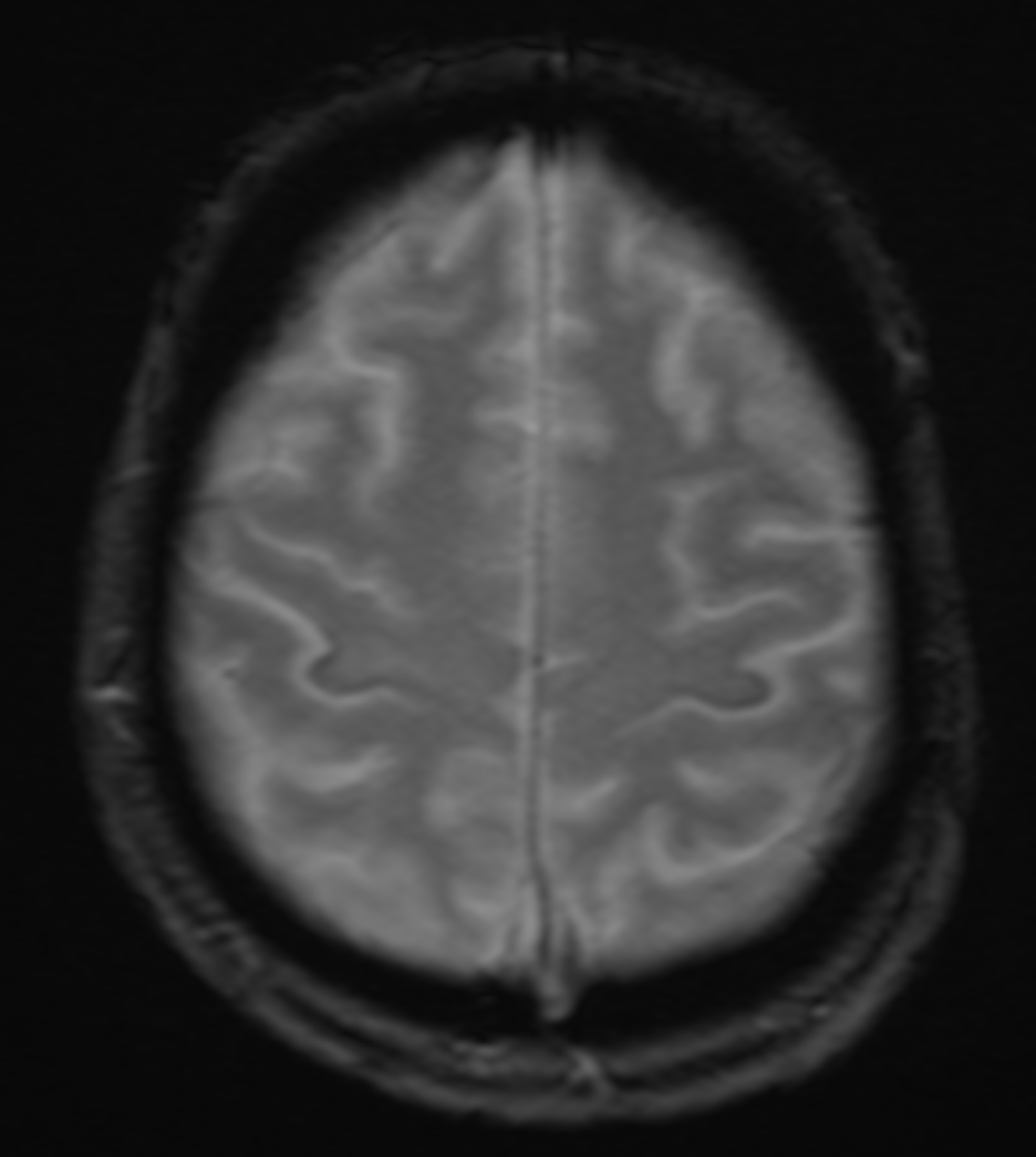
 Neither the El Escorial nor the Awaji criteria include specific radiologic findings in their diagnostic models. Therefore, while the role of radiology in the evaluation of ALS is mainly the exclusion of other possible etiologies of a patient’s clinical picture, a few subtle imaging findings have been associated with the upper motor disease found in ALS. Neuroimaging of ALS relies solely on magnetic resonance imaging (MRI). Studies have shown patients with ALS to demonstrate iron accumulation within the precentral gyrus.[27] As a result, on susceptibility-weighted imaging, the decreased signal is visible across the precentral gyrus, which is known as the “motor band sign.”[28] On conventional MRI, decreased signal intensity within the motor cortex on T2 weighted images has been associated with ALS and may be used to support the diagnosis. In addition, well-defined lesions of increased signal intensity can be visible within the corticospinal tracts on T2WI.

Findings indicative of upper motor neuronal disease have also been elucidated utilizing advanced MRI techniques such as spectroscopy and diffusion tensor imaging (DTI).[29] MR spectroscopy can detect and quantify chemical concentrations, specifically of N-acetyl aspartate (NAA), choline, and creatine within imaged tissues. Multiple studies have demonstrated decreased absolute and relative quantities of NAA in patients with ALS.[30][31][30]

Once the diagnosis has been established, genetic testing is encouraged, particularly for the SOD1 and C9ORF72 genotypes, due to emerging genotype-specific therapies in clinical trials. For patients who have a family history of the autosomal dominant disease, genetic testing can be used both as a screening tool and to provide prognostic information. For patients who have a family history of unclear inheritance, genetic testing can be useful to elucidate reduced penetrance or when information about family members is limited.

Treatment / Management

Unfortunately, symptomatic management is the main treatment in patients with amyotrophic lateral sclerosis, and it is recommended that their care team be interprofessional, with caregivers from neurology, physical therapy, occupational therapy, respiratory therapy, dietary, social work and nursing.[32]

Patients with ALS commonly suffer from chronic respiratory failure due to weakness of the diaphragmatic and intercostal muscles. As this weakness is progressive, it is recommended to have an early, careful discussion regarding respiratory management and future options such as tracheostomy, chronic ventilatory support, and noninvasive positive pressure ventilation (NIV). This discussion should be continuous, as it is expected that patients will change their opinion during their disease. Noninvasive ventilation is considered when orthopnea, nocturnal hypoxia, or a forced vital capacity (FVC) <50% present. Invasive ventilation is considered when NIV is not tolerated, or when the patient remains hypoxic and/or hypercarbic despite NIV. It is also important to offer appropriate information about terminal stage ALS when dyspnea and nocturnal hypoventilation begin, or when vital capacity (VC)<50% of predicted values. Patients should be made aware of palliative options for symptom control like anxiolytics, opiates, and/or sedation, as fears of “choking to death” are common.[33] Patients usually become symptomatic when VC <50%. When VS reaches <25% to 30%, there is a high risk of respiratory failure or sudden death. Hypoxia and hypercarbia are late findings that physicians should not rely on to predict the need for respiratory support.[34]

Progressive weakening of the muscles of mastication and swallowing results in dysphagia, which should be managed initially with diet modifications. As weight loss (particularly within the first two years after diagnosis) is associated with worse prognosis, emphasis should be placed on the intake of calorically dense foods and supplemental nutritional beverages.[35] As the dysphagia worsens, the risks of aspiration and weight loss increase. Numerous studies have demonstrated the efficacy of enteral nutrition via percutaneous gastrostomy, and it should be considered in patients with impaired oral intake. However, as there is no significant evidence regarding the impact of gastrostomy tube (G-tube) placement on quality of life, it should be an individualized discussion and decision. If the patient decides to have a G-tube placed, it should be done before the VC becomes <50% predicted value to avoid increased morbidity with the procedure itself.[36]

As the disease progresses, there will be increased dysarthria and the eventual impossibility of communication. As this can be frustrating to patients, with limited benefits found in speech therapy, the patient should choose an appropriate alternative communication method (writing, alphabet boards, electronic assistive communication devices).[37]

Frequent and painful muscle spasms can be treated with mexiletine, which was well-tolerated and demonstrated good symptomatic response at a dose of 150 mg BID. in a small sample study.[38][39] Other options are levetiracetam and with less efficacy gabapentin, baclofen, and tizanidine.[40] The latter two of which have shown efficacy in the management of spasticity. When oral therapy is not effective or well-tolerated, botulinum toxin injections into the spastic muscles can be useful. As weakness and functional decline inevitably progress, patients should be provided assistive devices (canes, orthoses, crutches, and eventually wheelchairs), removable headrests in those with neck weakness, specialized utensils, and holders, and eventually a pressure-relieving mattress with frequent repositioning to prevent pressure ulcers.[33]

Sialorrhea is very common and can be treated with atropine (0.4 mg q4-6h), hyoscyamine, amitriptyline (10 to 150 mg QHS), glycopyrrolate (1 mg TID), botulinum toxin injections into salivary glands, and even low-dose radiation therapy in those with refractory symptoms.[32][41][32] 

Pseudobulbar affect (episodes of uncontrollable laughter or crying) can affect up to 50% of patients with ALS, especially those with the bulbar form. In these patients, AVP-923 (dextromethorphan-quinidine 20mg/10mg) has proven to be effective in a multicenter, randomized, double-blind, controlled clinical trial.[42] Less rigorous evidence has supported the use of amitriptyline and fluvoxamine in smaller trials.

Riluzole (50mg bid) is thought to reduce glutamate-induced excitotoxicity and is the only drug that has proven improved overall survival (74% vs. 58% placebo at 12 months) and survival without tracheostomy (57% vs. 50% at 18 months) in two multicenter randomized trials done in the late 90s. Similar to these studies exclusion criteria, AAN guidelines recommended using it in patients with definite or probable ALS by El Escorial criteria in whom other causes of progressive muscle atrophy have been ruled out, symptoms have been present less than 5 years, VC >60% of predicted, and who do not have a tracheostomy. Expert opinion has also suggested a potential benefit in patients who do not meet these criteria, specifically limited to aspiration prevention. Since the original trials, multiple class I trials have shown a modest effect with prolonged survival of 2-3 months, and multiple other lower-class trials showed that riluzole might be associated with prolonged survival from 6 to even 21 months. Overall, it is considered a safe medication, and AAN guidelines recommend offering it to patients.[36] It is important to note that the elimination of riluzole will be affected by CYP1A2 inhibitors like caffeine and theophylline. Transaminase elevation is expected in about 50% of patients, and it is recommended to monitor transaminases monthly for the first three months and every three months afterward.

Edaravone is a free radical scavenger that is thought to reduce oxidative stress and has proven to be beneficial on a subset of patients with early-stage (less than two years of symptoms, independently living, VC >80% and scores of 2 or more in all items of ALSFRS-R) probable or definite ALS. One study showed approximately 33% slower functional decline in patients at 24 weeks follow up.[43] It has the limitation of being very expensive at around $146,000 per year and is administered in 60 mg daily infusions for 14 days followed by 14 days off for the first cycle, and then 60 mg/day for 10 days and 14 days off for the subsequent cycles. Edaravone should be used with caution in patients with asthma as it can cause serious asthmatic reactions in up to 5%.

Pain is a commonly reported symptom of patients with ALS, arising from numerous causes including muscle cramping, spasticity, and as a result of decreased mobility. Spasticity should be treated specifically, as outlined above. Assistive devices such as special mattresses, pillows, and wheelchairs may help to prevent pain. Ultimately many patients require nonopioid analgesics and anti-inflammatory drugs, and when these fail, opioids become the mainstay of pain treatment.[44]

Depression has a significant effect on the quality of life in patients with ALS, and studies have shown that treatment can improve quality of life. While no controlled trials have evaluated the treatment of depression in patients with ALS, Amitriptyline is commonly used as it can also treat other symptoms such as insomnia, sialorrhea, and pseudobulbar affect.

The issue of end-of-life care, and advanced directives, should be broached soon after diagnosis. One of the most important discussions revolves around tracheostomy placement and mechanical ventilation. Due to increased caregiver burden and significant associated costs, very few patients with ALS in the united states (less than 10%) opt for invasive ventilation. Hospice care can provide many resources that are not available, or as readily available in the home and can increase the likelihood of a peaceful death.[45] Dyspnea and anxiety are two common complaints that can be treated with morphine, and lorazepam or diazepam, respectively.

Differential Diagnosis

Multiple diseases can mimic amyotrophic lateral sclerosis including, but not limited to: cervical myelopathy, multifocal motor neuropathy, myasthenia gravis, Lambert-Eaton syndrome, inclusion body myositis, benign fasciculation, monomelic amyotrophy, spinal muscular atrophy, spinal bulbar muscular atrophy, poliomyelitis or post-poliomyelitis syndrome, late-onset Tay-Sachs disease, motor neuron syndromes secondary to lymphoproliferative disorders and other cancers, intraspinal lesions, radiation spinal myelopathy, hexosaminidase A deficiency, cramp-fasciculation syndrome, neuromyotonia, radiculoplexopathy, thyrotoxicosis, and myopathies.[46][47][48][49]

  • Benign fasciculation
  • Cramp-fasciculation syndrome
  • Multifocal motor neuropathy
  • Myasthenia gravis
  • Myopathies
  • Lambert-Eaton syndrome
  • Hexosaminidase A deficiency
  • Inclusion body myositis
  • Neuromyotonia
  • Poliomyelitis
  • Radiation spinal myelopathy
  • Spinal bulbar muscular atrophy
  • Spinal muscular atrophy
  • Thyrotoxicosis

Prognosis

The median survival is of 3 to 5 years; however, around 30% of patients are alive after five years of diagnosis and 10% to 20% after ten years. Factors associated with better survival include increased weight at diagnosis (mild obesity by BMI), younger age at onset, higher ALS functional rating scale score, FVC at presentation, and limb rather than bulbar symptoms.[50][51]

Complications

As mentioned in the treatment and management section, there are numerous complications associated with amyotrophic lateral sclerosis, including respiratory decline with eventual need for ventilatory support, dysphagia, dysarthria, malnutrition, spasms, spasticity, fatigue, functional decline due to muscular weakness, sialorrhea, thick mucus secretions and pseudobulbar affect. In addition, some complications may arise due to the less-common medications meant to manage symptoms of the disease itself. These side effects include gastrointestinal upset and arrhythmias in mexiletine, transaminitis and asthenia in Riluzole, and gait disturbance and headache in edaravone.

Postoperative and Rehabilitation Care

Rehabilitation

Rehabilitating individuals with ALS requires an interdisciplinary approach to address the broad scope of therapeutic needs. Early intervention is crucial to maximize functional mobility and longevity. Comprehensive initial assessments of balance, gait, muscle strength and tone, activity tolerance, and performance of activities of daily living (ADLs) guide interventions targeting specific deficits. Fall prevention strategies (including assistive devices and environmental modifications) and energy conservation techniques are needed as the weakness and fatigue that comes with the progression of ALS increase the risk for falls and further injury in these patients. Orthoses and adaptive equipment may also be utilized for these patients to further their independence with ADLs.

Collaboration among healthcare providers, therapists, and caregivers is vital for comprehensive disease management. Throughout the course of care, it is important to not only be mindful of the trajectory of the disease but to focus on problem-solving and maximizing the patient’s comfort, functionality, and quality of life.[52][53]

Deterrence and Patient Education

Patients need to know that this disease causes the muscles to weaken, eventually to the point of paralysis. Patients should also be aware that the disease will get worse and ultimately lead to death. Unfortunately, there is no cure; however, numerous medications can help lessen the associated symptoms. Patients may begin to notice difficulty with fine motor skills, from speaking to writing, as well as with walking, and eventually breathing.

Enhancing Healthcare Team Outcomes

ALS is a disease that necessarily progresses to significant, multifactorial debilitation and eventually death. As the clinical entity of ALS is broad in its subclassifications, involving many potential separate systems and symptoms, there is no single, standard model of patient care. Rather, the care plan of each patient needs to take into account the degree of lower motor involvement and weakness, especially whether it has progressed to the respiratory muscles, upper motor neuron involvement/spasticity, degrees of dysarthria and dysphagia, as well as the presence of other potentially associated symptoms such as frontotemporal dementia and depression. While the composition of these care teams will undoubtedly change based on the presence and degree of these symptoms, the interprofessional team will usually include the following practitioners: Physician, occupational and physical therapists, respiratory therapists, dietician, speech pathologist, social worker, and case manager. When patients with ALS are cared for by such an interprofessional team, there have been significant, demonstrable benefits in both the quality of life, as well as the length of survival.[54][55] [Level 2]


(Click Image to Enlarge)
MRI GRE sequence demonstrating iron deposition in the precentral gyrus (motor band sign) in a patient with amyotrophic lateral sclerosis
MRI GRE sequence demonstrating iron deposition in the precentral gyrus (motor band sign) in a patient with amyotrophic lateral sclerosis.
Contributed by Ryan Brotman, DO
Details

Author

Joe Joseph

Editor:

Gauri Pawar

Updated:

6/20/2023 7:01:44 AM

References


[1]

Polymenidou M, Cleveland DW. The seeds of neurodegeneration: prion-like spreading in ALS. Cell. 2011 Oct 28:147(3):498-508. doi: 10.1016/j.cell.2011.10.011. Epub     [PubMed PMID: 22036560]


[2]

Philips T, Robberecht W. Neuroinflammation in amyotrophic lateral sclerosis: role of glial activation in motor neuron disease. The Lancet. Neurology. 2011 Mar:10(3):253-63. doi: 10.1016/S1474-4422(11)70015-1. Epub     [PubMed PMID: 21349440]


[3]

Byrne S, Elamin M, Bede P, Shatunov A, Walsh C, Corr B, Heverin M, Jordan N, Kenna K, Lynch C, McLaughlin RL, Iyer PM, O'Brien C, Phukan J, Wynne B, Bokde AL, Bradley DG, Pender N, Al-Chalabi A, Hardiman O. Cognitive and clinical characteristics of patients with amyotrophic lateral sclerosis carrying a C9orf72 repeat expansion: a population-based cohort study. The Lancet. Neurology. 2012 Mar:11(3):232-40. doi: 10.1016/S1474-4422(12)70014-5. Epub 2012 Feb 3     [PubMed PMID: 22305801]


[4]

Zou ZY, Zhou ZR, Che CH, Liu CY, He RL, Huang HP. Genetic epidemiology of amyotrophic lateral sclerosis: a systematic review and meta-analysis. Journal of neurology, neurosurgery, and psychiatry. 2017 Jul:88(7):540-549. doi: 10.1136/jnnp-2016-315018. Epub 2017 Jan 5     [PubMed PMID: 28057713]

Level 1 (high-level) evidence

[5]

Turner MR, Swash M. The expanding syndrome of amyotrophic lateral sclerosis: a clinical and molecular odyssey. Journal of neurology, neurosurgery, and psychiatry. 2015 Jun:86(6):667-73. doi: 10.1136/jnnp-2014-308946. Epub 2015 Feb 2     [PubMed PMID: 25644224]


[6]

Mehta P, Kaye W, Raymond J, Punjani R, Larson T, Cohen J, Muravov O, Horton K. Prevalence of Amyotrophic Lateral Sclerosis - United States, 2015. MMWR. Morbidity and mortality weekly report. 2018 Nov 23:67(46):1285-1289. doi: 10.15585/mmwr.mm6746a1. Epub 2018 Nov 23     [PubMed PMID: 30462626]


[7]

Jordan H, Rechtman L, Wagner L, Kaye WE. Amyotrophic lateral sclerosis surveillance in Baltimore and Philadelphia. Muscle & nerve. 2015 Jun:51(6):815-21. doi: 10.1002/mus.24488. Epub 2015 Feb 11     [PubMed PMID: 25298019]


[8]

Jordan H, Fagliano J, Rechtman L, Lefkowitz D, Kaye W. Population-based surveillance of amyotrophic lateral sclerosis in New Jersey, 2009-2011. Neuroepidemiology. 2014:43(1):49-56. doi: 10.1159/000365850. Epub 2014 Oct 16     [PubMed PMID: 25323440]


[9]

Worms PM. The epidemiology of motor neuron diseases: a review of recent studies. Journal of the neurological sciences. 2001 Oct 15:191(1-2):3-9     [PubMed PMID: 11676986]


[10]

Cronin S, Hardiman O, Traynor BJ. Ethnic variation in the incidence of ALS: a systematic review. Neurology. 2007 Mar 27:68(13):1002-7     [PubMed PMID: 17389304]

Level 1 (high-level) evidence

[11]

Chiò A, Logroscino G, Traynor BJ, Collins J, Simeone JC, Goldstein LA, White LA. Global epidemiology of amyotrophic lateral sclerosis: a systematic review of the published literature. Neuroepidemiology. 2013:41(2):118-30. doi: 10.1159/000351153. Epub 2013 Jul 11     [PubMed PMID: 23860588]

Level 1 (high-level) evidence

[12]

Armon C. Smoking may be considered an established risk factor for sporadic ALS. Neurology. 2009 Nov 17:73(20):1693-8. doi: 10.1212/WNL.0b013e3181c1df48. Epub     [PubMed PMID: 19917993]


[13]

Saberi S, Stauffer JE, Schulte DJ, Ravits J. Neuropathology of Amyotrophic Lateral Sclerosis and Its Variants. Neurologic clinics. 2015 Nov:33(4):855-76. doi: 10.1016/j.ncl.2015.07.012. Epub     [PubMed PMID: 26515626]


[14]

Piao YS, Wakabayashi K, Kakita A, Yamada M, Hayashi S, Morita T, Ikuta F, Oyanagi K, Takahashi H. Neuropathology with clinical correlations of sporadic amyotrophic lateral sclerosis: 102 autopsy cases examined between 1962 and 2000. Brain pathology (Zurich, Switzerland). 2003 Jan:13(1):10-22     [PubMed PMID: 12580541]

Level 3 (low-level) evidence

[15]

Geser F, Martinez-Lage M, Robinson J, Uryu K, Neumann M, Brandmeir NJ, Xie SX, Kwong LK, Elman L, McCluskey L, Clark CM, Malunda J, Miller BL, Zimmerman EA, Qian J, Van Deerlin V, Grossman M, Lee VM, Trojanowski JQ. Clinical and pathological continuum of multisystem TDP-43 proteinopathies. Archives of neurology. 2009 Feb:66(2):180-9. doi: 10.1001/archneurol.2008.558. Epub     [PubMed PMID: 19204154]

Level 2 (mid-level) evidence

[16]

Zarei S, Carr K, Reiley L, Diaz K, Guerra O, Altamirano PF, Pagani W, Lodin D, Orozco G, Chinea A. A comprehensive review of amyotrophic lateral sclerosis. Surgical neurology international. 2015:6():171. doi: 10.4103/2152-7806.169561. Epub 2015 Nov 16     [PubMed PMID: 26629397]


[17]

Couratier P, Truong C, Khalil M, Devière F, Vallat JM. Clinical features of flail arm syndrome. Muscle & nerve. 2000 Apr:23(4):646-8     [PubMed PMID: 10716778]


[18]

Wijesekera LC, Mathers S, Talman P, Galtrey C, Parkinson MH, Ganesalingam J, Willey E, Ampong MA, Ellis CM, Shaw CE, Al-Chalabi A, Leigh PN. Natural history and clinical features of the flail arm and flail leg ALS variants. Neurology. 2009 Mar 24:72(12):1087-94. doi: 10.1212/01.wnl.0000345041.83406.a2. Epub     [PubMed PMID: 19307543]


[19]

Rowland LP. Progressive muscular atrophy and other lower motor neuron syndromes of adults. Muscle & nerve. 2010 Feb:41(2):161-5. doi: 10.1002/mus.21565. Epub     [PubMed PMID: 20082312]


[20]

Gordon PH, Cheng B, Katz IB, Pinto M, Hays AP, Mitsumoto H, Rowland LP. The natural history of primary lateral sclerosis. Neurology. 2006 Mar 14:66(5):647-53     [PubMed PMID: 16534101]


[21]

Singer MA, Statland JM, Wolfe GI, Barohn RJ. Primary lateral sclerosis. Muscle & nerve. 2007 Mar:35(3):291-302     [PubMed PMID: 17212349]


[22]

McCluskey LF, Elman LB, Martinez-Lage M, Van Deerlin V, Yuan W, Clay D, Siderowf A, Trojanowski JQ. Amyotrophic lateral sclerosis-plus syndrome with TAR DNA-binding protein-43 pathology. Archives of neurology. 2009 Jan:66(1):121-4. doi: 10.1001/archneur.66.1.121. Epub     [PubMed PMID: 19139310]


[23]

Brooks BR, Miller RG, Swash M, Munsat TL, World Federation of Neurology Research Group on Motor Neuron Diseases. El Escorial revisited: revised criteria for the diagnosis of amyotrophic lateral sclerosis. Amyotrophic lateral sclerosis and other motor neuron disorders : official publication of the World Federation of Neurology, Research Group on Motor Neuron Diseases. 2000 Dec:1(5):293-9     [PubMed PMID: 11464847]


[24]

Krivickas LS. Amyotrophic lateral sclerosis and other motor neuron diseases. Physical medicine and rehabilitation clinics of North America. 2003 May:14(2):327-45     [PubMed PMID: 12795519]


[25]

Costa J, Swash M, de Carvalho M. Awaji criteria for the diagnosis of amyotrophic lateral sclerosis:a systematic review. Archives of neurology. 2012 Nov:69(11):1410-6     [PubMed PMID: 22892641]

Level 1 (high-level) evidence

[26]

Gooch CL, Shefner JM. ALS surrogate markers. MUNE. Amyotrophic lateral sclerosis and other motor neuron disorders : official publication of the World Federation of Neurology, Research Group on Motor Neuron Diseases. 2004 Sep:5 Suppl 1():104-7     [PubMed PMID: 15512887]


[27]

Kwan JY, Jeong SY, Van Gelderen P, Deng HX, Quezado MM, Danielian LE, Butman JA, Chen L, Bayat E, Russell J, Siddique T, Duyn JH, Rouault TA, Floeter MK. Iron accumulation in deep cortical layers accounts for MRI signal abnormalities in ALS: correlating 7 tesla MRI and pathology. PloS one. 2012:7(4):e35241. doi: 10.1371/journal.pone.0035241. Epub 2012 Apr 17     [PubMed PMID: 22529995]


[28]

Chakraborty S, Gupta A, Nguyen T, Bourque P. The "Motor Band Sign:" Susceptibility-Weighted Imaging in Amyotrophic Lateral Sclerosis. The Canadian journal of neurological sciences. Le journal canadien des sciences neurologiques. 2015 Jul:42(4):260-3. doi: 10.1017/cjn.2015.40. Epub 2015 May 14     [PubMed PMID: 25971894]


[29]

Wang S, Melhem ER, Poptani H, Woo JH. Neuroimaging in amyotrophic lateral sclerosis. Neurotherapeutics : the journal of the American Society for Experimental NeuroTherapeutics. 2011 Jan:8(1):63-71. doi: 10.1007/s13311-010-0011-3. Epub     [PubMed PMID: 21274686]


[30]

Verma G, Woo JH, Chawla S, Wang S, Sheriff S, Elman LB, McCluskey LF, Grossman M, Melhem ER, Maudsley AA, Poptani H. Whole-brain analysis of amyotrophic lateral sclerosis by using echo-planar spectroscopic imaging. Radiology. 2013 Jun:267(3):851-7. doi: 10.1148/radiol.13121148. Epub 2013 Jan 29     [PubMed PMID: 23360740]


[31]

Govind V, Sharma KR, Maudsley AA, Arheart KL, Saigal G, Sheriff S. Comprehensive evaluation of corticospinal tract metabolites in amyotrophic lateral sclerosis using whole-brain 1H MR spectroscopy. PloS one. 2012:7(4):e35607. doi: 10.1371/journal.pone.0035607. Epub 2012 Apr 23     [PubMed PMID: 22539984]


[32]

Miller RG, Jackson CE, Kasarskis EJ, England JD, Forshew D, Johnston W, Kalra S, Katz JS, Mitsumoto H, Rosenfeld J, Shoesmith C, Strong MJ, Woolley SC, Quality Standards Subcommittee of the American Academy of Neurology. Practice parameter update: the care of the patient with amyotrophic lateral sclerosis: multidisciplinary care, symptom management, and cognitive/behavioral impairment (an evidence-based review): report of the Quality Standards Subcommittee of the American Academy of Neurology. Neurology. 2009 Oct 13:73(15):1227-33. doi: 10.1212/WNL.0b013e3181bc01a4. Epub     [PubMed PMID: 19822873]

Level 2 (mid-level) evidence

[33]

Borasio GD, Voltz R, Miller RG. Palliative care in amyotrophic lateral sclerosis. Neurologic clinics. 2001 Nov:19(4):829-47     [PubMed PMID: 11854102]


[34]

Rabinstein AA, Wijdicks EF. Warning signs of imminent respiratory failure in neurological patients. Seminars in neurology. 2003 Mar:23(1):97-104     [PubMed PMID: 12870111]


[35]

Kasarskis EJ, Mendiondo MS, Matthews DE, Mitsumoto H, Tandan R, Simmons Z, Bromberg MB, Kryscio RJ, ALS Nutrition/NIPPV Study Group. Estimating daily energy expenditure in individuals with amyotrophic lateral sclerosis. The American journal of clinical nutrition. 2014 Apr:99(4):792-803. doi: 10.3945/ajcn.113.069997. Epub 2014 Feb 12     [PubMed PMID: 24522445]


[36]

Miller RG, Jackson CE, Kasarskis EJ, England JD, Forshew D, Johnston W, Kalra S, Katz JS, Mitsumoto H, Rosenfeld J, Shoesmith C, Strong MJ, Woolley SC, Quality Standards Subcommittee of the American Academy of Neurology. Practice parameter update: the care of the patient with amyotrophic lateral sclerosis: drug, nutritional, and respiratory therapies (an evidence-based review): report of the Quality Standards Subcommittee of the American Academy of Neurology. Neurology. 2009 Oct 13:73(15):1218-26. doi: 10.1212/WNL.0b013e3181bc0141. Epub     [PubMed PMID: 19822872]

Level 2 (mid-level) evidence

[37]

Körner S, Sieniawski M, Kollewe K, Rath KJ, Krampfl K, Zapf A, Dengler R, Petri S. Speech therapy and communication device: impact on quality of life and mood in patients with amyotrophic lateral sclerosis. Amyotrophic lateral sclerosis & frontotemporal degeneration. 2013 Jan:14(1):20-5. doi: 10.3109/17482968.2012.692382. Epub 2012 Aug 7     [PubMed PMID: 22871079]

Level 2 (mid-level) evidence

[38]

Weiss MD, Macklin EA, Simmons Z, Knox AS, Greenblatt DJ, Atassi N, Graves M, Parziale N, Salameh JS, Quinn C, Brown RH Jr, Distad JB, Trivedi J, Shefner JM, Barohn RJ, Pestronk A, Swenson A, Cudkowicz ME, Mexiletine ALS Study Group. A randomized trial of mexiletine in ALS: Safety and effects on muscle cramps and progression. Neurology. 2016 Apr 19:86(16):1474-81. doi: 10.1212/WNL.0000000000002507. Epub 2016 Feb 24     [PubMed PMID: 26911633]

Level 1 (high-level) evidence

[39]

Oskarsson B, Moore D, Mozaffar T, Ravits J, Wiedau-Pazos M, Parziale N, Joyce NC, Mandeville R, Goyal N, Cudkowicz ME, Weiss M, Miller RG, McDonald CM. Mexiletine for muscle cramps in amyotrophic lateral sclerosis: A randomized, double-blind crossover trial. Muscle & nerve. 2018 Mar 6:():. doi: 10.1002/mus.26117. Epub 2018 Mar 6     [PubMed PMID: 29510461]

Level 1 (high-level) evidence

[40]

Bedlack RS, Pastula DM, Hawes J, Heydt D. Open-label pilot trial of levetiracetam for cramps and spasticity in patients with motor neuron disease. Amyotrophic lateral sclerosis : official publication of the World Federation of Neurology Research Group on Motor Neuron Diseases. 2009 Aug:10(4):210-5. doi: 10.1080/17482960802430773. Epub     [PubMed PMID: 18821142]

Level 3 (low-level) evidence

[41]

Stone CA, O'Leary N. Systematic review of the effectiveness of botulinum toxin or radiotherapy for sialorrhea in patients with amyotrophic lateral sclerosis. Journal of pain and symptom management. 2009 Feb:37(2):246-58. doi: 10.1016/j.jpainsymman.2008.02.006. Epub 2008 Aug 3     [PubMed PMID: 18676117]

Level 1 (high-level) evidence

[42]

Brooks BR, Thisted RA, Appel SH, Bradley WG, Olney RK, Berg JE, Pope LE, Smith RA, AVP-923 ALS Study Group. Treatment of pseudobulbar affect in ALS with dextromethorphan/quinidine: a randomized trial. Neurology. 2004 Oct 26:63(8):1364-70     [PubMed PMID: 15505150]

Level 1 (high-level) evidence

[43]

Writing Group, Edaravone (MCI-186) ALS 19 Study Group. Safety and efficacy of edaravone in well defined patients with amyotrophic lateral sclerosis: a randomised, double-blind, placebo-controlled trial. The Lancet. Neurology. 2017 Jul:16(7):505-512. doi: 10.1016/S1474-4422(17)30115-1. Epub 2017 May 15     [PubMed PMID: 28522181]

Level 1 (high-level) evidence

[44]

Chiò A, Mora G, Lauria G. Pain in amyotrophic lateral sclerosis. The Lancet. Neurology. 2017 Feb:16(2):144-157. doi: 10.1016/S1474-4422(16)30358-1. Epub 2016 Dec 8     [PubMed PMID: 27964824]


[45]

Ganzini L, Johnston WS, Silveira MJ. The final month of life in patients with ALS. Neurology. 2002 Aug 13:59(3):428-31     [PubMed PMID: 12177378]


[46]

Hardiman O, Al-Chalabi A, Chio A, Corr EM, Logroscino G, Robberecht W, Shaw PJ, Simmons Z, van den Berg LH. Amyotrophic lateral sclerosis. Nature reviews. Disease primers. 2017 Oct 5:3():17071. doi: 10.1038/nrdp.2017.71. Epub 2017 Oct 5     [PubMed PMID: 28980624]


[47]

Pradhan S. Bilaterally symmetric form of Hirayama disease. Neurology. 2009 Jun 16:72(24):2083-9. doi: 10.1212/WNL.0b013e3181aa5364. Epub     [PubMed PMID: 19528514]


[48]

Tiryaki E, Horak HA. ALS and other motor neuron diseases. Continuum (Minneapolis, Minn.). 2014 Oct:20(5 Peripheral Nervous System Disorders):1185-207. doi: 10.1212/01.CON.0000455886.14298.a4. Epub     [PubMed PMID: 25299277]


[49]

Kiernan MC, Vucic S, Cheah BC, Turner MR, Eisen A, Hardiman O, Burrell JR, Zoing MC. Amyotrophic lateral sclerosis. Lancet (London, England). 2011 Mar 12:377(9769):942-55. doi: 10.1016/S0140-6736(10)61156-7. Epub 2011 Feb 4     [PubMed PMID: 21296405]


[50]

Limousin N, Blasco H, Corcia P, Gordon PH, De Toffol B, Andres C, Praline J. Malnutrition at the time of diagnosis is associated with a shorter disease duration in ALS. Journal of the neurological sciences. 2010 Oct 15:297(1-2):36-9. doi: 10.1016/j.jns.2010.06.028. Epub 2010 Jul 31     [PubMed PMID: 20673675]


[51]

Westeneng HJ, Debray TPA, Visser AE, van Eijk RPA, Rooney JPK, Calvo A, Martin S, McDermott CJ, Thompson AG, Pinto S, Kobeleva X, Rosenbohm A, Stubendorff B, Sommer H, Middelkoop BM, Dekker AM, van Vugt JJFA, van Rheenen W, Vajda A, Heverin M, Kazoka M, Hollinger H, Gromicho M, Körner S, Ringer TM, Rödiger A, Gunkel A, Shaw CE, Bredenoord AL, van Es MA, Corcia P, Couratier P, Weber M, Grosskreutz J, Ludolph AC, Petri S, de Carvalho M, Van Damme P, Talbot K, Turner MR, Shaw PJ, Al-Chalabi A, Chiò A, Hardiman O, Moons KGM, Veldink JH, van den Berg LH. Prognosis for patients with amyotrophic lateral sclerosis: development and validation of a personalised prediction model. The Lancet. Neurology. 2018 May:17(5):423-433. doi: 10.1016/S1474-4422(18)30089-9. Epub 2018 Mar 26     [PubMed PMID: 29598923]

Level 1 (high-level) evidence

[52]

Majmudar S, Wu J, Paganoni S. Rehabilitation in amyotrophic lateral sclerosis: why it matters. Muscle & nerve. 2014 Jul:50(1):4-13. doi: 10.1002/mus.24202. Epub 2014 May 17     [PubMed PMID: 24510737]


[53]

Paganoni S, Karam C, Joyce N, Bedlack R, Carter GT. Comprehensive rehabilitative care across the spectrum of amyotrophic lateral sclerosis. NeuroRehabilitation. 2015:37(1):53-68. doi: 10.3233/NRE-151240. Epub     [PubMed PMID: 26409693]


[54]

Chiò A, Bottacchi E, Buffa C, Mutani R, Mora G, PARALS. Positive effects of tertiary centres for amyotrophic lateral sclerosis on outcome and use of hospital facilities. Journal of neurology, neurosurgery, and psychiatry. 2006 Aug:77(8):948-50     [PubMed PMID: 16614011]


[55]

Traynor BJ, Alexander M, Corr B, Frost E, Hardiman O. Effect of a multidisciplinary amyotrophic lateral sclerosis (ALS) clinic on ALS survival: a population based study, 1996-2000. Journal of neurology, neurosurgery, and psychiatry. 2003 Sep:74(9):1258-61     [PubMed PMID: 12933930]