Continuing Education Activity
Aphthous stomatitis is a common ailment, idiopathic in nature, with recurrent painful aphthous ulcers (commonly termed “canker sores”) on the non-keratinized oral mucous membranes. This activity reviews the evaluation and treatment of aphthous stomatitis, and the role of interprofessional teams caring for patients afflicted with this condition.
Objectives:
- Review the risk factors for developing aphthous stomatitis.
- Describe the typical exam findings of aphthous stomatitis and identify characteristics to differentiate aphthous stomatitis from other causes of oral ulcerations.
- Outline the management considerations for patients with aphthous stomatitis.
- Review interprofessional team strategies for improving the care of patients with aphthous stomatitis to improve outcomes.
Introduction
Aphthous stomatitis is a common ailment, idiopathic in nature, with recurrent painful aphthous ulcers (commonly termed “canker sores”) on the non-keratinized oral mucous membranes.[1][2][3]
Etiology
The cause of aphthous stomatitis is idiopathic and multifactorial, but likely involves activation of the cell-mediated immune system. Aphthous ulcers are not caused by acute infections and are therefore not contagious. Aphthous stomatitis may be triggered by local trauma, emotional or physiologic stress, allergy or sensitivity (such as to sodium lauryl sulfate present in toothpaste and oral hygiene products, foods such as cinnamon, cheese, citrus, figs or pineapple), toxin exposure (nitrates in drinking water), menstruation, or alterations in the oral microbiome. Malabsorption, enteropathy, or celiac disease may be present. As many as 20% of cases are related to hematinic deficiencies (iron, folate, vitamin B6 and B12), although other deficiencies such as vitamin D, zinc, or thiamine may also be present. Aphthous ulcers are more prevalent in nonsmokers and smokers who quit and less common in individuals with good oral hygiene practices.[4][5][6]
Epidemiology
Aphthous stomatitis affects approximately 20% of the general population. It is slightly more common in girls and women as well as among affluent socioeconomic classes and countries. Race does not appear to be a factor in the disease. Age of onset may be during childhood, but more commonly in the second and third decade of life, becoming less common with advancing age. Aphthous stomatitis can be a manifestation of Behcet syndrome, systemic lupus erythematosus, reactive arthritis, or inflammatory bowel disease (especially Crohn disease). These disorders may be excluded based on systemic signs and symptoms. [7][8]
Pathophysiology
Aphthous ulcerations are initially and primarily the result of T cell-mediated immune dysfunction but also may involve neutrophil and mast cell-mediated destruction of the mucosal epithelium. Lesions can have alterations in several intercellular mediators, such as elevations in interferon gamma, tumor necrosis factor-alpha, and interleukins (IL)-2, IL-4 and IL-5, as well as various adhesion molecules involved in cell communication and epithelial integrity. This inflammatory process results in a pseudomembrane containing fibrinous exudate, bacteria, inflammatory cells, and necrotic mucosal cells.
Aphthous ulcers occur on non-keratinized oral mucosae such as along the labial or buccal surfaces, soft palate, the floor of the mouth, the ventral or lateral surface of the tongue, tonsillar fauces, free (marginal or unattached) gingiva adjacent to teeth, and alveolar gingiva in the maxillary and mandibular sulci. In contrast, ulcerations from herpes simplex virus (HSV) involve the keratinized mucosal surfaces such as the attached gingival and dorsum of the tongue, lips, and hard palate.
History and Physical
Patients may notice a prodrome of burning discomfort a day or two before the onset of ulcerations. Fever, rash, headache, or lymphadenopathy are typically absent and would suggest a different diagnosis such as herpangina or PFAPA syndrome (periodic fever, pharyngitis, adenitis, and oral ulceration). A history of prior ulceration is typical.
On physical exam, patients with aphthous stomatitis are well-appearing and afebrile. Assess for clinical signs of dehydration, especially in infants and children. Involvement of the eye (uveitis) or genitalia suggest other diagnoses, such as Behçet syndrome or MAGIC syndrome (mouth and genital ulcers with inflamed cartilage).
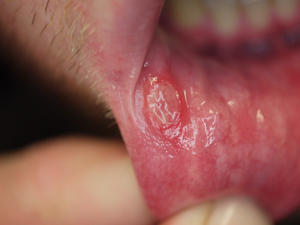
The ulcers of aphthous stomatitis are present as well-circumscribed lesions with central necrotic ulcer with gray, fibrinous exudate surrounded by an erythematous halo on the non-keratinized oral mucosa. Typical locations include the buccal (cheek) and labial (lip) mucosae, the floor of the mouth, the ventral surface of the tongue, and the soft palate. Minor aphthous ulcerations, the most common form of aphthous stomatitis, are less than 1 cm in diameter, round or oval in shape, with yellow or gray pseudomembrane surrounded by an inflammatory red halo, and heal typically within 7-14 days. Major aphthous ulcers are deeper, larger (often 2-3 cm in diameter), may have irregular raised borders, and can take many weeks or months to heal, sometimes with scarring. Much less common are herpetiform recurrent aphthous ulcers, 1 to 2 mm in diameter in clusters of 10 to 100 in groups or throughout the mouth, which usually heal within a few weeks.
Evaluation
Diagnosis of aphthous stomatitis is clinical, and laboratory testing is usually unnecessary, although diagnostic testing might be considered in persistent, severe, or recurrent cases. [9][8]
A complete blood count demonstrating anemia might suggest hematinic deficiency such as iron, folate, or vitamin B12. Neutropenia might prompt consideration of cyclic neutropenia as a cause of ulcerations.
Gluten-sensitive enteropathy (celiac disease) present in fewer than 5% of recurrent aphthous stomatitis cases and can be identified with serum anti-endomysium antibody and transglutaminase assay.
Consider HIV testing in cases with complex or severe involvement, persistent herpetiform or major aphthous stomatitis, or those involving keratinized mucosa (adherent gingival, dorsum of the tongue, hard palate).
Treatment / Management
The goals of treatment are to reduce pain (allowing adequate hydration and nutrition), enhance healing, and prevent recurrence. Many treatment options are available for aphthous stomatitis, including topical agents such as local anesthetics (benzocaine), coating or occlusive agents (bismuth subsalicylate, sucralfate, 2-octyl cyanoacrylate, and various bioadherent emollient pastes), antiseptics (chlorhexidine gluconate and hydrogen peroxide), anti-inflammatory agents such as glucocorticosteroids (clobetasol, dexamethasone, fluocinonide, and triamcinolone), amlexanox and metalloprotease inhibitors (antimicrobials tetracycline, doxycycline, or minocycline), honey, and immunomodulatory agents (amlexanox, colchicine, cyclosporine, cyclophosphamide, dapsone, methotrexate, montelukast, thalidomide, or retinoids). [10][11][12]
A step-wise approach to the treatment of aphthous stomatitis involves initial topical anesthetic and occlusive or antiseptic agents for symptom relief of minor cases. First-line treatment of major or minor aphthous stomatitis with significant symptoms is typical with topical steroids in gel or emollient paste (e.g., Orabase) to shorten the duration. Another option would be a one-time local steroid injection, such as triamcinolone. Severe refractory or persistent cases may further be treated with systemic steroids (dexamethasone or prednisone), immunomodulatory agents (listed above), pentoxifylline, or quercetin.
The experimental treatment may include various herbal products or local desiccation (such as with tincture of benzoin), cautery (such as the application of silver nitrate), or even biopsy, all after local anesthesia. Laser therapy may be effective for severe or recurrent cases. Good oral hygiene may prevent recurrences. Dietary supplementation with iron, zinc, or vitamins B1, B2, B6, B12, or C may be useful in individuals with deficiencies of these. A gluten-free diet is important only for those individuals diagnosed with celiac disease.
Differential Diagnosis
- Contact dermatitis
- Oral cancer
- Herpes simplex
- Drug induced lesions
- Lupus
- Lichen planus
Deterrence and Patient Education
Patient education is important to prevent recurrence of aphthous ulcerations.
- Practice good oral hygiene and avoidance of local trauma or oral hygiene products of known sensitivity.
- Consider taking dietary supplements with iron, zinc, or vitamins B1, B2, B6, B12, or C if a vitamin or mineral deficiency is identified.
- Only individuals diagnosed with celiac disease should choose a gluten-free diet.
- Avoid known trigger foods, emotional or physiologic stress whenever possible.
Enhancing Healthcare Team Outcomes
Aphthous ulcers or canker sores are very common oral mucosal lesions, whose etiology remains a mystery. These shallow ulcers while benign have a very high morbidity and thus an interprofessional team is recommended for management. In most people, aphthous ulcers are recurrent and often take 1-3 weeks to resolve. However, the ulcer-free period varies from individual to individual. There are many treatments for these oral mucosal lesions and the choice depends on the severity and personal experience. Unfortunately, there is no single therapy that works in everyone and no treatment is better than the other. Patient education by a team of clinicians, nurses, and pharmacists is the key to prevent morbidity. Smoking should be discontinued and one may have to avoid the triggers- mostly certain foods. Patients should be educated that in most cases, these ulcers resolve without any treatment.[13][14] (Level V)