Continuing Education Activity
Torsion of testicular appendages is considered one of the most common causes of acute scrotal pain in prepubertal children. Therefore, it should be included in the differential for any male with an acute scrotum. This activity reviews the evaluation and treatment of testicular appendage torsion and explains the role of the interprofessional team in quickly diagnosing and treating patients with this condition.
Objectives:
Differentiate between torsion of the appendix testis and other causes of acute scrotal pain, such as testicular torsion or epididymo-orchitis.
Assess the severity and duration of symptoms in patients with torsion of the appendix testis to guide appropriate management decisions.
Apply evidence-based conservative treatment measures, such as rest, scrotal elevation, ice, and analgesics, for patients diagnosed with torsion of the appendix testis.
Collaborate with radiologists and urologists to interpret imaging findings and formulate optimal management plans for patients with torsion of the appendix testis.
Introduction
Torsion of the testicular appendages is considered the most common cause of acute scrotal pain in prepubertal children and may even be the single most prevalent cause of pediatric orchalgia.[1] Therefore, it should be included in the differential diagnosis for any male presenting with an acute scrotum, particularly in the pediatric age group.[1] Two testicular appendages can undergo torsion and become symptomatic: the appendix testis and the appendix epididymis.
The appendix testis, sometimes called hydatid of Morgagni, is a vestigial remnant of the Mullerian duct and is present in 76% to 83% of testes.[2] When present, it is located on the superior pole of the testicle between the testis and epididymis and is the most common testicular appendage to undergo torsion.[3] It is homologous to the female's fimbriated end of the Fallopian tube.
The appendix epididymis is a vestigial Wolffian (mesonephric) duct remnant in 22% to 28% of testes.[2] When present, it occurs along the head of the epididymis. It is sometimes considered to be a detached efferent epididymal duct.
Etiology
Both testicular appendages are commonly pedunculated, which increases their susceptibility to torsion.[4] The exact cause of torsion remains unknown, but it is believed to be associated with factors such as trauma and prepubertal enlargement, which may be why torsion occurs most frequently in boys aged 7 to 12 years.
Some authors have proposed a seasonal etiology for spermatic cord (testicular) and testicular appendage torsion, suggesting that low temperatures during the winter contribute to increased torsion episodes.[5][6]
Epidemiology
Torsion of a testicular appendage is most commonly observed in boys between the ages of 7 and 12, although it can occur at any age. It is important to note that more than 50% of boys presenting with acute scrotal pain are diagnosed with torsion of a testicular appendage.[1][7] In summary, the torsion of the appendix testis or epididymis is the leading cause of a painful scrotum in prepubertal boys.[1][7]
A study involving 238 boys aged 19 years and younger, who presented to a children's hospital with acute scrotal pain, revealed that 46% were diagnosed with torsion of the appendix testis, 35% had epididymitis, and only 16% exhibited testicular torsion.[7]
History and Physical
The initial diagnosis of torsion of the testicular appendages is primarily based on clinical evaluation. However, it can be challenging due to the variable presentation, leading to a high rate of misdiagnosis, with 45% of general practitioners making an incorrect initial diagnosis.[8] Due to the high rate of misdiagnosis, imaging is recommended for all cases of acute scrotal pain to aid in the accurate diagnosis of torsion of the testicular appendages.
Torsion of either testicular appendage typically causes pain similar to testicular torsion, although the onset is usually more gradual. In addition, the pain is commonly more localized to the upper pole of the testis or epididymis. It is not typically associated with urinary symptoms or systemic signs such as fever, nausea, or vomiting.
A comprehensive physical examination should include a thorough but gentle evaluation and palpation of the scrotum, testes, epididymis, inguinal area, and abdomen.[3]
On initial physical examination of the condition, tenderness is often localized to the upper pole of the testis or epididymis. There may be a palpable, localized mass in the area of maximum tenderness. The scrotum typically appears normal, and the cremasteric reflex is usually intact.
With a normal cremasteric reflex, there would not be any "angel wing" or "bell clapper deformity" of the opposite testicle. This deformity refers to a horizontal position of the testicle, which widens the scrotum inferiorly and creates an appearance resembling angel wings. This is typically caused by inadequate fixation of the testicle's inferior pole to the tunica vaginalis by the gubernaculum, increasing the risk of testicular torsion.
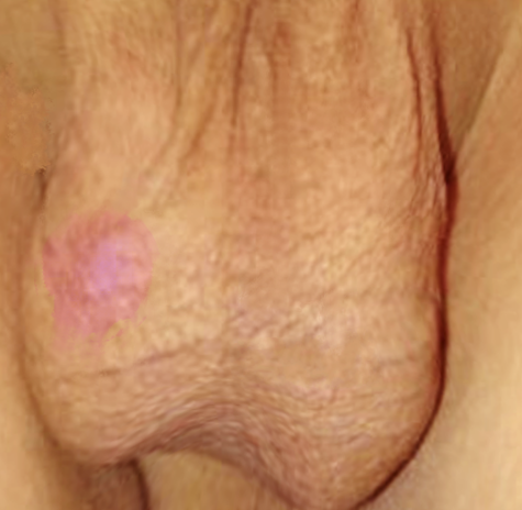
A "blue dot sign" may also be present as a para-testicular nodule noted on the superior aspect of the testicle; this can be identified by stretching the scrotal skin overlying the superior pole of the testicle and is representative of an ischemic testicular appendage. While the "blue dot" sign is worth noting, it is not consistently present in all cases of torsed testicular appendages. Studies have reported that the sign is found in approximately 21% (ranging from 0%-52%) of all torsed testicular appendages. It is essential to be aware that a false positive "blue dot" sign has been reported in the literature, even in cases of testicular torsion.[9]
The inflammatory response may lead to less specific physical examination findings as the condition progresses. These findings include scrotal erythema, edema, and nonspecific tenderness involving the entire testicle and epididymis.
A urinalysis should be obtained in all cases of acute scrotal pain.[3]
A summary of physical findings of torsion of the testicular appendages include the following:[10][11][12]
- The patient is afebrile and has normal vital signs.
- The scrotum usually looks normal and has the expected vertical orientation, but some scrotal erythema or edema may present.
- The cremasteric reflex is usually present and intact, often ruling out testicular torsion.
- The testicle will not be "high riding" or elevated in the scrotum, as seen in testicular torsion.
- The testis may exhibit tenderness typically localized to the the upper pole on the affected side. However, the tenderness is usually not as pronounced or diffuse compared to testicular torsion, which produces diffuse scrotal tenderness.
- A small para-testicular nodule may be palpable at the upper pole of the affected testis. While a "blue-dot" finding is considered pathognomonic of a torsed testicular appendage, it is present in approximately 20% of patients.
- The combination of a "blue-dot" sign, along with the ability to palpate a non-tender testis underneath, can effectively make the diagnosis of torsion of the testicular appendage. In addition, these findings help exclude the diagnosis of testicular torsion.
- In patients with scrotal pain, epididymitis can be differentiated by dysuria, epididymal pain on palpation, increased echogenicity, and greater peri-testicular perfusion on ultrasound compared to a torsed testicular appendage.
- Clinical signs alone are generally not considered sufficiently reliable to make a definitive diagnosis.
Evaluation
Color Doppler ultrasonography is the preferred imaging modality for evaluating the acute scrotum in patients of all age groups. It is superior to radionuclide imaging in terms of diagnostic accuracy. Additionally, it is readily available in emergency settings and allows for faster imaging and evaluation of blood flow to the scrotum.
A testicular appendage's normal ultrasonic appearance will typically reveal minimal or no detectable vascular flow.[10][13] Ultrasound imaging of a testicular appendage can rarely visualize the structure itself. However, it commonly reveals normal blood flow to the affected testicle, excluding testicular torsion. Additionally, hypoperfusion of the associated epididymis is often observed on ultrasound. A normal appendix testis will be less than 5.6 mm when a testicular appendage is visualized on ultrasound.
In contrast, a torsed testicular appendage will have a size exceeding 5.6 mm and may exhibit different ultrasound characteristics depending upon the duration of torsion. In boys presenting within 24 hours, it may appear as an ovoid hypoechoic nodule, while after 24 hours, it is more likely to appear as a hyperechoic or heterogeneous nodule.[14][15] A torsed appendix testis may also present as an avascular lesion surrounded by a hyperemic epididymis, often exhibiting posterior enhancement on ultrasound imaging.[16] Based on ultrasound findings alone, the hyperemia of the surrounding structures can make it challenging to differentiate a torsed testicular appendage from epididymitis.[16] Additional clinical information and assessment may be required to make an accurate diagnosis.
A large torsed appendage may even give the sonographic appearance of a pyocele, further emphasizing the importance of the clinical history and conducting a thorough physical examination.[17] The echogenicity of a torsed testicular appendage on ultrasound can vary based on factors such as the size of the appendage and the time interval between the onset of symptoms and the imaging study.[18]
In cases of testicular torsion, the affected testicle is often found to be "high riding" due to the torsion of the spermatic cord. However, in the case of a torsed testicular appendage, the testicle typically does not exhibit the "high riding" characteristic. Instead, the position of the testicle may appear relatively normal or slightly elevated, but it does not show the significant upward displacement characteristic of testicular torsion.
Radionuclide imaging of the scrotum can be utilized to detect a "hot dot" sign at the site of the torsed testicular appendage. However, this imaging modality is most useful when the symptoms and torsion have been present for at least five hours. Even after five hours, the "hot dot" sign is observed in approximately only 45% of patients diagnosed with a torsed testicular appendage.[19] Radionuclide scrotal imaging also takes much longer to perform. For these reasons, ultrasound is usually the preferred imaging modality for the initial evaluation of all acute scrotal pathologies.[20]
If a patient with scrotal pain presents with voiding symptoms like dysuria, urgency, or frequency, obtaining a urinalysis with culture is crucial. A torsed testicular appendage is less likely to cause urinary symptoms or show abnormalities in the urinalysis than other disorders.
If the diagnosis remains uncertain after evaluation, urgent surgical exploration is recommended.[12][21]
Treatment / Management
Torsion of a testicular appendage is generally a self-limiting condition, and most cases require only conservative therapy.[22] Conservative management includes bed rest, scrotal elevation, ice application, nonsteroidal anti-inflammatory medications, and analgesics. The inflammation and pain usually resolve within one week but may persist longer. Surgical excision can be considered for cases of prolonged pain.
Surgery is rarely indicated for a torsed testicular appendage. Scrotal exploration should be considered only in the following circumstances: difficulty differentiating from testicular torsion, severe pain not responding to analgesics, or if the pain persists or recurs despite conservative management.[22]
If there is any reasonable doubt about the diagnosis, a scrotal exploration should take place to exclude testicular torsion definitively.[12][21] If surgical intervention is necessary for a torsed testicular appendage, there is no absolute requirement to explore the opposite side as is typically done for testicular torsion. However, it is optional to consider the exploration and removal of the testicular appendages on the contralateral side during the surgery. Some experts recommend this optional procedure due to a future likelihood of testicular appendage torsion in the opposite testis, estimated at around 4.2%.[23]
Differential Diagnosis
In a patient presentation of acute scrotal pain, the differential includes ischemic conditions (testicular torsion, torsion of a testicular appendage), infections (acute epididymal-orchitis), or trauma-related injuries (scrotal contusion, testis rupture). However, the acute scrotum should be considered a surgical emergency until a testicular torsion is ruled out due to the potential catastrophic loss of a testicle. Timely intervention is essential for testicular salvage, with most testicles remaining viable if surgically detorsed within 6 hours of symptom onset.
Testicular torsion typically presents with a more sudden and severe onset of symptoms than torsion of a testicular appendage, although the presentation can vary. The absence of a cremasteric reflex of the affected side and an abnormal transverse lie of the unaffected testicle may be observed during physical examination. Pain relief upon lifting the affected testicle (Prehn sign) is not considered a reliable indicator in distinguishing between the two conditions.
Doppler ultrasound imaging often reveals absent or minimal arterial flow to the affected testicle. It is worth noting that the onset of symptoms during sleep has been identified as an indicator of testicular torsion.[20] The affected testicle is often found to be "high riding" in testicular torsion but not in a torsed testicular appendage.
Epididymo-orchitis, like torsion of a testicular appendage, will show hyperemia to the affected epididymis on color Doppler ultrasound imaging, but the degree of hyperemia may be more pronounced. In addition, epididymo-orchitis is commonly associated with voiding symptoms such as dysuria, frequency, and urgency. There may also be a history of urinary tract infections.
Patients may also present with systemic signs and symptoms of fever, nausea, or vomiting, although this is less likely. On physical exam, the epididymis and testis on the affected side may be enlarged and tender, but diffuse tenderness of the testicle is usually lacking. Occasionally, elevating the affected testicle will relieve pain (positive Prehn sign). No "angel wing" or "bell clapper deformity" will exist.[10]
In cases where the diagnosis is uncertain and there is suspicion of testicular torsion or other severe conditions, it is crucial to proceed with an emergency scrotal exploration rather than delay treatment. Prompt surgical intervention is necessary to prevent the potential loss of the testicle and mitigate any adverse outcomes.[24]
Prognosis
The prognosis for torsion of either testicular appendage is generally favorable, as these appendages are considered vestigial remnants without a known function. In addition, the pain and inflammation associated with the torsion are self-limiting, and the condition typically resolves within one week without requiring surgical intervention.
Complications
The primary complication of torsion of a testicular appendage is a misdiagnosis leading to the loss of a testis due to a missed testicular torsion. To avoid such misdiagnoses, ultrasonography is the recommended diagnostic approach in all cases of acute scrotal emergencies.
Deterrence and Patient Education
Parents and patients diagnosed with testicular appendix torsion should be encouraged to adhere to all treatment recommendations. This includes following a regimen of bed rest, scrotal elevation, ice application, nonsteroidal anti-inflammatory drugs, and analgesics as prescribed.
Additionally, they should be advised to closely monitor their symptoms and seek reevaluation if symptoms worsen or if they do not improve within one week.
Pearls and Other Issues
- A torse testicular appendage is the most common cause of an acute scrotum in a prepubertal boy.
- The "blue dot" sign is a classic physical exam finding unique to testicular appendix torsion. However, it is often absent and can be falsely positive in cases of true testicular torsion. Therefore, the absence of a "blue dot" sign is not diagnostic.
- Because of their lack of function and potential to torse, the appendix testes and epididymal appendix are commonly removed if encountered during an elective scrotal exploration for other purposes.
- Testicular appendage torsion in prepubertal boys is often misdiagnosed as epididymitis based on scrotal ultrasound imaging, as both can demonstrate epididymal hypervascularity.
- Epididymitis should be considered highly unlikely in a pre-sexual boy with no urologic abnormalities, recent catheterization, or history of urinary tract infections.
- If a testicular appendage is more than 5.6 mm on ultrasound, it should be considered suspicious for torsion of the appendage.
- Patients with testicular appendage torsion are likely younger than those with testicular torsion. They also will lack the "angel wing deformity" and not demonstrate the "high riding" testicle position usually associated with testicular torsion.[20]
- Rare causes of an acute scrotum can include incarcerated hernias and traumatic testicular ruptures.
- There have been reported cases of simultaneous testicular torsion with testicular appendage torsion.
- Removing the testicular appendages in the contralateral testis during a scrotal exploration for torsion of the testicular appendages is now considered optional but is not required.
- It is possible to have torsion of a testicular appendage in an undescended testicle.
Enhancing Healthcare Team Outcomes
Management of the acute scrotum should be treated as a surgical emergency until a diagnosis is confirmed. Given the situation's urgency, patients with this condition often present to the emergency department, where a triage nurse initially assesses them. The triage nurse must be aware of the potential for testicular torsion and the need for immediate action.
Upon recognizing the possibility of testicular torsion, the triage nurse should promptly contact the clinical interprofessional team, including the attending physician, urologist, or surgeon, to ensure timely evaluation and management.
An early history and physical exam should give the clinician some clear guidance and direction, usually necessitating a color Doppler ultrasound to be promptly read by a radiologist.
This collaborative approach allows for a swift response and facilitates appropriate diagnostic procedures, such as ultrasound or surgical exploration, to confirm the diagnosis and initiate treatment as needed. Early involvement of the clinical interprofessional team is vital in maximizing the chances of testicular salvage and minimizing the risk of complications associated with a delayed intervention. [Level 5]