Catheter Management of Mitral Regurgitation

Earn CME/CE in your profession:


Continuing Education Activity

Mitral regurgitation (MR) is one of the most common valvular abnormalities. Medical management, along with regular surveillance, is the typical recommendation for mild MR. Severe cases have traditionally been treated with invasive surgery. Recently, studies have shown increased utilization of a minimally invasive transcatheter approach for mitral regurgitation repair. The edge-to-edge leaflet repair device is a catheter-based therapy that uses a clip to bring together the leaflets, which lessens the regurgitation. This activity details primary and secondary mitral regurgitation, non-invasive catheter management options and their indications and contraindications, procedural techniques, and complications. This activity highlights the inter-professional team's role in caring for patients with mitral valve regurgitation.

Objectives:

  • Describe the pathophysiology of mitral regurgitation.
  • Discuss the indications for percutaneous catheter-based management of mitral regurgitation.
  • Describe the technique, echocardiographic guidance, and steps for FDA-approved catheter-based management of mitral regurgitation.
  • Discuss the procedural complications from catheter-based management of mitral regurgitation.

Introduction

Mitral regurgitation (MR) is one of the most common valvular abnormalities, second only to aortic valve stenosis.[1][2] Management options depend on the duration and severity of the lesion. Acute severe MR resulting from papillary muscle rupture or leaflet perforation from infective endocarditis can cause severe hemodynamic impairment, acute volume overload, and congestion and requires urgent surgical intervention.[3] Chronic MR can result from Primary (degenerative pathology, disease of mitral valve apparatus)(media 11) or secondary (functional due to left ventricle or Left atrial pathology) (media12). Chronic MR can be medically managed along with chronic surveillance if it's mild and asymptomatic. However, symptomatic chronic MR patients should be evaluated for surgical intervention.[3][4] Asymptomatic patients with Chronic MR can be recommended surgery if they have depressed LV function, LV dilatation, atrial fibrillation, or pulmonary HTN.[5][6]

Transthoracic echocardiography (TTE) is the initial imaging modality for screening and evaluating mitral valve morphology and pathology, determining the mechanism of Mitral regurgitation, and quantifying MR severity, assessment of Left ventricular function, size, and Left atrial size.[3] Parameters used to assess qualitatively and to quantify MR includes - a two-dimensional assessment of mitral valve leaflet thickness, motion, coaptation, MR jet to Left atrial (LA) area ratio, Vena contracta, Effective regurgitant orifice area (EROA), Regurgitant volume (RV), Regurgitant area, LV ejection fraction (LVEF) and Left ventricular end-diastolic area (LVEDA). When TTE images are inadequate, TEE can provide the information and assessment.[7][8] Three-dimensional TEE can provide an "enface" view of the mitral valve resembling surgical inspection, facilitating discussion and pre-procedure planning (Image 1). If there is a contraindication to TEE, Cardiac MRI can provide highly accurate data on MR assessment and evaluation of Left Ventricular dimensions.

[Table 1] Etiology of Mitral Regurgitation

Primary MR ( Problem with valve)  Secondary MR ( Ventricular remodeling)
Mitral Valve prolapse -Myxomatous changes (Prolapse, filial, ruptured, elongated Chordae) Ischemia (2/2 Coronary Artery disease )
Degenerative MR (Calcification, thickening) Non-Ischemic Cardiomyopathy 

Infectious (Infective endocarditis, vegetation, perforation)

Inflammatory (Rheumatic, collagen vascular disease)

Drug-induced,

Radiation-induced

Annular Dilation 
Congenital  (Parachute Mitral valve, Cleft)   

[Table 2] Stages of Primary Mitral Regurgitation [3]

Stage  Definition  Valve Anatomy Echocardiographic Mitral Regurgitation quantification  Left ventricle (LV), Left atrial (LA) echo assessment Symptoms
A At the risk of MR Mild valve prolapse, thickening, or restriction  No MR / small MR, Vena Contracta <0.3 cm None  Absent
B Progressive MR Moderate to severe mitral valve prolapse, leaflet restriction, loss of coaptation  Central MR <40% LA area, vena contracta <0.7cm, regurgitant volvume <60ml, Regurgitant fraction <50%, Effective regurgitant oriface area (EROA) <0.4 cm^2 Mild LA enlargement, LV normal Absent
C Asymptomatic severe MR Severe prolapse or flail. Severe leaflet restriction or severe leaflet thickening, loss of central coaptation, prior Infective endocarditis Central MR >40% LA area, holosystolic eccentric jet, vena contracta >0.7 cm, Regurgitant volume >60 ml, regurgitant fraction >50%, EROA >0.4 cm^2

Moderate - Severe LA, LV enlargement

C1  LVEF >60%, LVESD <40 mm

C2  LVEF <60%, LVESD >/=40 mm

Absent
D Symptomatic Severe MR Severe prolapse or flail, severe leaflet restriction, loss of central coaptation, prior history of  infective endocarditis  Central MR >40% LA area, holosystolic eccentric jet, vena contracta >0.7 cm, Regurgitant volume >60 ml, Regurgitant fraction >50%, EROA >0.4 cm^2  Moderate to severe LA, LV enlargement, presence of Pulmonary HTN Dyspnea and reduced exercise tolerance

Recent studies have shown Percutaneous MV repair as an alternative option for high-surgical-risk patients with severe symptomatic Mitral regurgitation. The procedure can be accomplished with low morbidity and mortality rates in a substantial portion.[9] EVEREST 1 trial established the safety and feasibility of edge-to-edge repair. EVEREST 2 Trail was a randomized control trial comparing percutaneous edge-to-edge repair with surgical mitral valve repair/replacement and suggested the superiority of surgery in reducing MR but supported the long-term safety of edge-to-edge repair device and durability of MR reduction.[10][11] The edge-to-edge leaflet repair device is a minimally invasive catheter-based therapy that is based on the principle of the "Alfieri stitch," a surgical technique first used by Dr. Ottavio Alfieri, an Italian cardiothoracic surgeon, which involves bringing together the two flailing leaflets of the mitral valve, resulting in decreased or reduced regurgitation. This repair typically leads to the creation of a double orifice and is based on surgical edge-to-edge Alfieri repair.[12][13]

Many percutaneous options exist for patients with MR, multi-system dysfunction, and comorbidities, which may put them at high risk for surgical interventions.[14] Percutaneous techniques can be classified based on the site of the mitral apparatus they work on  -  leaflets(edge-to-edge repair), annulus (indirect or direct annuloplasty), chordae (neo-chords, percutaneous chord implantation), or LV (percutaneous LV remodeling).[15][14][16]

This article will discuss primary and secondary mitral regurgitation and non-invasive catheter management options, including indications, contraindications, procedural techniques, and complications. We will discuss the FDA-approved edge-to-edge repair devices as the primary focus. 

Anatomy and Physiology

Andreas Vesalius first suggested the name " mitral " for the left-sided atrioventricular valve, given its resemblance with bishops' "miter."[17][18] The mitral valve (MV) apparatus is a complex anatomic structure that includes the anterior and posterior mitral leaflets, mitral annulus, subvalvular structures (chordae tendineae, papillary muscles), and Left ventricle (Image 2). The mitral annulus is a saddle-shaped structure; the competence of the mitral valve depends on the correct interaction of different components of the MV apparatus.[2][18][19] The anterior and posterior leaflets of the mitral valve should "coapt" to prevent MR during systole; alteration in the functioning of any component of the mitral valve can lead to the development of mitral regurgitation (MR).[20]  The two types of MR are primary and secondary. Primary mitral regurgitation is a degenerative valve disease, while secondary mitral valve regurgitation is characteristically a functional myocardial disease. [Table 1]

MR can further classify as mild, moderate, and severe. Severe (or moderate-severe) MR is currently only recommended for catheter management. The echocardiogram is the primary tool used to assess the structure and function of the mitral valve- systolic competence and non-restriction during diastole. Severe MR is described as having a color flow jet that may be central and large (>6 cm or >30 percent of the left atrial area) or smaller, if eccentric, encircling the left atrium. Pulmonary vein flow may show systolic blunting or systolic flow reversal, vena contracta width >/=0.5 cm measured in the parasternal long-axis view, a regurgitant volume of >/=45 mL/beat, regurgitant fraction >/=40%, and/or regurgitant orifice area >/=0.30 cm^2 according to the American College of Cardiology and American Heart Association.[21] Several interventions exist to treat severe MR, including surgical and non-surgical. If untreated, severe MR can lead to fatal sequelae, including heart failure.[22] 

[Table 3] Mitral Valve Pathology Based on Echocardiography[23]

Type  Leaflet Motion  Example 
I Normal  leaflet motion

Annular dilation without leaflet tethering,

Cleft or indentation

Perforation

II Excessive leaflet motion Billowing, prolapse, flail leaflet
III Restricted leaflet motion

Systolic restriction

  - Symmetric systolic (dilated or ischemic cardiomyopathy with leaflet tethering)

   - Asymmetric systolic (segmental ischemia )


Systolic and diastolic restriction (rheumatic)

IV Systolic anterior motion  Systolic anterior motion of mitral valve- Hypertrophic cardiomyopathy associated or Post mitral valve repair
V Mixed conditions  Prolapse of one leaflet with restriction of another leaflet

Pre-procedural Anatomical Considerations

The successful grasp of mitral valve leaflets during the procedure requires certain conditions to be met. The leaflets must be pliable and non-calcified at the grasping site, and there must be no significant clefts or perforations. Additionally, the shorter MitraClip NT and NTW require a minimal posterior leaflet length of 6 mm, while the longer MitraClip XT and XTW require a minimal posterior leaflet length of 9 mm.[24] Furthermore, a transmitral gradient (MG) of less than 5 mmHg and a mitral valve area (MVA) of at least 4 cm^2 is desirable to minimize the risk of mitral stenosis. If the MVA is ≤ 3 cm2, it is considered a contraindication for TEER, and the decision to proceed in borderline cases can be individualized based on the severity and location of MR and the anticipated number of devices needed. To avoid overestimation errors, the MVA should be measured using 3D multiplanar reformatting (MPR).

Patients with an extensive flail, defined as a flail segment width of 15 mm or greater or a flail gap of 10 mm or greater, were not included in the initial clinical trials for TEER therapy, such as EVEREST II.[10] Despite this, treating degenerative mitral valve (MV) disease with a flail leaflet is an important application of TEER therapy. It can be associated with higher mortality risk in elderly individuals.[25] The presence of a flail leaflet segment can predict a greater acute improvement in mean left atrial pressure after TEER. Improvement in left atrial pressure after TEER has been linked to improved functional status.[26] With the availability of longer and wider TEER devices, treating wider and larger flail gaps is now possible. The development of independent leaflet grasping technology has enabled the treatment of larger and wider flail gaps. This technology is available with both the MitraClip G4 and the PASCAL repair system, and it allows for the initial capture of the flail segment, followed by the steering of the delivery system to the non-flail leaflet. This ensures that both leaflets have a sufficient and stable grasp, thus facilitating the treatment of larger and wider flail gaps.

The EVEREST II trial only included patients with a primary regurgitant mitral jet originating from the center of the mitral valve (A2-P2 segments).[10] Excluding patients with non-central MR from treatment with TEER would mean that many patients would go untreated. Approximately a third of all patients with significant MR have non-central MR jets, often originating from the commissures and involving the edges of the leaflets.[27][28] Deploying TEER devices in these cases can be more challenging, especially when dealing with large prolapsing leaflets and flail segments closer to the medial and lateral commissures. This is due to the increased risk of device entanglement and chordal disruption caused by the additional number and complexity of the chordae tendineae in the commissures. To reduce the risk of engagement with the chordal apparatus and the LV wall, some operators choose to avoid larger TEER devices, as their longer arms increase the risk of entanglement. However, due to the shorter posterior leaflet length in the commissures, short device arms are often sufficient for an adequate tissue grasp (i.e., <9 mm). To visualize the full extent of the pathology and the anticipated device choice and orientation, extensive use of 3D TEE and unconventional imaging planes are useful.[29] The presence of severe bileaflet prolapse, or Barlow's disease, prevented patients from being included in the EVEREST trials. This patient population presents a challenge regarding successful TEER, as the hypermobile leaflets make it difficult to grasp. To attain a significant height reduction of the redundant leaflets tissue and achieve a longer-lasting reduction of mitral regurgitation, multiple large TEER devices are required, making it an even greater challenge.[30]

When considering TEER in secondary MR, it is important to differentiate patients with preserved LV function and annular dilation (atrial functional MR) from patients with LV dysfunction and leaflet tethering. A subgroup analysis of the COAPT trial demonstrated that patients with atrial fibrillation who underwent TEER maintained a clinical benefit; however, they had a worse prognosis compared to patients without atrial fibrillation.[31]

Mitral Annular Calcification (MAC) is a degenerative process that predominantly affects the mitral valve annulus and is frequently associated with mitral regurgitation[32]. Grasping the thickened and non-pliable leaflets can be difficult. In addition, there is an elevated risk of high diastolic gradients across the mitral annulus in patients with reduced Mitral Valve Annulus (MVA) at baseline. Recent studies suggest that in select patients with MAC and severe MR, TEER therapy might be safe and feasible with comparable midterm outcomes.[33]

 Up to 35% of patients who have undergone surgical MV repair at high volume centers of excellence develop moderate to severe MR 10 yrs. after initial repair.[34][35][36] Re-do sternotomy in these patients is often associated with a high risk of morbidity and mortality.[37]  The safety and feasibility of TEER in this situation have been demonstrated in some studies, but further research is needed to fully understand its efficacy.[38][39][40] It is crucial to understand that people who have had surgical annuloplasty in the past frequently have lower MVA introduced by the annuloplasty ring. Since more than one TEER device is needed to properly lower MR, the structural team must exercise caution and prevent a subsequent rise in the diastolic inflow gradients. Post-surgical MV repair, the posterior MV leaflet is frequently resected, leaving a shorter and tiny posterior leaflet making it more challenging to grab during the TEER process. A different strategy is to grip the anterior leaflet and the posterior portion of the annuloplasty ring itself when the posterior leaflet tissue length is insufficient. However, expertise with this approach is limited. The visibility of the posterior leaflet is often limited, due to the presence of an annuloplasty ring, thereby making the leaflet grasping very challenging during TEER. The risk of entanglement of the TEER device is elevated in the presence of artificial chords.[41]

Pre-procedural TEE examination also helps in predicting the difficulty of repair. The following table [Table4] summarizes the predictors[42][43][8]

 Echocardiographic Criterion  Ideal Anatomic Features  Challenging Anatomic features  Relative contraindications 
 Anatomic location of pathology  Regurgitation jet in A2 - P2 segment   Involving medial or lateral commissure, segments A1-P1, A3-P3 Perforation or cleft or severe calcification of leaflet
 Mitral valve area and gradient   area >4 cm^2 gradient <4mmHG  area >3.5 cm^2, gradient >/=4mmhg  area <3.5 cm^2, gradient >/=4mmHG
 Leaflet grasping length  >10 mm  7 - 10 mm   <7mm
 Secondary MR   Coaptation depth <11 mm  Coaptation depth >11 mm   Rheumatic thickening and restriction 
 Primary MR  Flail width <15 mm, flail gap <10 mm  Flail width >15 mm with a large valve area   Barlow disease with multisegment involvement 

Indications

Currently, the edge-to-edge leaflet repair device is the only recommended intervention to treat MR, but there are many emerging technologies, including but not limited to neo-cords, transcatheter mitral valve repair, and rings. At present, the following are the indications[44][45] for the edge to edge repair:

  • Moderate to severe primary MR (3 to 4+)
  • Moderate to severe secondary MR (3 to 4+)
  • Symptomatic heart failure
  • High or prohibitive risk for surgery 
  • Favorable anatomy
  • Life expectancy >1 year

Contraindications

There are very few contraindications to catheter intervention with the edge-to-edge leaflet repair device.[46][47][48] The following are the contraindications for the procedures:

  • Unable to tolerate anticoagulation
  • Active endocarditis of the mitral valve
  • Rheumatic mitral valve disease 
  • Intracardiac, inferior vena cava (IVC), or femoral venous thrombus
  • Severe mitral annular calcification involving leaflets
  • Presence of significant cleft or perforation in mitral valve leaflets
  • Mitral valve stenosis

Equipment

The transcatheter edge-to-edge leaflet repair (TEER) device procedure requires the supplies listed below:

  • The edge-to-edge leaflet repair device
  • Transeptal puncture kit including catheters, needle, or radiofrequency wire)
  • Fluoroscopy machine
  • Code cart 
  • Sterile gown
  • Sterile drape
  • Anesthetic
  • Transesophageal Echocardiography, preferably with 3-D imaging capability
  • Transducers and equipment for invasive hemodynamic monitoring 
  • Defibrillator in case of arrhythmia
  • Availability of perfusionist and heart-lung machine in case of device embolization

For the treatment of both primary mitral regurgitation (PMR) and secondary mitral regurgitation (SMR), the MitraClip device is the first transcatheter technology to get FDA and CE approval[49][50]. The fourth-generation MitraClip has four implant sizes available as of 2020 [51]. A wider implant size of 6 mm is available with both arm lengths in addition to the "traditional" NT and XT clip sizes (4 mm width; 9 [NT] and 12 [XT] mm arm length) (6 mm width; 9 [NTW] and 12 [XTW] mm arm length). The MitraClip is made up of two rigid arms (cobalt-chromium alloy) with flexible nitinol-based "grippers," each of which has four (NT/NTW) or six (XT/XTW) tiny hooks (referred to as "frictional elements") aligned longitudinally. In addition to treating bigger coaptation gaps and leaflet flails, the longer clip arms (XT/XTW) go beyond the strict anatomic and morphologic EVEREST inclusion criteria[52]. Concerns about the risk of leaflet injuries and single leaflet device attachment (SLDA) due to increased leaflet tension following the grasping of more tissue with the XT/XTW devices, the active locking mechanism, and device stiffness are raised by the expansion of the technique to patients with more complex anatomy. Increased tension forces have been described to cause leaflet injury in a variety of anatomies, including in patients with calcified leaflets[52]. The long-arm XTR clip system did not, however, exhibit increased rates of adverse leaflet events when compared to the smaller NTR device in a structured examination of the EXPAND registry[24]. The fourth-generation MitraClip device now enables autonomous and controlled gripper actuation (CGA) to confirm and/or optimize leaflet gripping in addition to enabling continuous left atrial pressure monitoring through the guiding catheter.

The PASCAL transcatheter MV repair technology was originally deployed in 2016 and has since been studied in a compassionate-use cohort of 23 patients with difficult anatomical features for TEER[53]. The PASCAL system's second version is now available, and it includes three embedded catheters: a 22 Fr steerable guide sheath, a steerable catheter, and an implant catheter with the device preattached at the distal end. This design provides a wide range of motion and improves maneuverability in the left atrium. The nitinol-based PASCAL P10 implant is made up of a central spacer and two spring-loaded, curved paddles with a grabbing length of 26 mm when opened to 180°, and two clasps (10 mm each). The middle spacer is considered to fill some of the coaptation gaps within the main MR jet area, reducing stresses on the MV leaflets. The nitinol clasps, which have a horizontal line of small hooks ("retention elements") at the distal end, can be adjusted separately, allowing for simultaneous or independent leaflet capture. A second lower-size PASCAL Ace has been available, offering a similar gripping breadth compared to the PASCAL P10 implant, while the paddles are just 6 mm wide to fit smaller anatomies and permit multiple implant techniques. Both PASCAL implants provide separate leaflet gripping, allowing for either "leaflet optimization" or "staged leaflet capture". In August 2022, the second-generation PASCAL Precision platform was announced, featuring improvements to the catheter system to improve device stability and steerability[54].

Personnel

A Heart team concept involving key stakeholders is necessary to establish the structural heart program and maintain quality assurance. The key personnel required to adequately and safely perform catheter-based MR treatment with the edge-to-edge leaflet repair device:

  • Interventional cardiologist
  • Echocardiographer (either cardiac anesthesiologist or cardiologist)
  • Cardiac anesthesiologist
  • First-assist for proceduralist (if available)
  • Nursing and technical staff for the procedure
  • Cardiac surgeon and OR staff in case of emergent complication
  • Perfusionist if there is a need for cardiopulmonary bypass

Preparation

Mitral valve edge-to-edge repair procedure is typically performed in a catheterization lab or hybrid operating room with fluoroscopic capability and requires real-time TEE. TEE is essential to confirm the pathology, guide the procedure and ensure adequate repair. This procedure is usually performed under general anesthesia (GA) to facilitate TEE evaluation and avoid patient movement, which can be catastrophic. Pre-operative echocardiography (usually TEE) should be done to assess the lesion and ease of repair. Pre-anesthesia evaluation should be done to optimize patients for general Anesthesia. Typically structural heart team discussion involves the entire peri-procedure team - interventional cardiologist, cardiac anesthesiologist, operating room personnel, and nursing staff. The equipment and devices should be available and preferably pre-procedure time out to ensure appropriate communication between the team.

Before undergoing a catheter-based approach to MR, management requires a thorough assessment of the mitral valve apparatus by echocardiogram and sometimes other forms of cardiac imaging by a trained cardiovascular interventionalist. Sometimes a cardiovascular imaging specialist is used for the consult. The principal preparation for any procedure is to obtain a sterile field. As in most catheterizations, the site of sterilization is the site of catheter insertion. All personnel close to the sterile field should scrub and be fully gowned. Clean and drape the area before beginning the procedure.[55] 

Device Selection

The primary criteria to be taken into account while choosing a device are shown in [Table 5]. Using 3D echocardiography, it is crucial to carefully assess the underlying etiology, baseline mitral valve area, mean transmitral gradient, and anatomical complexity before selecting the appropriate device.

Mitral valve anatomy NT NTW XT XTW PASCAL P10 PASCAL ACE
Length of the leaflet grasping zone < 9mm + +     + +
Length of the leaflet grasping zone > 9mm     + + + +
Barlow’s disease     + + + +
Thin leaflet structure + +     + +
Broad Gap size   +   + +  
Commissural jet  +  +        +
Mitral valve area < 4.0 cm2  +  +        +

Multiplanar reconstruction on precisely collected high-resolution 3D volumes of the mitral valve should ideally be utilized to quantify the mitral valve area. The deployment of a PASCAL P10 device reduces the mitral valve area by roughly 47%. The baseline mitral valve area is anticipated to be more significantly impacted by the use of rigid implants with extended arms (XTW/XT).  The mitral valve area reduction achieved with the NTR and XTR implants was  52% and 57%, respectively[56]. The reduction of the mitral valve area relies on where the device is located along the line of coaptation, with the A2/P2 position experiencing the greatest reduction and commissural placement experiencing the least[56].

Two key variables that affect device selection are treatment strategy and jet localization. In fact, a greater baseline mitral valve area of around 6 cm2 is needed in individuals with discrete jets, in whom the implantation of two distant clips is anticipated, to prevent causing significant mitral valve stenosis[56]. Devices with extended arms (XTW, XT, or PASCAL) appear to be more effective in lowering MR in patients with significant flail gaps or wide prolapses, especially if numerous implants are utilized for stabilization[57]. The PASCAL P10 should not be used if a multiple clip strategy is planned since the paddles' concave design may make it difficult to fit two implants perfectly. Implants with tiny arms (like NTW/NT) and steady steering should be chosen for treating isolated commissural lesions[57]. It is crucial to do a thorough analysis of the length and thickness of the leaflet tissue. The use of smaller, more flexible devices should be encouraged by the identification of annular calcifications with leaflet infiltration as a predictor of a higher transmitral gradient after TEER[26][58]. Devices with extended arms, like the MitraClip XT and XTW, should be avoided in secondary mitral regurgitation with a short and/or thin tethered posterior leaflet in order to prevent single leaflet device attachment (SLDA) or leaflet injury. Due to the flexible nitinol construction and horizontal positioning of the grabbing parts, the usage of the PASCAL devices seems less troublesome, especially when there is a short posterior leaflet, as the gripping force is applied at the leaflet base (the "hinge point" with the mitral annulus)[54].

Technique or Treatment

The Edge-to-Edge Leaflet Repair Device

The structural heart team is comprised of an interventional cardiologist, cardiac surgeon, cardiac anesthesiologist, and operating room nurse; this is who is required to perform the transcatheter mitral valve repair with the edge-to-edge leaflet repair device. Procedural rooms are specialized with fluoroscopic capability. The procedure involves using fluoroscopic and transesophageal echocardiographic (TEE) guidance to image the heart before, during, and after the procedure.[59] The patient will be under general anesthesia for ease and comfort and to avoid patient movement during the procedure.

The following table [Table 6] demonstrates the major steps of the procedure; TEE views to guide those steps, and potential complications with each step. 

Major Steps in the Procedure Things to Consider for a Successful Procedure TEE View  Image  Potential Complications [60]

Pre-procedure  cardiac assessment and evaluation of mitral valve [61]


_________________________________________ 


Femoral venous access 


Confirm the valvular pathology. Rule out Intracardiac thrombus before starting the procedure.


Favorable echocardiographic features[9]

Coaptation length >/=2 mm, coaptation depth </=11mm. 

Flail gap <10mm , Flail width <15 mm


 ________________________________________

Ultrasound guidance for femoral venous access, follow the wire in fluoroscopic view to ensure no kinking/malposition. 

Assess the size of the vessel to be able to accommodate the catheter and delivery system.

Compressibility and color doppler to rule out femoral venous thrombus. 

For femoral venous cannulation, consider a micropuncture kit followed by the sheath and percutaneous suture devices. 


Before femoral venous cannulation - 

Mid esophageal 4-chamber, 2-chamber, modified bicaval, mid-esophageal long axis, and left atrial appendage focussed views to rule out intracardiac thrombus. Color and pulse wave doppler of the left atrial appendage. Evaluate inter-atrial septum anatomy. 

Determine baseline MR severity, mechanism, Doppler gradients across the mitral valve, mitral valve area measurement, presence of any pericardial effusion, and Pulmonary vein doppler profile.[62]

Determine if any pericardial effusions are present before starting the procedure and quantify the effusion.

 ________________________________________


After femoral vein cannulation- 

Transgastric IVC views in short and long axis to visualize the wire as it advances into IVC to the Right atrium.

Mid esophageal bicaval view to guide trans septal puncture needle into the right atrium


Media 4  Bleeding, retroperitoneal hemorrhage, femoral arterial injury, and injury to surrounding structures. 

Transseptal puncture --->


Insertion of steerable guide catheter (SGC)---->


Introducing the edge-to-edge repair device (clip) delivery system (CDS) into the left atrium (LA)

Septal puncture is performed in the posterior superior portion of Inter atrial septum (IAS).

The tip of the transeptal puncture needle on the Interatrial septum appears like an indentation (tenting). The tip should point toward the Left atrium and confirm the Anterior-posterior and superior-inferior orientation as well as the height of the puncture above the mitral annular plane before crossing the septum. 

Typically the height of the transeptal puncture should be  4-5 cm from the mitral annular plane. A septal puncture with less height may limit the maneuverability of the edge-to-edge repair device delivery system and may lead to adjustments made towards the ventricular side, thus risking chordae entanglement. A septal puncture too high can limit the ability of the edge-to-edge repair device delivery system to pass below leaflets and make leaflet capture difficult.


Higher height of transeptal puncture for medial jets and lower height for lateral jets.[63]


De-airing of device delivery system to avoid air embolism. 


Consider a radiofrequency transeptal needle if the spetum is very "floppy," fibrotic, or lipomatous. 


Real-time TEE monitoring during transeptal puncture to ensure the puncture needle is not directed towards the aorta or posterior left atrium. Consider orthogonal plane imaging and Wide sector full-volume view of the left atrium. 


Activated clotting time >250 to decrease the risk of clot formation. Heparinize and monitor ACT q 15 -30 min. 


Amplatz extra stiff wire is parked in the left upper pulmonary vein under TEE and fluoro guidance. Over this, a steerable guide catheter and dilator are advanced.  

The dilator is identified by its cone-shaped tip, and the double ring echo bright radiopaque appearance characterizes the guide catheter. 

Once the steerable guide catheter is placed in the left atrium, Amplatz extra stiff wire is retrieved first, followed by the dilator. 

The edge-to-edge repair device delivery system is advanced through the steerable guide catheter under TEE and fluoro guidance.[64]


TO position the edge-to-edge repair device delivery system above the mitral valve, posterior torque on SGC and medial deflection of the edge-to-edge repair device delivery system with retraction of the whole system is required. 

Edge-to-edge repair device alignment and adjustments are monitored medial-lateral and anterior-posterior under TEE and Flouro guidance.


Direct tip of edge-to-edge repair device towards the largest regurgitant area. 

Mid-esophageal bicaval 120' to orient superior-inferior orientation with tenting of the fossa -  Superior towards  superior vena cava (SVC), inferior towards Inferior vena cava (IVC )

Mid-esophageal Aortic valve short axis - anterior-posterior portion of Inter atrial septum. The anterior part is towards the aorta/ aortic valve.

Mid-esophageal 4-chamber view - Distance from the point of septal puncture to the mitral annular plane.

Real-time 3D imaging from aortic valve short axis or mitral commissural views to get a 3-dimensional assessment of the guide catheter and delivery system in the left atrium. 

Media 5


Media 15,


Media 16

If a transseptal puncture it's too anterior can cause injury to the aortic valve, ascending aorta, and aortic root. 

Cardiac tamponade

Air embolism.

Axial alignment of the edge-to-edge repair device (Clip) delivery system  The guide catheter is advanced into the left atrium oriented perpendicular to the coaptation line of the mitral valve. 

Mid-esophageal mitral commissural view - medial, lateral orientation on mitral valve

Mid-esophageal long-axis view - Anteroposterior orientation on mitral valve

Three-dimensional en-face view of the mitral valve to help guide edge-to-edge repair device delivery system and orient it in relation to MR jet.

Real-time 3d Enface can help guide the trajectory and motion of the edge-to-edge repair device delivery system from the Left partial through the mitral valve into LV.

 Media 6 Arrhythmias, injury to the Atrial wall, tamponade, injury to surrounding structures. Injury to mitral valve apparatus, air embolism.
Advancement of the device into the left ventricle and Leaflet grasping 

The device should be continuously visualized, especially the delivery system's tip, to avoid injury.

The delivery device should be perpendicular to the mitral valve's regurgitant orifice and coaptation line.

Confirm the position in relation to the regurgitant jet and mitral valve leaflets as the delivery system and device enter the left ventricle.

Ensure no entanglement exists in the subvalvular mitral valve apparatus and chordae tendinae.

May need rapid pacing or holding ventilation to add precision to the procedure.

3D en-face view of the mitral valve, looking both from the left atrial (LA) and left ventricle perspective.

Biplane imaging of mid-esophageal mitral commissural view and mid-esophageal long axis view, with and without color Doppler. To evaluate the orientation of mitral valve anterior and posterior leaflets in relation to the edge-to-edge repair device arms. 

Fluoroscopy to aid orientation along with TEE

 Media 7,8  Arrhythmias, injury to the mitral valve, the entanglement of the device delivery system, and device in subvalvular apparatus. 
Assessment of leaflet capture and edge-to-edge repair device deployment

Integrate echo images and fluoroscopic images.

Orient before entering into LV and avoid too much manipulation of the device in LV, as it can lead to edge-to-edge repair device entanglement and injury to the subvalvular apparatus. 

Consider holding mechanical ventilation or rapid ventricular pacing to decrease motion and achieve a better grasp.[43]

If the coaptation defect width is significant, we may consider using the "zip and clip" technique, where the first edge-to-edge repair device can be deployed immediately medial or lateral to the largest coaptation defect and thus facilitate leaflet grip in the area of bigger coaptation defect. 

Monitor for spontaneous echo contrast and monitor ACT to ensure no thrombus formation in the left atrium. 

Simultaneous 2D Mid esophageal mitral commissural view and long axis view, visualization of the length of leaflets within the edge-to-edge repair device arms, and restriction of leaflet motion with edge-to-edge repair device grasping the leaflets.

Fluoroscopy to confirm the position and stability of the device.


Edge-to-edge repair device detachment, injury to mitral valve apparatus, Mitral valve leaflet injury causing severe MR not amenable to percutaneous repair, 

Mitral stenosis 

Post-edge-to-edge repair device deployment assessment

Assess for any complications and stability of leaflet capture and device stability.

Assess for residual MR, mitral stenosis, and edge-to-edge repair device dislodgement. 

The transmitral gradient should be less than 5 mmHg.

Simultaneous assessment of 2 D mid esophageal commissural view and long axis view, 3 D  enface view from both Left atrial and ventricular perspective.

Doppler assessment of gradient at Mitral valve post deployment of edge-to-edge repair device.


 Media 9 Leaflet injury, leaflet perforation, creation of Mitral stenosis, injury to subvalvular mitral apparatus. Residual MR necessitates the placement of an additional edge-to-edge repair device. 
Withdrawal of the edge-to-edge repair device delivery system and steerable guide from the patient. 

Evaluate for iatrogenic atrial septal defect (ASD)

Reverse heparin with protamine to avoid bleeding and help with hemostasis

Monitor for protamine reaction

Monitor femoral venous sites for bleeding

Post-procedure complete echocardiographic evaluation to assess the repair and rule out complications

Mid-esophageal 4-chamber view, mid-esophageal commissural view, mid-esophageal long axis view, with and without color Doppler.

Mid esophageal view assessing pulmonary venous flow to assess the reduction in mitral regurgitation.

Continuous wave doppler assessment of mitral valve to assess for mitral stenosis. 

The 3D enface view of the repaired mitral valve from the LA and LV view ensures leaflet grip and device stability. 

3-D reconstruction and utilizing multiplanar reconstruction to assess the area of repaired double orifice Mitral valve[65][66]

Mid-esophageal bicaval view, 3D imaging of Interatrial septum to assess for iatrogenic ASD[67]

 Media 13 Tamponade, iatrogenic ASD, injury to IVC, femoral veins, protamine reaction. 
 Post-procedural Monitoring  Monitor for procedural complications, bleeding from femoral access sites, post-anesthesia care, or ICU.     Post-procedure complications include post-anesthesia complications like respiratory issues, post-op nausea, vomiting, severe hemodynamic instability, tamponade, bleeding from the femoral site, etc.

Complications

Despite the significant load of comorbidities among the patients being treated, TEER is a very safe operation with a low likelihood of major consequences.

Table 7 provides a summary of the most common complications and their relative occurrence rates.

Complication Rate of complication References
Single leaflet device attachment 1.5%-5.1% [11][68]
Injury to the mitral valve leaflet 0%-2% [51][52][51]
Device embolization 0.05%-0.70%  [11][69]
Transmitral gradient >5 mmHg Up to 15%  [52]
Residual mitral regurgitation >2+ 3.4%-17.0% [51] [52]
Pericardial effusion or tamponade 0%-0.5%  [70]
Major vascular complications 1.4%-4.0%  [70]
Severe bleeding requiring blood transfusion 0%-17%  [70]
Stroke 0%-1%  [70]
Myocardial infarction  0%-3%  [70]

The increased risk of leaflet perforation, tear, or SLDA in patients with long-standing secondary mitral regurgitation, and calcified leaflets is a particular cause for worry. Percutaneous retrieval of embolized devices may be difficult, especially if larger clips are involved[71]. Although though afterload mismatch can happen in patients with reduced LV function, it is an uncommon, transient occurrence that can be treated with inotropic medications and typically does not require mechanical support. Afterload mismatch may have a deleterious effect on long-term results, which is likely indicative of the advanced stage of heart failure.   Rarely, thrombus development in the left atrium/ventricle is seen in patients with secondary mitral regurgitation and significantly impaired LV function. Early and intensive anticoagulant therapy may be necessary for these patients[72].

The indication for MV surgery or re-intervention must be reviewed by the multidisciplinary team in the event of residual or recurrent mitral regurgitation. In order to comprehend the underlying disease, identify the residual leaflet anatomy for the implantation of further devices, and determine the likelihood that the patient would experience significant mitral stenosis, a transesophageal echocardiogram must typically be repeated. In case series with limited safety information, alternative interventional strategies for the therapy of considerable para-clip or inter-clip residual mitral regurgitation have been described. Examples include inserting an Amplatzer vascular plug (Abbott), which was initially intended to embolize the peripheral vasculature[38], or an enlarged polytetrafluoroethylene double-disk occluder[73], which was initially intended to close atrial septal defects, respectively. According to a large multicenter registry, implant failure caused by SLDA or loss of leaflet insertion affects 3.5% of patients and is linked to greater in-hospital and longer-term mortality[74]. Redo TEER is viable and might be preferable over surgery in anatomically acceptable primary/secondary mitral regurgitation patients, in whom surgical outcomes are appalling[75]

Clinical Significance

Catheter management of mitral regurgitations is a relatively recent innovation. At the risk of an understatement, it is a considerable advancement in cardiology. It offers a correction option for patients with severe MR and prohibitive surgical risks. Recently studies have shown catheter management to be superior to surgical intervention in some circumstances.[20]

Post MV repair with the edge-to-edge device, left ventricular contractility and cardiac output remain constant, although total ejection fraction and global strain may decrease. This is likely due to a decrease in regurgitant volume after a repair, which reduces the Left ventricular end-diastolic volume, thereby decreasing myocardial Oxygen demand and can lead to improved NYHA functional class after three months.[76]

Enhancing Healthcare Team Outcomes

Catheter management of mitral valve regurgitation is a serious procedure and can carry some potentially serious complications. Fortunately, most complications are rare, but the procedure should be taken seriously and performed by a highly trained cardiovascular interventionalist working with an interprofessional team. [Level 1] In addition to being a highly trained physician, it is important to have a team approach when assessing these patients. Before the intervention, the following should take place:

  • Explanation of the risks and benefits of catheter management of mitral valve regurgitation as pertains to the specific intervention
  • Evaluation by the pulmonary and cardiologist to optimize lung and heart function
  • Obtain imaging such as an echocardiogram to assess the function and structure of the heart before planning the procedure
  • Consult by a cardiovascular imaging specialist or structuralist
  • Pharmaceutical consultation for post-op pain management, antiemetics, and use of blood thinners
  • Consult by the anesthesiologist to ensure that the patient is optimized for general anesthesia
  • Specialty-trained cardiology nurse to assist with the pre-operative, operative, and post-operative monitoring and education of the patient and coordination of follow-up care with the team

An interprofessional team approach that includes clinicians, nurses, and other ancillary support personnel will lead to the best outcomes. [Level 5]

Nursing, Allied Health, and Interprofessional Team Monitoring

Standard post-anesthesia care unit monitoring, and if the patient has multisystem comorbidities monitor in the ICU setting post-procedure. 


(Click Image to Enlarge)
Inferior Vena Cava, Internal Iliac Vein, Common Iliac Vein, External Iliac Vein, Femoral Vein, Deep Circumflex Iliac Vein, Mi
Inferior Vena Cava, Internal Iliac Vein, Common Iliac Vein, External Iliac Vein, Femoral Vein, Deep Circumflex Iliac Vein, Middle Sacral, Lateral Sacral
Contributed by Gray's Anatomy Plates

(Click Image to Enlarge)
Mitral valve anatomy
Image courtesy S Bhimji MD

(Click Image to Enlarge)
Mitral valve anatomy
Image courtesy S Bhimji MD

(Click Image to Enlarge)
En Face view of mitral valve by 3 D TEE
Contributed by Dr. Vaibhav Bora

(Click Image to Enlarge)
Trans septal Puncture for Mitra Clip Procedure
Contributed by Dr. Vaibhav Bora
Details

Author

Vaibhav Bora

Editor:

Michael J. Lim

Updated:

2/25/2023 4:52:11 PM

References


[1]

Iung B, Baron G, Butchart EG, Delahaye F, Gohlke-Bärwolf C, Levang OW, Tornos P, Vanoverschelde JL, Vermeer F, Boersma E, Ravaud P, Vahanian A. A prospective survey of patients with valvular heart disease in Europe: The Euro Heart Survey on Valvular Heart Disease. European heart journal. 2003 Jul:24(13):1231-43     [PubMed PMID: 12831818]

Level 3 (low-level) evidence

[2]

Quader N, Rigolin VH. Two and three dimensional echocardiography for pre-operative assessment of mitral valve regurgitation. Cardiovascular ultrasound. 2014 Oct 25:12():42. doi: 10.1186/1476-7120-12-42. Epub 2014 Oct 25     [PubMed PMID: 25344779]


[3]

Writing Committee Members, Otto CM, Nishimura RA, Bonow RO, Carabello BA, Erwin JP 3rd, Gentile F, Jneid H, Krieger EV, Mack M, McLeod C, O'Gara PT, Rigolin VH, Sundt TM 3rd, Thompson A, Toly C, ACC/AHA Joint Committee Members, O'Gara PT, Beckman JA, Levine GN, Al-Khatib SM, Armbruster A, Birtcher KK, Ciggaroa J, Deswal A, Dixon DL, Fleisher LA, de Las Fuentes L, Gentile F, Goldberger ZD, Gorenek B, Haynes N, Hernandez AF, Hlatky MA, Joglar JA, Jones WS, Marine JE, Mark D, Palaniappan L, Piano MR, Spatz ES, Tamis-Holland J, Wijeysundera DN, Woo YJ. 2020 ACC/AHA guideline for the management of patients with valvular heart disease: A report of the American College of Cardiology/American Heart Association Joint Committee on Clinical Practice Guidelines. The Journal of thoracic and cardiovascular surgery. 2021 Aug:162(2):e183-e353. doi: 10.1016/j.jtcvs.2021.04.002. Epub 2021 May 8     [PubMed PMID: 33972115]

Level 1 (high-level) evidence

[4]

Guarracino F, Baldassarri R, Ferro B, Giannini C, Bertini P, Petronio AS, Di Bello V, Landoni G, Alfieri O. Transesophageal echocardiography during MitraClip® procedure. Anesthesia and analgesia. 2014 Jun:118(6):1188-96. doi: 10.1213/ANE.0000000000000215. Epub     [PubMed PMID: 24842173]


[5]

Vahanian A, Beyersdorf F, Praz F, Milojevic M, Baldus S, Bauersachs J, Capodanno D, Conradi L, De Bonis M, De Paulis R, Delgado V, Freemantle N, Gilard M, Haugaa KH, Jeppsson A, Jüni P, Pierard L, Prendergast BD, Sádaba JR, Tribouilloy C, Wojakowski W, ESC/EACTS Scientific Document Group. 2021 ESC/EACTS Guidelines for the management of valvular heart disease: Developed by the Task Force for the management of valvular heart disease of the European Society of Cardiology (ESC) and the European Association for Cardio-Thoracic Surgery (EACTS). Revista espanola de cardiologia (English ed.). 2022 Jun:75(6):524. doi: 10.1016/j.rec.2022.05.006. Epub     [PubMed PMID: 35636831]


[6]

Kang DH, Kim JH, Rim JH, Kim MJ, Yun SC, Song JM, Song H, Choi KJ, Song JK, Lee JW. Comparison of early surgery versus conventional treatment in asymptomatic severe mitral regurgitation. Circulation. 2009 Feb 17:119(6):797-804. doi: 10.1161/CIRCULATIONAHA.108.802314. Epub 2009 Feb 2     [PubMed PMID: 19188506]


[7]

Sidebotham DA, Allen SJ, Gerber IL, Fayers T. Intraoperative transesophageal echocardiography for surgical repair of mitral regurgitation. Journal of the American Society of Echocardiography : official publication of the American Society of Echocardiography. 2014 Apr:27(4):345-66. doi: 10.1016/j.echo.2014.01.005. Epub 2014 Feb 15     [PubMed PMID: 24534653]


[8]

Bonow RO, O'Gara PT, Adams DH, Badhwar V, Bavaria JE, Elmariah S, Hung JW, Lindenfeld J, Morris AA, Satpathy R, Whisenant B, Woo YJ. 2020 Focused Update of the 2017 ACC Expert Consensus Decision Pathway on the Management of Mitral Regurgitation: A Report of the American College of Cardiology Solution Set Oversight Committee. Journal of the American College of Cardiology. 2020 May 5:75(17):2236-2270. doi: 10.1016/j.jacc.2020.02.005. Epub 2020 Feb 14     [PubMed PMID: 32068084]

Level 3 (low-level) evidence

[9]

Feldman T, Kar S, Rinaldi M, Fail P, Hermiller J, Smalling R, Whitlow PL, Gray W, Low R, Herrmann HC, Lim S, Foster E, Glower D, EVEREST Investigators. Percutaneous mitral repair with the MitraClip system: safety and midterm durability in the initial EVEREST (Endovascular Valve Edge-to-Edge REpair Study) cohort. Journal of the American College of Cardiology. 2009 Aug 18:54(8):686-94. doi: 10.1016/j.jacc.2009.03.077. Epub     [PubMed PMID: 19679246]


[10]

Feldman T, Kar S, Elmariah S, Smart SC, Trento A, Siegel RJ, Apruzzese P, Fail P, Rinaldi MJ, Smalling RW, Hermiller JB, Heimansohn D, Gray WA, Grayburn PA, Mack MJ, Lim DS, Ailawadi G, Herrmann HC, Acker MA, Silvestry FE, Foster E, Wang A, Glower DD, Mauri L, EVEREST II Investigators. Randomized Comparison of Percutaneous Repair and Surgery for Mitral Regurgitation: 5-Year Results of EVEREST II. Journal of the American College of Cardiology. 2015 Dec 29:66(25):2844-2854. doi: 10.1016/j.jacc.2015.10.018. Epub     [PubMed PMID: 26718672]

Level 1 (high-level) evidence

[11]

Feldman T, Foster E, Glower DD, Kar S, Rinaldi MJ, Fail PS, Smalling RW, Siegel R, Rose GA, Engeron E, Loghin C, Trento A, Skipper ER, Fudge T, Letsou GV, Massaro JM, Mauri L, EVEREST II Investigators. Percutaneous repair or surgery for mitral regurgitation. The New England journal of medicine. 2011 Apr 14:364(15):1395-406. doi: 10.1056/NEJMoa1009355. Epub 2011 Apr 4     [PubMed PMID: 21463154]


[12]

Alfieri O, Maisano F, De Bonis M, Stefano PL, Torracca L, Oppizzi M, La Canna G. The double-orifice technique in mitral valve repair: a simple solution for complex problems. The Journal of thoracic and cardiovascular surgery. 2001 Oct:122(4):674-81     [PubMed PMID: 11581597]


[13]

Mack MJ, Lindenfeld J, Abraham WT, Kar S, Lim DS, Mishell JM, Whisenant BK, Grayburn PA, Rinaldi MJ, Kapadia SR, Rajagopal V, Sarembock IJ, Brieke A, Rogers JH, Marx SO, Cohen DJ, Weissman NJ, Stone GW, COAPT Investigators. 3-Year Outcomes of Transcatheter Mitral Valve Repair in Patients With Heart Failure. Journal of the American College of Cardiology. 2021 Mar 2:77(8):1029-1040. doi: 10.1016/j.jacc.2020.12.047. Epub     [PubMed PMID: 33632476]


[14]

Khatib D, Neuburger PJ, Pan S, Rong LQ. Transcatheter Mitral Valve Interventions for Mitral Regurgitation: A Review of Mitral Annuloplasty, Valve Replacement, and Chordal Repair Devices. Journal of cardiothoracic and vascular anesthesia. 2022 Oct:36(10):3887-3903. doi: 10.1053/j.jvca.2022.05.005. Epub 2022 May 8     [PubMed PMID: 35871885]


[15]

De Backer O, Piazza N, Banai S, Lutter G, Maisano F, Herrmann HC, Franzen OW, Søndergaard L. Percutaneous transcatheter mitral valve replacement: an overview of devices in preclinical and early clinical evaluation. Circulation. Cardiovascular interventions. 2014 Jun:7(3):400-9. doi: 10.1161/CIRCINTERVENTIONS.114.001607. Epub     [PubMed PMID: 24944303]

Level 3 (low-level) evidence

[16]

Ooms JF, Van Mieghem NM. Transcatheter Repair and Replacement Technologies for Mitral Regurgitation: a European Perspective. Current cardiology reports. 2021 Jul 16:23(9):125. doi: 10.1007/s11886-021-01556-6. Epub 2021 Jul 16     [PubMed PMID: 34269914]

Level 3 (low-level) evidence

[17]

Condado JA, Vélez-Gimón M. Catheter-based approach to mitral regurgitation. Journal of interventional cardiology. 2003 Dec:16(6):523-34     [PubMed PMID: 14632950]


[18]

Perloff JK, Roberts WC. The mitral apparatus. Functional anatomy of mitral regurgitation. Circulation. 1972 Aug:46(2):227-39     [PubMed PMID: 5046018]


[19]

Blanke P, Naoum C, Webb J, Dvir D, Hahn RT, Grayburn P, Moss RR, Reisman M, Piazza N, Leipsic J. Multimodality Imaging in the Context of Transcatheter Mitral Valve Replacement: Establishing Consensus Among Modalities and Disciplines. JACC. Cardiovascular imaging. 2015 Oct:8(10):1191-1208. doi: 10.1016/j.jcmg.2015.08.004. Epub     [PubMed PMID: 26481845]

Level 3 (low-level) evidence

[20]

El Sabbagh A, Reddy YNV, Nishimura RA. Mitral Valve Regurgitation in the Contemporary Era: Insights Into Diagnosis, Management, and Future Directions. JACC. Cardiovascular imaging. 2018 Apr:11(4):628-643. doi: 10.1016/j.jcmg.2018.01.009. Epub     
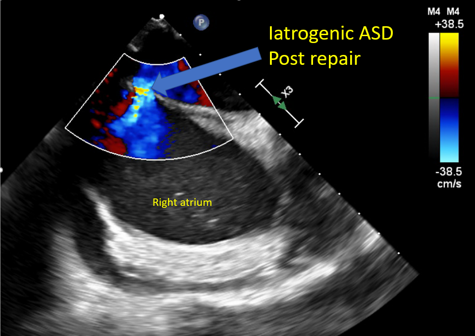
[PubMed PMID: 29622181]

Level 3 (low-level) evidence

[21]

Shiota T. Role of echocardiography for catheter-based management of valvular heart disease. Journal of cardiology. 2017 Jan:69(1):66-73. doi: 10.1016/j.jjcc.2016.09.015. Epub 2016 Nov 15     [PubMed PMID: 27863908]


[22]

Dziadzko V, Clavel MA, Dziadzko M, Medina-Inojosa JR, Michelena H, Maalouf J, Nkomo V, Thapa P, Enriquez-Sarano M. Outcome and undertreatment of mitral regurgitation: a community cohort study. Lancet (London, England). 2018 Mar 10:391(10124):960-969. doi: 10.1016/S0140-6736(18)30473-2. Epub     [PubMed PMID: 29536860]


[23]

Shah PM, Raney AA. Echocardiography in mitral regurgitation with relevance to valve surgery. Journal of the American Society of Echocardiography : official publication of the American Society of Echocardiography. 2011 Oct:24(10):1086-91. doi: 10.1016/j.echo.2011.08.017. Epub     [PubMed PMID: 21933744]


[24]

Asch FM, Little SH, Mackensen GB, Grayburn PA, Sorajja P, Rinaldi MJ, Maisano F, Kar S. Incidence and standardised definitions of mitral valve leaflet adverse events after transcatheter mitral valve repair: the EXPAND study. EuroIntervention : journal of EuroPCR in collaboration with the Working Group on Interventional Cardiology of the European Society of Cardiology. 2021 Dec 3:17(11):e932-e941. doi: 10.4244/EIJ-D-21-00012. Epub 2021 Dec 3     [PubMed PMID: 34031024]


[25]

Avierinos JF, Tribouilloy C, Grigioni F, Suri R, Barbieri A, Michelena HI, Ionico T, Rusinaru D, Ansaldi S, Habib G, Szymanski C, Giorgi R, Mahoney DW, Enriquez-Sarano M, Mitral regurgitation International DAtabase (MIDA) Investigators. Impact of ageing on presentation and outcome of mitral regurgitation due to flail leaflet: a multicentre international study. European heart journal. 2013 Sep:34(33):2600-9. doi: 10.1093/eurheartj/eht250. Epub 2013 Jul 12     [PubMed PMID: 23853072]


[26]

Thaden JJ, Malouf JF, Nkomo VT, Pislaru SV, Holmes DR Jr, Reeder GS, Rihal CS, Eleid MF. Mitral Valve Anatomic Predictors of Hemodynamic Success With Transcatheter Mitral Valve Repair. Journal of the American Heart Association. 2018 Jan 13:7(2):. doi: 10.1161/JAHA.117.007315. Epub 2018 Jan 13     [PubMed PMID: 29331957]


[27]

Estévez-Loureiro R, Franzen O, Winter R, Sondergaard L, Jacobsen P, Cheung G, Moat N, Ihlemann N, Ghione M, Price S, Duncan A, Streit Rosenberg T, Barker S, Di Mario C, Settergren M. Echocardiographic and clinical outcomes of central versus noncentral percutaneous edge-to-edge repair of degenerative mitral regurgitation. Journal of the American College of Cardiology. 2013 Dec 24:62(25):2370-2377. doi: 10.1016/j.jacc.2013.05.093. Epub 2013 Sep 4     [PubMed PMID: 24013059]

Level 2 (mid-level) evidence

[28]

Lapenna E, De Bonis M, Sorrentino F, La Canna G, Grimaldi A, Torracca L, Maisano F, Alfieri O. Commissural closure for the treatment of commissural mitral valve prolapse or flail. The Journal of heart valve disease. 2008 May:17(3):261-6     [PubMed PMID: 18592922]


[29]

Katz WE, Conrad Smith AJ, Crock FW, Cavalcante JL. Echocardiographic evaluation and guidance for MitraClip procedure. Cardiovascular diagnosis and therapy. 2017 Dec:7(6):616-632. doi: 10.21037/cdt.2017.07.04. Epub     [PubMed PMID: 29302467]


[30]

Khan F, Winkel M, Ong G, Brugger N, Pilgrim T, Windecker S, Praz F, Fam N. Percutaneous Mitral Edge-to-Edge Repair: State of the Art and a Glimpse to the Future. Frontiers in cardiovascular medicine. 2019:6():122. doi: 10.3389/fcvm.2019.00122. Epub 2019 Sep 18     [PubMed PMID: 31620446]


[31]

Gertz ZM, Herrmann HC, Lim DS, Kar S, Kapadia SR, Reed GW, Puri R, Krishnaswamy A, Gersh BJ, Weissman NJ, Asch FM, Grayburn PA, Kosmidou I, Redfors B, Zhang Z, Abraham WT, Lindenfeld J, Stone GW, Mack MJ. Implications of Atrial Fibrillation on the Mechanisms of Mitral Regurgitation and Response to MitraClip in the COAPT Trial. Circulation. Cardiovascular interventions. 2021 Apr:14(4):e010300. doi: 10.1161/CIRCINTERVENTIONS.120.010300. Epub 2021 Mar 15     [PubMed PMID: 33719505]


[32]

Abramowitz Y, Jilaihawi H, Chakravarty T, Mack MJ, Makkar RR. Mitral Annulus Calcification. Journal of the American College of Cardiology. 2015 Oct 27:66(17):1934-41. doi: 10.1016/j.jacc.2015.08.872. Epub     [PubMed PMID: 26493666]


[33]

Fernández-Peregrina E, Pascual I, Freixa X, Tirado-Conte G, Estévez-Loureiro R, Carrasco-Chinchilla F, Benito-González T, Asmarats L, Sanchís L, Jiménez-Quevedo P, Avanzas P, Caneiro-Queija B, Molina-Ramos AI, Fernández-Vázquez F, Li CH, Flores-Umanzor E, Sans-Roselló J, Nombela-Franco L, Arzamendi D. Transcatheter edge-to-edge mitral valve repair in patients with mitral annulus calcification. EuroIntervention : journal of EuroPCR in collaboration with the Working Group on Interventional Cardiology of the European Society of Cardiology. 2022 Mar 18:17(16):1300-1309. doi: 10.4244/EIJ-D-21-00205. Epub     [PubMed PMID: 34483091]


[34]

Flint N, Raschpichler M, Rader F, Shmueli H, Siegel RJ. Asymptomatic Degenerative Mitral Regurgitation: A Review. JAMA cardiology. 2020 Mar 1:5(3):346-355. doi: 10.1001/jamacardio.2019.5466. Epub     [PubMed PMID: 31995124]


[35]

Flameng W, Herijgers P, Bogaerts K. Recurrence of mitral valve regurgitation after mitral valve repair in degenerative valve disease. Circulation. 2003 Apr 1:107(12):1609-13     [PubMed PMID: 12668494]


[36]

Kim JH, Lee SH, Joo HC, Youn YN, Yoo KJ, Chang BC, Lee S. Effect of Recurrent Mitral Regurgitation After Mitral Valve Repair in Patients With Degenerative Mitral Regurgitation. Circulation journal : official journal of the Japanese Circulation Society. 2017 Dec 25:82(1):93-101. doi: 10.1253/circj.CJ-17-0380. Epub 2017 Jul 20     [PubMed PMID: 28724839]


[37]

Mehaffey HJ, Hawkins RB, Schubert S, Fonner C, Yarboro LT, Quader M, Speir A, Rich J, Kron IL, Ailawadi G. Contemporary outcomes in reoperative mitral valve surgery. Heart (British Cardiac Society). 2018 Apr:104(8):652-656. doi: 10.1136/heartjnl-2017-312047. Epub 2017 Oct 5     [PubMed PMID: 28982718]


[38]

Niikura H, Bae R, Gössl M, Lin D, Jay D, Sorajja P. Transcatheter therapy for residual mitral regurgitation after MitraClip therapy. EuroIntervention : journal of EuroPCR in collaboration with the Working Group on Interventional Cardiology of the European Society of Cardiology. 2019 Aug 9:15(6):e491-e499. doi: 10.4244/EIJ-D-18-01148. Epub 2019 Aug 9     [PubMed PMID: 31217145]


[39]

Braun D, Frerker C, Körber MI, Gaemperli O, Patzelt J, Schaefer U, Hammerstingl C, Boekstegers P, Ott I, Ince H, Thiele H, Hausleiter J. Percutaneous Edge-to-Edge Repair of Recurrent Severe Mitral Regurgitation After Surgical Mitral Valve Repair. Journal of the American College of Cardiology. 2017 Jul 25:70(4):504-505. doi: 10.1016/j.jacc.2017.05.045. Epub     [PubMed PMID: 28728696]


[40]

Grasso C, Ohno Y, Attizzani GF, Cannata S, Immè S, Barbanti M, Pistritto AM, Ministeri M, Caggegi A, Chiarandà M, Dipasqua F, Ronsivalle G, Mangiafico S, Scandura S, Capranzano P, Capodanno D, Tamburino C. Percutaneous mitral valve repair with the MitraClip system for severe mitral regurgitation in patients with surgical mitral valve repair failure. Journal of the American College of Cardiology. 2014 Mar 4:63(8):836-8. doi: 10.1016/j.jacc.2013.09.045. Epub 2013 Oct 23     [PubMed PMID: 24161329]


[41]

Kanda BS, Jay D, Farivar RS, Sorajja P. Leaflet-to-Annuloplasty Ring Clipping for Severe Mitral Regurgitation. JACC. Cardiovascular interventions. 2016 Apr 11:9(7):e63-4. doi: 10.1016/j.jcin.2015.12.272. Epub 2016 Mar 4     [PubMed PMID: 26952908]


[42]

Wu IY, Barajas MB, Hahn RT. The MitraClip Procedure-A Comprehensive Review for the Cardiac Anesthesiologist. Journal of cardiothoracic and vascular anesthesia. 2018 Dec:32(6):2746-2759. doi: 10.1053/j.jvca.2018.05.020. Epub 2018 Sep 27     [PubMed PMID: 30268642]


[43]

Hahn RT. Transcathether Valve Replacement and Valve Repair: Review of Procedures and Intraprocedural Echocardiographic Imaging. Circulation research. 2016 Jul 8:119(2):341-56. doi: 10.1161/CIRCRESAHA.116.307972. Epub     [PubMed PMID: 27390336]


[44]

Hahn RT, Chan V, Adams DH. Current Indications for Transcatheter Edge-to-Edge Repair in a Patient With Primary Mitral Regurgitation. Circulation. 2022 Oct 25:146(17):1263-1265. doi: 10.1161/CIRCULATIONAHA.122.061495. Epub 2022 Oct 24     [PubMed PMID: 36279415]


[45]

Lee G, Chikwe J, Milojevic M, Wijeysundera HC, Biondi-Zoccai G, Flather M, Gaudino MFL, Fremes SE, Tam DY. ESC/EACTS vs. ACC/AHA guidelines for the management of severe aortic stenosis. European heart journal. 2023 Mar 7:44(10):796-812. doi: 10.1093/eurheartj/ehac803. Epub     [PubMed PMID: 36632841]


[46]

Russell EA, Walsh WF, Costello B, McLellan AJA, Brown A, Reid CM, Tran L, Maguire GP. Medical Management of Rheumatic Heart Disease: A Systematic Review of the Evidence. Cardiology in review. 2018 Jul/Aug:26(4):187-195. doi: 10.1097/CRD.0000000000000185. Epub     [PubMed PMID: 29608495]

Level 1 (high-level) evidence

[47]

Van Praet KM, Stamm C, Sündermann SH, Meyer A, Unbehaun A, Montagner M, Nazari Shafti TZ, Jacobs S, Falk V, Kempfert J. Minimally Invasive Surgical Mitral Valve Repair: State of the Art Review. Interventional cardiology (London, England). 2018 Jan:13(1):14-19. doi: 10.15420/icr.2017:30:1. Epub     [PubMed PMID: 29593831]


[48]

Taramasso M, Gaemperli O, Maisano F. Treatment of degenerative mitral regurgitation in elderly patients. Nature reviews. Cardiology. 2015 Mar:12(3):177-83. doi: 10.1038/nrcardio.2014.210. Epub 2014 Dec 23     [PubMed PMID: 25533801]


[49]

Asch FM, Grayburn PA, Siegel RJ, Kar S, Lim DS, Zaroff JG, Mishell JM, Whisenant B, Mack MJ, Lindenfeld J, Abraham WT, Stone GW, Weissman NJ, COAPT Investigators. Echocardiographic Outcomes After Transcatheter Leaflet Approximation in Patients With Secondary Mitral Regurgitation: The COAPT Trial. Journal of the American College of Cardiology. 2019 Dec 17:74(24):2969-2979. doi: 10.1016/j.jacc.2019.09.017. Epub 2019 Sep 28     [PubMed PMID: 31574303]


[50]

Nishimura RA, Vahanian A, Eleid MF, Mack MJ. Mitral valve disease--current management and future challenges. Lancet (London, England). 2016 Mar 26:387(10025):1324-34. doi: 10.1016/S0140-6736(16)00558-4. Epub     [PubMed PMID: 27025438]


[51]

Chakravarty T, Makar M, Patel D, Oakley L, Yoon SH, Stegic J, Singh S, Skaf S, Nakamura M, Makkar RR. Transcatheter Edge-to-Edge Mitral Valve Repair With the MitraClip G4 System. JACC. Cardiovascular interventions. 2020 Oct 26:13(20):2402-2414. doi: 10.1016/j.jcin.2020.06.053. Epub 2020 Sep 30     [PubMed PMID: 33011141]


[52]

Praz F, Braun D, Unterhuber M, Spirito A, Orban M, Brugger N, Brinkmann I, Spring K, Moschovitis A, Nabauer M, Blazek S, Pilgrim T, Thiele H, Lurz P, Hausleiter J, Windecker S. Edge-to-Edge Mitral Valve Repair With Extended Clip Arms: Early Experience From a Multicenter Observational Study. JACC. Cardiovascular interventions. 2019 Jul 22:12(14):1356-1365. doi: 10.1016/j.jcin.2019.03.023. Epub 2019 May 22     [PubMed PMID: 31129091]

Level 2 (mid-level) evidence

[53]

Praz F, Spargias K, Chrissoheris M, Büllesfeld L, Nickenig G, Deuschl F, Schueler R, Fam NP, Moss R, Makar M, Boone R, Edwards J, Moschovitis A, Kar S, Webb J, Schäfer U, Feldman T, Windecker S. Compassionate use of the PASCAL transcatheter mitral valve repair system for patients with severe mitral regurgitation: a multicentre, prospective, observational, first-in-man study. Lancet (London, England). 2017 Aug 19:390(10096):773-780. doi: 10.1016/S0140-6736(17)31600-8. Epub     [PubMed PMID: 28831993]


[54]

Hausleiter J, Stocker TJ, Adamo M, Karam N, Swaans MJ, Praz F. Mitral valve transcatheter edge-to-edge repair. EuroIntervention : journal of EuroPCR in collaboration with the Working Group on Interventional Cardiology of the European Society of Cardiology. 2023 Jan 23:18(12):957-976. doi: 10.4244/EIJ-D-22-00725. Epub     [PubMed PMID: 36688459]


[55]

Huang EY, Chen C, Abdullah F, Aspelund G, Barnhart DC, Calkins CM, Cowles RA, Downard CD, Goldin AB, Lee SL, St Peter SD, Arca MJ, 2011 American Pediatric Surgical Association Outcomes and Clinical Trials Committee. Strategies for the prevention of central venous catheter infections: an American Pediatric Surgical Association Outcomes and Clinical Trials Committee systematic review. Journal of pediatric surgery. 2011 Oct:46(10):2000-11. doi: 10.1016/j.jpedsurg.2011.06.017. Epub     [PubMed PMID: 22008341]

Level 1 (high-level) evidence

[56]

Kassar M, Praz F, Hunziker L, Pilgrim T, Windecker S, Seiler C, Brugger N. Anatomical and Technical Predictors of Three-Dimensional Mitral Valve Area Reduction After Transcatheter Edge-To-Edge Repair. Journal of the American Society of Echocardiography : official publication of the American Society of Echocardiography. 2022 Jan:35(1):96-104. doi: 10.1016/j.echo.2021.08.021. Epub 2021 Oct 11     [PubMed PMID: 34506920]


[57]

Singh GD, Smith TW, Rogers JH. Multi-MitraClip therapy for severe degenerative mitral regurgitation: "anchor" technique for extremely flail segments. Catheterization and cardiovascular interventions : official journal of the Society for Cardiac Angiography & Interventions. 2015 Aug:86(2):339-46. doi: 10.1002/ccd.25811. Epub 2015 Feb 12     [PubMed PMID: 25559345]


[58]

Oguz D, Padang R, Rashedi N, Pislaru SV, Nkomo VT, Mankad SV, Malouf JF, Guerrero M, Reeder GS, Eleid MF, Rihal CS, Thaden JJ. Risk for Increased Mean Diastolic Gradient after Transcatheter Edge-to-Edge Mitral Valve Repair: A Quantitative Three-Dimensional Transesophageal Echocardiographic Analysis. Journal of the American Society of Echocardiography : official publication of the American Society of Echocardiography. 2021 Jun:34(6):595-603.e2. doi: 10.1016/j.echo.2021.01.018. Epub 2021 Jan 29     [PubMed PMID: 33524491]


[59]

Naqvi TZ. Echocardiography in percutaneous valve therapy. JACC. Cardiovascular imaging. 2009 Oct:2(10):1226-37. doi: 10.1016/j.jcmg.2009.08.004. Epub     [PubMed PMID: 19833314]


[60]

Gheorghe L, Ielasi A, Rensing BJWM, Eefting FD, Timmers L, Latib A, Swaans MJ. Complications Following Percutaneous Mitral Valve Repair. Frontiers in cardiovascular medicine. 2019:6():146. doi: 10.3389/fcvm.2019.00146. Epub 2019 Oct 18     [PubMed PMID: 31681798]


[61]

Hahn RT, Saric M, Faletra FF, Garg R, Gillam LD, Horton K, Khalique OK, Little SH, Mackensen GB, Oh J, Quader N, Safi L, Scalia GM, Lang RM. Recommended Standards for the Performance of Transesophageal Echocardiographic Screening for Structural Heart Intervention: From the American Society of Echocardiography. Journal of the American Society of Echocardiography : official publication of the American Society of Echocardiography. 2022 Jan:35(1):1-76. doi: 10.1016/j.echo.2021.07.006. Epub 2021 Jul 17     [PubMed PMID: 34280494]


[62]

Lancellotti P, Tribouilloy C, Hagendorff A, Popescu BA, Edvardsen T, Pierard LA, Badano L, Zamorano JL, Scientific Document Committee of the European Association of Cardiovascular Imaging. Recommendations for the echocardiographic assessment of native valvular regurgitation: an executive summary from the European Association of Cardiovascular Imaging. European heart journal. Cardiovascular Imaging. 2013 Jul:14(7):611-44. doi: 10.1093/ehjci/jet105. Epub 2013 Jun 3     [PubMed PMID: 23733442]


[63]

Alkhouli M, Rihal CS, Holmes DR Jr. Transseptal Techniques for Emerging Structural Heart Interventions. JACC. Cardiovascular interventions. 2016 Dec 26:9(24):2465-2480. doi: 10.1016/j.jcin.2016.10.035. Epub     [PubMed PMID: 28007198]


[64]

Sherif MA, Paranskaya L, Yuecel S, Kische S, Thiele O, D'Ancona G, Neuhausen-Abramkina A, Ortak J, Ince H, Öner A. MitraClip step by step; how to simplify the procedure. Netherlands heart journal : monthly journal of the Netherlands Society of Cardiology and the Netherlands Heart Foundation. 2017 Feb:25(2):125-130. doi: 10.1007/s12471-016-0930-7. Epub     [PubMed PMID: 27933588]


[65]

Swaans MJ, Van den Branden BJ, Van der Heyden JA, Post MC, Rensing BJ, Eefting FD, Plokker HW, Jaarsma W. Three-dimensional transoesophageal echocardiography in a patient undergoing percutaneous mitral valve repair using the edge-to-edge clip technique. European journal of echocardiography : the journal of the Working Group on Echocardiography of the European Society of Cardiology. 2009 Dec:10(8):982-3. doi: 10.1093/ejechocard/jep101. Epub 2009 Aug 4     [PubMed PMID: 19654135]


[66]

Maslow A, Mahmood F, Poppas A, Singh A. Three-dimensional echocardiographic assessment of the repaired mitral valve. Journal of cardiothoracic and vascular anesthesia. 2014 Feb:28(1):11-17. doi: 10.1053/j.jvca.2013.05.007. Epub 2013 Sep 25     [PubMed PMID: 24075641]


[67]

Saitoh T, Izumo M, Furugen A, Tanaka J, Miyata-Fukuoka Y, Gurudevan SV, Tolstrup K, Siegel RJ, Kar S, Shiota T. Echocardiographic evaluation of iatrogenic atrial septal defect after catheter-based mitral valve clip insertion. The American journal of cardiology. 2012 Jun 15:109(12):1787-91. doi: 10.1016/j.amjcard.2012.02.023. Epub 2012 Apr 3     [PubMed PMID: 22475361]


[68]

Writing Committee Members, Otto CM, Nishimura RA, Bonow RO, Carabello BA, Erwin JP 3rd, Gentile F, Jneid H, Krieger EV, Mack M, McLeod C, O'Gara PT, Rigolin VH, Sundt TM 3rd, Thompson A, Toly C. 2020 ACC/AHA Guideline for the Management of Patients With Valvular Heart Disease: Executive Summary: A Report of the American College of Cardiology/American Heart Association Joint Committee on Clinical Practice Guidelines. Journal of the American College of Cardiology. 2021 Feb 2:77(4):450-500. doi: 10.1016/j.jacc.2020.11.035. Epub 2020 Dec 17     [PubMed PMID: 33342587]


[69]

Nickenig G, Estevez-Loureiro R, Franzen O, Tamburino C, Vanderheyden M, Lüscher TF, Moat N, Price S, Dall'Ara G, Winter R, Corti R, Grasso C, Snow TM, Jeger R, Blankenberg S, Settergren M, Tiroch K, Balzer J, Petronio AS, Büttner HJ, Ettori F, Sievert H, Fiorino MG, Claeys M, Ussia GP, Baumgartner H, Scandura S, Alamgir F, Keshavarzi F, Colombo A, Maisano F, Ebelt H, Aruta P, Lubos E, Plicht B, Schueler R, Pighi M, Di Mario C, Transcatheter Valve Treatment Sentinel Registry Investigators of the EURObservational Research Programme of the European Society of Cardiology. Percutaneous mitral valve edge-to-edge repair: in-hospital results and 1-year follow-up of 628 patients of the 2011-2012 Pilot European Sentinel Registry. Journal of the American College of Cardiology. 2014 Sep 2:64(9):875-84. doi: 10.1016/j.jacc.2014.06.1166. Epub     [PubMed PMID: 25169171]

Level 3 (low-level) evidence

[70]

Schnitzler K, Hell M, Geyer M, Kreidel F, Münzel T, von Bardeleben RS. Complications Following MitraClip Implantation. Current cardiology reports. 2021 Aug 13:23(9):131. doi: 10.1007/s11886-021-01553-9. Epub 2021 Aug 13     [PubMed PMID: 34387748]


[71]

Sticchi A, Bartkowiak J, Brugger N, Weiss S, Windecker S, Praz F. Retrograde Retrieval of a Novel Large Mitral Clip After Embolization Into the Left Ventricle. JACC. Case reports. 2021 Oct 20:3(14):1561-1568. doi: 10.1016/j.jaccas.2021.08.024. Epub 2021 Oct 20     [PubMed PMID: 34729501]

Level 3 (low-level) evidence

[72]

Hamm K, Barth S, Diegeler A, Kerber S. Stroke and thrombus formation appending to the MitraClip: what is the appropriate anticoagulation regimen? The Journal of heart valve disease. 2013 Sep:22(5):713-5     [PubMed PMID: 24383386]


[73]

Nakajima Y, Kar S. First experience of the usage of a GORE CARDIOFORM Septal Occluder device for treatment of a significant residual commissural mitral regurgitation jet following a MitraClip procedure. Catheterization and cardiovascular interventions : official journal of the Society for Cardiac Angiography & Interventions. 2018 Sep 1:92(3):607-610. doi: 10.1002/ccd.27438. Epub 2017 Dec 8     [PubMed PMID: 29219253]


[74]

Mangieri A, Melillo F, Montalto C, Denti P, Praz F, Sala A, Winkel MG, Taramasso M, Tagliari AP, Fam NP, Rubbio AP, De Marco F, Bedogni F, Toggweiler S, Schofer J, Brinkmann C, Sievert H, Van Mieghem NM, Ooms JF, Paradis JM, Rodés-Cabau J, Brochet E, Himbert D, Perl L, Kornowski R, Ielasi A, Regazzoli D, Baldetti L, Masiero G, Tarantini G, Latib A, Laricchia A, Gattas A, Tchetchè D, Dumonteil N, Francesco G, Agricola E, Montorfano M, Lurz P, Crimi G, Maisano F, Colombo A. Management and Outcome of Failed Percutaneous Edge-to-Edge Mitral Valve Plasty: Insight From an International Registry. JACC. Cardiovascular interventions. 2022 Feb 28:15(4):411-422. doi: 10.1016/j.jcin.2021.11.040. Epub     [PubMed PMID: 35210047]


[75]

Alessandrini H, Dreher A, Harr C, Wohlmuth P, Meincke F, Hakmi S, Ubben T, Kuck KH, Hassan K, Willems S, Schmoeckel M, Geidel S. Clinical impact of intervention strategies after failed transcatheter mitral valve repair. EuroIntervention : journal of EuroPCR in collaboration with the Working Group on Interventional Cardiology of the European Society of Cardiology. 2021 Apr 20:16(17):1447-1454. doi: 10.4244/EIJ-D-20-01008. Epub     [PubMed PMID: 33074154]


[76]

Lavall D, Reil JC, Segura Schmitz L, Mehrer M, Schirmer SH, Böhm M, Laufs U. Early Hemodynamic Improvement after Percutaneous Mitral Valve Repair Evaluated by Noninvasive Pressure-Volume Analysis. Journal of the American Society of Echocardiography : official publication of the American Society of Echocardiography. 2016 Sep:29(9):888-98. doi: 10.1016/j.echo.2016.05.012. Epub 2016 Jun 29     [PubMed PMID: 27372560]