Hypertrophic Cardiomyopathy

Earn CME/CE in your profession:


Continuing Education Activity

Hypertrophic cardiomyopathy (HCM) is an autosomal dominant disease caused by a mutation in sarcomere protein genes which effect contraction of the heart. HCM causes alterations of heart structure that affect function. An increase in left ventricular wall thickness (hypertrophy) causes left ventricular outflow obstruction, diastolic dysfunction, myocardial ischemia, and mitral regurgitation. This can lead to fatigue, dyspnea, chest pain, palpitations, and syncope. Sudden cardiac death represents the most severe and devastating presentation of HCM, and is usually seen in young athletes. This activity reviews the evaluation and management of hypertrophic cardiomyopathy and highlights the role of the interprofessional team in the care of patients with this condition.

Objectives:

  • Review the pathophysiology of hypertrophic cardiomyopathy.
  • Outline the typical presentation of a patient with hypertrophic cardiomyopathy.
  • Explain the recommended management for hypertrophic cardiomyopathy.
  • Explain the interprofessional team strategies for improving care coordination and communication regarding the management of patients with hypertrophic cardiomyopathy.

Introduction

Hypertrophic cardiomyopathy (HCM) is a genetic (autosomal dominant) heart muscle disease caused by a mutation in sarcomere protein genes which encodes for elements of the contractile machinery of the heart. It is characterized by an increase in left ventricular wall thickness (hypertrophy) which causes left ventricular outflow obstruction, diastolic dysfunction, myocardial ischemia, and mitral regurgitation. 

These structural and function abnormalities can produce fatigue, dyspnea, chest pain, palpitations, and syncope. Sudden cardiac death can represent the most devastating presenting manifestation, especially in young athletes.[1][2][3]

Etiology

Familial Hypertrophic cardiomyopathy occurs as an autosomal dominant Mendelian-Inherited disorder in approximately 50% of cases. A mutation in the sarcomere protein gene encoding for contractile elements of the heart has been found, with 6 different genes on at least 4 chromosomes being associated with HCM. More than 50 different mutations have been identified. The phenotypic expression may vary according to the mutation involved, with a variability of symptoms and degree of severity of the hypertrophy expressed. Recent data has associated abnormal myocardial calcium kinetics to the cause of inappropriate myocardial hypertrophy and specific features of HCM, especially in patients with diastolic functional abnormalities. [4][5][6][7]

Epidemiology

The prevalence of HCM in the general population around the world is 0.2% (1 in 500 adults), determined from echocardiographic studies. The morphologic evidence is found in approximately 25% of first-degree relatives of patients with HCM. Newer genetic testing has been developed and can be used to identify asymptomatic family members with same sarcomere mutation. HCM is more common in males than females, however, since the condition is autosomal dominant, genetic inheritance does not follow sex predilection. The most common presentation is in the third decade of life but may be present at any age, from newborns to elderly patients.

Pathophysiology

Ventricular hypertrophy results in a dynamic pressure gradient across the left ventricular outflow tract (LVOT), which is associated with further narrowing during systole. During this cardiac cycle, the mitral valve is pulled towards the septum by several proposed mechanisms: contraction of the papillary muscles, abnormal location in the outflow tract, and low pressure that occurs as blood is ejected at high velocity through a narrowed outflow tract (Venturi effect).

Histopathology

Histology usually reveals myocardial hypertrophy with a gross disarray of the myofibrils and disorganization of the muscle architecture.

There are visible fibrous scars and abnormal internal coronary vessels.

The histological abnormality is most often seen in the ventricular septum.

History and Physical

The most common presenting symptom of hypertrophic cardiomyopathy is dyspnea. Patients also can develop syncope, palpitations, angina, orthopnea, paroxysmal nocturnal dyspnea, dizziness, congestive heart failure, and sudden cardiac death. The latter represents the most devastating presenting symptom.

Physical examination findings are characterized by a double apical impulse due to forceful left atrial contraction against a highly noncompliant left ventricle, normal s1 and split s2, s3 due to decompensated heart failure, prominent "a wave" of jugular venous pressure, laterally displaced apical impulse, and double carotid pulse.

A systolic ejection murmur is present as follows: diminished intensity with increase preload (squatting) or afterload (handgrip) and increased intensity with a decrease in preload (Valsalva maneuver, standing), and with any decrease in afterload (vasodilator administration).

Evaluation

All patients should have a comprehensive cardiac history and physical examination. An electrocardiogram (ECG) and cardiac imaging to identify left ventricular hypertrophy should be performed in all patients. [8][9][10]

  • ECG: the most sensitive diagnostic test. Findings include localized or widespread repolarization changes, prominent Q waves in the inferior (II, III, and aVF) and lateral leads (I, aVL, and V4-V6), left atrial or biatrial enlargement, left axis deviation, deeply inverted T waves. The combination of left ventricular hypertrophy (LVH) with right atrial enlargement is strongly suggestive of HCM.
  • Transthoracic echocardiogram (TTE): Demonstrates cardiac morphology, systolic and diastolic function, the presence and severity of any LVOT gradient, and the degree of mitral regurgitation.
  • Ambulatory ECG monitoring: Should be performed for 24 to 48 hours in all patients diagnosed with HCM for risk assessment of ventricular arrhythmias and sudden death.
  • Exercise stress testing: For risk stratification and assessment of LV outflow tract (LVOT) gradient. Exercise stress is the preferred method.

Some patients may also require the following:

  • Cardiac catheterization: Determines cardiac hemodynamic, the degree of left ventricular outflow obstruction, and anatomy of the coronary vessels.

  • Electrophysiological studies: Determine the origin of the arrhythmias.

Treatment / Management

Treatment strategies for HCM are based upon observational data and clinical experience since no large randomized trials have been performed. Pharmacological therapy is the first-line approach to symptomatic HCH. The best initial medications include negative inotropic agents, including beta blockers, nondihydropyridine calcium channel blockers (verapamil), and disopyramide.[11][12]

  • Diuretics can be given in patients without LVOT obstruction and refractory heart failure symptoms presenting with volume overload. Volume depletion decreases stroke volume and worsens the LVOT gradient. This can lead to hypotension, lightheadedness, and syncope.
  • Nonpharmacologic therapies include surgical myomectomy and alcohol septal ablation (if NYHA III/IV class persist despite optimal medical therapy or syncope related to hemodynamic compromise from LVOT obstruction, an LVOT gradient >50mmHg).
  • Alcohol septal ablation reduces LVOT obstruction, improves symptoms, increases exercise capacity, and may improve long-term survival.

Surgical therapeutic options include the following:

  • A heart transplant is not the first treatment option and usually recommended only in patients who have failed all medical and surgical treatments.

  • Left ventricular myomectomy

  • Mitral valve replacement

  • Permanent pacemaker implantation

  • Placement of an implantable cardioverter defibrillator is an option in patients with ventricular fibrillation who do not want lifelong medical therapy.

Differential Diagnosis

  • Aortic stenosis
  • Athlete’s heart
  • Amyloidosis
  • Genetics of Fabry disease
  • Glycogen storage disease
  • Hypertensive heart disease
  • Mucopolysaccharide storage disease
  • Right ventricular hypertrophy
  • Restrictive cardiomyopathy
  • Sarcoidosis

Prognosis

  • Mortality rates of 1% to 4% have been reported in patients with HCM, but these numbers have greatly improved in the past two decades.
  • Even though most patients with HCM have no symptoms, the first clinical presentation is often sudden death from malignant arrhythmias. The highest mortality is in young people.
  • Early diagnosis is important as it allows the healthcare provider is to prescribe an appropriate level of safe physical activity
  • Besides sudden death, patients can have atrial and ventricular arrhythmias. 
  • Those who have concomitant mitral regulation and diastolic dysfunction are also prone to recurrent episodes of heart failure.

Pearls and Other Issues

  • Individuals diagnosed with hypertrophic cardiomyopathy should avoid strenuous exercise.
  • Family members with a history of sudden death in a family member should be screened for the disorder.
  • Individuals with abnormal blood pressure in response to exercise should also be screened.
  • Low-level physical activity is not a risk factor for sudden death.

Enhancing Healthcare Team Outcomes

The diagnosis and management of patients with hypertrophic cardiomyopathy is with an interprofessional team consisting of a cardiologist, cardiac surgeon, electrophysiologist, internist, primary care provider and the nurse practitioner. There are several treatments for the disorder. Besides pharmaological therapy, some patients may benefit from surgery. The surgical options include a mitral valve replacement, left ventricular myomectomy, pacemaker or AICD insertion or a heart transplant. The outcomes of these patients is good as long as they remain compliant with treatment. Unfortunately, even after treatment many continue to have atrial and ventricular arrhythmias. For those untreated, sudden death may be the first symptom.[13][7]


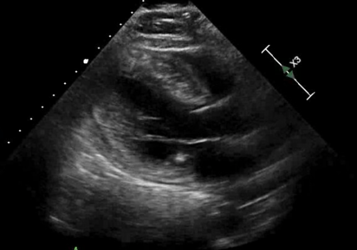
(Click Image to Enlarge)
Hypertrophic Cardiomyopathy
Contributed by Yamama Hafeez, DO
Details

Author

Daniel Brito

Author

Hajira Basit

Editor:

Saurabh Sharma

Updated:

4/7/2023 3:07:36 PM

References


[1]

Spudich JA. Three perspectives on the molecular basis of hypercontractility caused by hypertrophic cardiomyopathy mutations. Pflugers Archiv : European journal of physiology. 2019 May:471(5):701-717. doi: 10.1007/s00424-019-02259-2. Epub 2019 Feb 15     [PubMed PMID: 30767072]

Level 3 (low-level) evidence

[2]

van Driel B, Nijenkamp L, Huurman R, Michels M, van der Velden J. Sex differences in hypertrophic cardiomyopathy: new insights. Current opinion in cardiology. 2019 May:34(3):254-259. doi: 10.1097/HCO.0000000000000612. Epub     [PubMed PMID: 30747730]

Level 3 (low-level) evidence

[3]

Kraft T, Montag J. Altered force generation and cell-to-cell contractile imbalance in hypertrophic cardiomyopathy. Pflugers Archiv : European journal of physiology. 2019 May:471(5):719-733. doi: 10.1007/s00424-019-02260-9. Epub 2019 Feb 11     [PubMed PMID: 30740621]


[4]

Philipson DJ, Rader F, Siegel RJ. Risk factors for atrial fibrillation in hypertrophic cardiomyopathy. European journal of preventive cardiology. 2019 Feb 6:():2047487319828474. doi: 10.1177/2047487319828474. Epub 2019 Feb 6     [PubMed PMID: 30727760]


[5]

Mavrogeni SI, Tsarouhas K, Spandidos DA, Kanaka-Gantenbein C, Bacopoulou F. Sudden cardiac death in football players: Towards a new pre-participation algorithm. Experimental and therapeutic medicine. 2019 Feb:17(2):1143-1148. doi: 10.3892/etm.2018.7041. Epub 2018 Nov 30     [PubMed PMID: 30679986]


[6]

Maron MS, Wells S. Myocardial Strain in Hypertrophic Cardiomyopathy: A Force Worth Pursuing? JACC. Cardiovascular imaging. 2019 Oct:12(10):1943-1945. doi: 10.1016/j.jcmg.2018.09.026. Epub 2019 Jan 16     [PubMed PMID: 30660525]


[7]

Rigopoulos AG, Ali M, Abate E, Matiakis M, Melnyk H, Mavrogeni S, Leftheriotis D, Bigalke B, Noutsias M. Review on sudden death risk reduction after septal reduction therapies in hypertrophic obstructive cardiomyopathy. Heart failure reviews. 2019 May:24(3):359-366. doi: 10.1007/s10741-018-09767-w. Epub     [PubMed PMID: 30617667]


[8]

Walsh R, Mazzarotto F, Whiffin N, Buchan R, Midwinter W, Wilk A, Li N, Felkin L, Ingold N, Govind R, Ahmad M, Mazaika E, Allouba M, Zhang X, de Marvao A, Day SM, Ashley E, Colan SD, Michels M, Pereira AC, Jacoby D, Ho CY, Thomson KL, Watkins H, Barton PJR, Olivotto I, Cook SA, Ware JS. Quantitative approaches to variant classification increase the yield and precision of genetic testing in Mendelian diseases: the case of hypertrophic cardiomyopathy. Genome medicine. 2019 Jan 29:11(1):5. doi: 10.1186/s13073-019-0616-z. Epub 2019 Jan 29     [PubMed PMID: 30696458]

Level 3 (low-level) evidence

[9]

Afanasyev A, Bogachev-Prokophiev A, Lenko E, Sharifulin R, Ovcharov M, Kozmin D, Karaskov A. Myectomy with mitral valve repair versus replacement in adult patients with hypertrophic obstructive cardiomyopathy: a systematic review and meta-analysis. Interactive cardiovascular and thoracic surgery. 2019 Mar 1:28(3):465-472. doi: 10.1093/icvts/ivy269. Epub     [PubMed PMID: 30184144]

Level 1 (high-level) evidence

[10]

Robyns T, Nuyens D, Lu HR, Gallacher DJ, Vandenberk B, Garweg C, Ector J, Pagourelias E, Van Cleemput J, Janssens S, Willems R. Prognostic value of electrocardiographic time intervals and QT rate dependence in hypertrophic cardiomyopathy. Journal of electrocardiology. 2018 Nov-Dec:51(6):1077-1083. doi: 10.1016/j.jelectrocard.2018.09.005. Epub 2018 Sep 12     [PubMed PMID: 30497734]


[11]

Marrakchi S, Kammoun I, Bennour E, Laroussi L, Kachboura S. Risk stratification in hypertrophic cardiomyopathy. Herz. 2020 Feb:45(1):50-64. doi: 10.1007/s00059-018-4700-8. Epub 2018 Apr 25     [PubMed PMID: 29696341]


[12]

Daubert C, Gadler F, Mabo P, Linde C. Pacing for hypertrophic obstructive cardiomyopathy: an update and future directions. Europace : European pacing, arrhythmias, and cardiac electrophysiology : journal of the working groups on cardiac pacing, arrhythmias, and cardiac cellular electrophysiology of the European Society of Cardiology. 2018 Jun 1:20(6):908-920. doi: 10.1093/europace/eux131. Epub     [PubMed PMID: 29106577]

Level 3 (low-level) evidence

[13]

Yanagiuchi T,Tada N,Haga Y,Suzuki S,Sakurai M,Taguri M,Ootomo T, Utility of preprocedural multidetector computed tomography in alcohol septal ablation for hypertrophic obstructive cardiomyopathy. Cardiovascular intervention and therapeutics. 2019 Feb 6;     [PubMed PMID: 30725361]