Choledochal Cyst

Earn CME/CE in your profession:


Continuing Education Activity

A choledochal cyst (CC) or biliary cyst is a congenital or acquired anomaly affecting the biliary tree. It involves the dilation of the biliary tree that could affect the extrahepatic and/or the intrahepatic segments. Early diagnosis and management are critical. This activity discusses the evaluation and management of choledochal cysts and highlights the role of the interprofessional team in managing patients with this condition.

Objectives:

  • Explain the etiology of the choledochal cysts.
  • Describe the presentation of patients with choledochal cysts.
  • Review the treatment considerations for patients with choledochal cysts.
  • Outline the importance of improving care coordination among the interprofessional team to enhance the delivery of care for patients with choledochal cysts.

Introduction

A choledochal cyst (CC) has traditionally been considered as a cystic dilation of the extrahepatic bile duct. Choledochal cysts are now termed biliary cysts to include intrahepatic cysts also. Biliary cysts are defined as cystic dilations involving the biliary tree at single or multiple segments of both the extrahepatic as well as intrahepatic bile ducts. In 1959 Alonso-LEJ et al. were the first to classify CC into 3 types based on the site of the biliary duct dilation.[1] It was not until 1977 when Todani et al. modified the latter classification by adding 2 types of CCs.[2] This five-category classification is the most commonly used by clinicians today; however, it is in dispute by some experts who claim that each type of CC has its natural course, complications, and management. It is proposed that a classification that focuses more on the pathogenesis rather than the simple anatomy of the biliary tree is recommended.[3] 

Choledochal cyst is a rare anomaly that is, at times, considered a premalignant condition, which often poses a diagnostic dilemma. The typical presentation of this condition is non-specific. Healthcare teams must have a high clinical suspicion of choledochal cysts while investigating patients with jaundice, abdominal pain, and palpable abdominal mass. Due to the vague nature of these symptoms and physical findings, proper imaging studies are crucial for its diagnosis. Excision of the CC has shown excellent results with an 89% event-free rate and an overall 5-year survival rate above 90%. Hence early diagnosis and appropriate management are needed for optimal results and good prognosis.

Etiology

The exact etiology of a choledochal cyst remains unknown, which has led to the postulation of multiple theories. The most popular theory is Babbitt's, which states that the cyst develops from an anomalous pancreaticobiliary junction (APBJ). APBJ is when the biliary and the pancreatic duct join 1 to 2 cm proximal to the sphincter of Oddi. As a result, the long channel formed is not covered by the sphincter and allows for the backflow and mixing of pancreatic and biliary secretions, which leads to the activation of pancreatic enzymes. This results in a rise in pressure, which eventually causes dilation, inflammation, epithelial damage, dysplasia, and malignancy of the biliary tree.[4]

This theory is supported by the presence of a high level of amylase, and few studies reported elevated levels of phospholipase A2 and trypsinogen in CCs bile.[5][6] Animal studies support this theory in which the authors surgically created an APBJ in the models, which led to ductal dilation and cyst formation in the biliary tree.[7] Nonetheless, Babbitt's theory is challenged by authors who state that APBJ was present in only 50% to 80% of cases and that in CCs diagnosed antenatally, there wasn't the presence of reflux. In addition, they suggest that neonatal pancreatic acini are not capable of producing enough pancreatic enzymes to cause this damage.[8]

On the other hand, some theories postulate that the etiology of this condition is purely congenital. Kusunoki et al. suggest the presence of scantier ganglionic cells in the distal common bile duct (CBD) of patients with CCs when compared to controls. This leads to the dilation of the proximal segment of the CBD, which is comparable to the pathogenesis of achalasia and Hirschsprung's disease.[9][10]

These previous theories apply to CCs type I and IV. Regarding type II (true CBD diverticulum) and type III (choledochocele) CCs, it was suggested that the cause is related to biliary duplications cysts for type II and biliary or duodenal duplications cysts for type III CCs. It is suggested that a distal intramural bile duct localized dilation could be explained by a blockage at the ampulla of Vater.[11]

The etiology of Caroli disease, also known as type V CC, is presumed to be a halt in the remodeling of the ductal plates. Caroli disease is associated with biliary atresia since it might share the same pathogenesis of ductal plate malformation, but is also associated with autosomal recessive polycystic kidney disease (PKD) and less frequently with autosomal dominant PKD.[9]

Epidemiology

A clear regional variation exists for choledochal cysts, where two-thirds of the reported cases in Asia occur in Japan. This condition predominantly affects the Asian population with an incidence of 1 in 1000 live births, compared to an incidence of 1 in 100000-150000 live births in the western population. Although uncommon in the western population, the incidence of this disease in the U.S. and Australia was reported to be 1 in 13500 and 1 in 15000 live births, respectively.[12][13] CCs type I and IV occur more commonly in females and have a female to male ratio of 4:1 or 3:1.[14] The reason for the Asian and female predominance remains unknown. 

Pathophysiology

As previously discussed, there are multiple classifications for choledochal cysts; however, Todani’s modified classification implemented in 1977, which includes 5 types of CCs, is the most widely used by clinicians.[2][15]

Type I

Type I cysts represent 50% to 80% of CCs and are characterized by cystic dilation of the common bile duct. It is further divided into three subgroups. Type IA involves cystic dilation of the whole extrahepatic biliary tree, with APBJ. Type IB involves segmental dilation of the extrahepatic biliary tree, without APBJ. Type IC involves diffuse fusiform dilation of the whole extrahepatic biliary tree with APBJ. Todani suggested in 2003 that this dilation could continuously extend into the intrahepatic duct.[16]

Type II 

Type II cysts represent 2% of CCs. It involves diverticular dilation anywhere along the extrahepatic duct.

Type III

Type III cysts, also known as choledochocele, represent 1.4% to 4.5% of CCs. It involves Intraduodenal cystic dilation of the distal common bile duct.

Type IV

Type IV cysts represent 15% to 35% of CCs and are multiple. It is further divided into 2 subgroups. Type IVA involves multiple dilations affecting both the intrahepatic and extrahepatic biliary tree. Type IVB involves multiple dilations confined to the extrahepatic biliary tree 

Type V

Type V cysts, also known as Caroli’s disease, represent 20% of CCs. It involves multiple dilations confined to the intrahepatic biliary tree. Caroli syndrome refers to the presence of type V CC as well as congenital hepatic fibrosis. 

Histopathology

The histopathology of CC depends on the patient’s age. In younger patients, lymphocytic infiltration into the wall of the cyst, which is lined by columnar epithelium, is demonstrated. In addition, there is a presence of dense collagenous tissue along with bundles of smooth muscle within the wall, indicating cyst wall fibrosis. In contrast, adult cysts show evidence of mucosal inflammation and hyperplasia.[17][18]There is also a variation in the histologic appearance of each type of CC. Type I and type IV CCs may show absence or patchy distribution of the biliary mucosal layer. Type II CC is difficult to distinguish from gallbladder duplication. Type III CC is frequently lined by duodenal mucosa but could uncommonly be lined by biliary mucosa. Type V CC usually demonstrates hepatic fibrosis.[19]

History and Physical

Most cases of CCs are diagnosed during childhood. Only 25% of cases are discovered in adults, although this number has been increasing in recent years.[3][20] Abdominal pain, palpable abdominal mass, and jaundice, also known as the classic triad for CCs, are found in only 20% of cases. 85% of children present with 2 of these classical clinical features, which mostly include abdominal mass and jaundice, compared with only 25% in adults. Infants under the age of 12 months may present with jaundice, acholic stools, and vomiting.[2] The most common presenting feature in adults is abdominal pain.[3][21]

Evaluation

No laboratory studies are specific for choledochal cysts. 

Radiology

Ultrasonography (US) 

Prenatally, choledochal cysts can be identified on abdominal ultrasound as a cystic structure in the region of the porta hepatis.[22][23] Postnatally ultrasonography is usually the preferred initial imaging modality. It has a good sensitivity (71% to 97%) for identifying choledochal cysts and can give valuable information regarding cyst location, hepatic echotexture, and portal structures. Nonetheless, if CBD dilation is detected by ultrasound, it fails to identify the cause in around one-third of patients. It also fails to identify the presence of anomalous pancreaticobiliary junction (APBJ).[24]

Computed Tomography Cholangiography (CT Cholangiography)

CT cholangiography can identify dilatation of the intrahepatic ducts, the distal common bile duct and the pancreatic duct with more accuracy than ultrasound, making this modality better for type IV and V cysts. The sensitivity of CT cholangiography is 90% for diagnosing choledochal cysts but only 64% for characterizing the pancreatic duct. Due to its ability to detect intraductal dilation as well as the extent of dilation, CT cholangiography is also beneficial for planning the surgical approach, such as the need for segmental lobectomy.[25]

Technetium-99 Hepatobiliary Iminodiacetic Acid scan (Technectium-99 HIDA scan)

Technetium-99 HIDA scan can be used to distinguish between a choledochal cyst and biliary atresia in neonates. HIDA scan has a very high sensitivity (100%) to diagnose type I CCs, but this percentage drops to 67% when diagnosing type IVA CCs, because of the limited ability to detect intrahepatic duct dilation. This imaging study is also beneficial in detecting spontaneous rupture of the CCs where there would be leakage of the dye into the peritoneal cavity.[25]

Magnetic Resonance Cholangiopancreatography (MRCP)

MRCP is the gold standard for the diagnosis of choledochal cyst with a very high sensitivity of 90% to 100%.[25] MRCP had gained popularity over the years because it is noninvasive, does not expose the patient to ionizing radiation, and has no associated complications such as bleeding, perforation, cholangitis or acute pancreatitis as seen in ERCP. Furthermore, MRCP is useful in identifying APBJ, cholangiocarcinoma, and choledocholithiasis, in addition to CCs. Limitations of MRCP include its inability to detect minor ductal abnormalities or small Type III CCs (choledochoceles).[26][27]

Endoscopic Retrograde Cholangiopancreatography (ERCP)

ERCP has the highest diagnostic accuracy for CCs, but due to its complications, exposure to ionizing radiation, and its invasive nature, its use has been limited in suspected cases of CC. It is believed that patients with long common pancreaticobiliary channels, sphincter of Oddi dysfunction, or dilated ducts have higher risks of developing ERCP associated complications.[25][28] ERCP has the benefit of being both diagnostic and therapeutic, which is of particular interest in type III CCs (choledochoceles) where endoscopic sphincterotomy can be performed.[29]

Treatment / Management

The management approach of choledochal cysts depends on the cyst type and the extent of hepatobiliary pathology. As a rule, all cysts should be resected, and bile flow should be restored.

Early surgical excision of CCs is recommended as Diao et al. reported in their randomized clinical trial, where patients who received the diagnosis of CC antenatally and underwent early surgical excision of the cyst (surgery in less than one-month-old neonates) were found to have less risk of hepatic fibrosis and faster normalization of liver function tests compared to control.[30]

Type I and type IV CCs are managed with complete excision of the choledochus and restoration of bile flow by preferably Roux-en-Y hepaticojejunostomy (HJ) or hepaticoduodenostomy (HD). Which type of bilioenteric restoration is preferred was a topic of debate. Most published studies suggest a higher rate of gastric malignancy due to bile reflux and biliary malignancy in HD compared to HJ cases.[31] A meta-analysis noted a high rate of postoperative reflux and gastritis in those who had HD.[32] 

In the past, the treatment of CC consisted of either external or internal drainage along with cholecystectomy. The recommended approach to these cases is cyst excision due to the continued risk of malignancy as well as other complications such as pancreatitis, cholangitis, and stenosis.[2][33]

The risk of type II and type III cysts to transform into malignancies is lower. The management of type II consists of simple excision of the cyst or diverticulectomy along with occlusion of the diverticular neck. Type III choledochal cysts (choledochocele), can be treated in symptomatic cases or young patients without symptoms by sphincterotomy alone in most cases accompanied by a biopsy of the cyst epithelium to exclude dysplasia and identify the type of epithelium lining the cyst as the biliary mucosal lining is associated with a higher risk of malignancy. Cyst marsupialization during ERCP or by marsupialization of the cyst through a transduodenal approach if the cyst is too large.[34]

Caroli disease (Type V CCs) management depends on the extent of the disease. If it is localized or unilobular, then segmental hepatic resection is the best option. If the disease is diffuse or bilobar and symptomatic, then an orthotopic liver transplant is recommended.[35]

For patients with no cyst but an APJ, cholecystectomy is recommended due to the high risk for gallbladder cancer.

Differential Diagnosis

  • Hepatic cysts
  • Duodenal atresia 
  • Mesenteric or omental cysts 
  • Intestinal duplication
  • Gallbladder duplication 
  • Ovarian cysts

Prognosis

A meta-analysis conducted by Ten Hove et al. found that out of the 2904 cases of CC, 11% developed cancer. The prognosis of those who develop a malignancy is poor, with a 5-year survival rate of around 5%. Additionally, it is important to note that the risk of developing malignancy in cases who only underwent surgical drainage of the cysts without excision was four times higher than those who have undergone surgical excision of the cysts. Hence early diagnosis and appropriate management are needed for the best prognosis. It was also noted that the median age of cholangiocarcinoma in patients without choledochal cyst is 65 years compared to a median age of 49.5 years in patients with this condition. Consequently, it is recommended that adult patients with asymptomatic choledochal cysts must undergo complete surgical cyst excision.[36]

In general, the excision of the choledochal cyst has shown excellent results with an 89% event-free rate and an overall 5-year survival rate above 90%.[37] Other than the risks of postoperative morbidity and mortality, which is mostly seen in adults and is uncommon in children, type IVA cysts have the highest rate of complications, such as stones in the intrahepatic ducts and stricture formation at the site of anastomosis.[17] However, after cyst excision, the risk of biliary malignancy continues to be high, even after 15 years of treatment. Consequently, it is strongly recommended to follow-up long term with biochemical evaluation and abdominal ultrasound.[38][39]

Complications

Malignancy 

Draining the choledochal cyst without resecting it could be harmful because it carries a very high risk of malignant transformation. Even after cyst excision, there is a chance of malignancy, which warrants long term surveillance postoperatively.[38][39] Neoplasms mostly occur in type I and IV CCs and are uncommon in type II, III, and V CCs. The genetic events that precede biliary cancer include K-Ras and p53 mutations seen in 60% of carcinomas, followed by DPC-4 gene inactivation.[40][41] Since APBJ leads to the reflux of pancreatic juice and hepatocellular injury, it is suggested that it may be associated with carcinogenesis.[42] It is believed that inducible nitric oxide synthase (iNOS) may also have a role in mucosal hyperplasia and carcinogenesis.[43]

Stricture  

Postoperative stricture formation is associated with many complications, including bile stasis, stone formation, ascending cholangitis, acute pancreatitis, mucosal chronic inflammation, fibrosis, and eventual malignancy.[44] To reduce the risk of stricture formation postoperatively, it was recommended, during surgery, to make a wide anastomosis to allow free and smooth drainage of bile into the bowel.[45]

Ascending Cholangitis

Ascending cholangitis is a very common complication of choledochal cysts due to the reflux of the pancreatic juice and enteric content into the biliary tree. Even postoperatively, the formation of a stricture at the site of anastomosis or residual debris present in dilated intrahepatic ducts could cause bile stasis, cholelithiasis, and cholangitis.[46] To avoid these complications, a choledochoscopy is performed during surgery to reduce the risk of cholelithiasis and cholangitis by removing intrahepatic stones.[47]

Sludge and Stone Formation

Sludge and stone formation can occur after surgery due to either stricture formation at the site of the anastomosis or dilated hepatic ducts with residual debris. Chijiiwa et al. reported an incidence of postoperative choledocholithiasis of 25% and hepatolithiasis of 33%.[48][49]

Deterrence and Patient Education

Choledochal cyst is a rare anomaly that is considered to be premalignant. It often poses a diagnostic dilemma. The typical presentation of this condition is non-specific. Healthcare teams must have a high index of suspicion of choledochal cysts while investigating patients with jaundice, abdominal pain, and palpable abdominal mass. Due to the vague nature of these symptoms and physical findings, proper imaging studies are crucial for its diagnosis. Choledochal cysts are associated with several complications including cholangitis, pancreatitis, cholangiocarcinoma, biliary and hepatic cirrhosis. Excision of the CC has shown excellent results; hence early diagnosis and management are recommended.

Enhancing Healthcare Team Outcomes

Identifying the signs and symptoms and accurate radiological assessment of patients with choledochal cysts plays a vital role in the initial management of this condition. The treatment of choledochal cyst is almost always surgical excision with Roux-en-Y hepaticojejunostomy, exceptions being the rare type II and type III cysts. Most patients require long term post-operative monitoring for the development of malignancy. The largest metanalysis that included 18 observational studies showed that to reduce morbidity and mortality associated with CCs significantly and to achieve the best prognosis, early diagnosis, and early surgical removal of the cyst with Roux-en-Y hepaticojejunostomy (before the age of 1 month) dramatically lowered the risk of developing malignancy.[36] [Level 3]

To achieve the best results in the care and management of patients with choledochal cysts, it is important to form an interprofessional group of specialists including pediatric surgeons, pediatric gastroenterologists, pathologists, physiotherapists, nutritionists, oncologists, and radiologists. 


(Click Image to Enlarge)
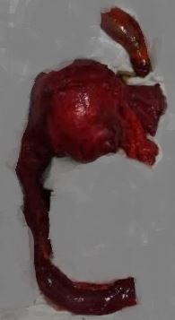
Radical excision of choledochal cyst
Contributed by Sunil Munakomi, MD
Details

Editor:

Savio John

Updated:

8/29/2022 7:13:07 PM

References


[1]

ALONSO-LEJ F,REVER WB Jr,PESSAGNO DJ, Congenital choledochal cyst, with a report of 2, and an analysis of 94, cases. International abstracts of surgery. 1959 Jan;     [PubMed PMID: 13625059]

Level 3 (low-level) evidence

[2]

Todani T,Watanabe Y,Narusue M,Tabuchi K,Okajima K, Congenital bile duct cysts: Classification, operative procedures, and review of thirty-seven cases including cancer arising from choledochal cyst. American journal of surgery. 1977 Aug;     [PubMed PMID: 889044]

Level 3 (low-level) evidence

[3]

Bhavsar MS,Vora HB,Giriyappa VH, Choledochal cysts : a review of literature. Saudi journal of gastroenterology : official journal of the Saudi Gastroenterology Association. 2012 Jul-Aug;     [PubMed PMID: 22824764]


[4]

Babbitt DP, [Congenital choledochal cysts: new etiological concept based on anomalous relationships of the common bile duct and pancreatic bulb]. Annales de radiologie. 1969;     [PubMed PMID: 5401505]


[5]

Sugiyama M,Haradome H,Takahara T,Izumisato Y,Abe N,Masaki T,Mori T,Hachiya J,Atomi Y, Biliopancreatic reflux via anomalous pancreaticobiliary junction. Surgery. 2004 Apr;     [PubMed PMID: 15041974]


[6]

Okada A,Hasegawa T,Oguchi Y,Nakamura T, Recent advances in pathophysiology and surgical treatment of congenital dilatation of the bile duct. Journal of hepato-biliary-pancreatic surgery. 2002;     [PubMed PMID: 12353145]

Level 3 (low-level) evidence

[7]

Tajiri T,Tate G,Inagaki T,Kunimura T,Inoue K,Mitsuya T,Yoshiba M,Morohoshi T, Mucinous cystadenoma of the pancreas 17 years after excision of gallbladder because of a choledochal cyst. Journal of gastroenterology. 2004;     [PubMed PMID: 15069627]


[8]

Imazu M,Iwai N,Tokiwa K,Shimotake T,Kimura O,Ono S, Factors of biliary carcinogenesis in choledochal cysts. European journal of pediatric surgery : official journal of Austrian Association of Pediatric Surgery ... [et al] = Zeitschrift fur Kinderchirurgie. 2001 Feb;     [PubMed PMID: 11370978]


[9]

Levy AD,Rohrmann CA Jr,Murakata LA,Lonergan GJ, Caroli's disease: radiologic spectrum with pathologic correlation. AJR. American journal of roentgenology. 2002 Oct;     [PubMed PMID: 12239064]


[10]

Kusunoki M,Yamamura T,Takahashi T,Kantoh M,Ishikawa Y,Utsunomiya J, Choledochal cyst. Its possible autonomic involvement in the bile duct. Archives of surgery (Chicago, Ill. : 1960). 1987 Sep;     [PubMed PMID: 3619630]


[11]

Kagiyama S,Okazaki K,Yamamoto Y,Yamamoto Y, Anatomic variants of choledochocele and manometric measurements of pressure in the cele and the orifice zone. The American journal of gastroenterology. 1987 Jul;     [PubMed PMID: 3605025]


[12]

O'Neill JA Jr, Choledochal cyst. Current problems in surgery. 1992 Jun;     [PubMed PMID: 1582241]


[13]

Moslim MA,Takahashi H,Seifarth FG,Walsh RM,Morris-Stiff G, Choledochal Cyst Disease in a Western Center: A 30-Year Experience. Journal of gastrointestinal surgery : official journal of the Society for Surgery of the Alimentary Tract. 2016 Aug;     [PubMed PMID: 27260526]


[14]

Liu CL,Fan ST,Lo CM,Lam CM,Poon RT,Wong J, Choledochal cysts in adults. Archives of surgery (Chicago, Ill. : 1960). 2002 Apr;     [PubMed PMID: 11926955]


[15]

Singham J,Yoshida EM,Scudamore CH, Choledochal cysts: part 1 of 3: classification and pathogenesis. Canadian journal of surgery. Journal canadien de chirurgie. 2009 Oct;     [PubMed PMID: 19865581]


[16]

Todani T,Watanabe Y,Toki A,Morotomi Y, Classification of congenital biliary cystic disease: special reference to type Ic and IVA cysts with primary ductal stricture. Journal of hepato-biliary-pancreatic surgery. 2003;     [PubMed PMID: 14598133]


[17]

Shah OJ,Shera AH,Zargar SA,Shah P,Robbani I,Dhar S,Khan AB, Choledochal cysts in children and adults with contrasting profiles: 11-year experience at a tertiary care center in Kashmir. World journal of surgery. 2009 Nov;     [PubMed PMID: 19701664]


[18]

Khandelwal C,Anand U,Kumar B,Priyadarshi RN, Diagnosis and management of choledochal cysts. The Indian journal of surgery. 2012 Feb;     [PubMed PMID: 23372304]


[19]

Masetti R,Antinori A,Coppola R,Coco C,Mattana C,Crucitti A,La Greca A,Fadda G,Magistrelli P,Picciocchi A, Choledochocele: changing trends in diagnosis and management. Surgery today. 1996;     [PubMed PMID: 8727951]


[20]

Nicholl M,Pitt HA,Wolf P,Cooney J,Kalayoglu M,Shilyansky J,Rikkers LF, Choledochal cysts in western adults: complexities compared to children. Journal of gastrointestinal surgery : official journal of the Society for Surgery of the Alimentary Tract. 2004 Mar-Apr;     [PubMed PMID: 15019916]


[21]

Lipsett PA,Pitt HA,Colombani PM,Boitnott JK,Cameron JL, Choledochal cyst disease. A changing pattern of presentation. Annals of surgery. 1994 Nov;     [PubMed PMID: 7979612]


[22]

Chen CP,Cheng SJ,Chang TY,Yeh LF,Lin YH,Wang W, Prenatal diagnosis of choledochal cyst using ultrasound and magnetic resonance imaging. Ultrasound in obstetrics     [PubMed PMID: 14971007]


[23]

Dewbury KC,Aluwihare AP,Birch SJ,Freeman NV, Prenatal ultrasound demonstration of a choledochal cyst. The British journal of radiology. 1980 Sep;     [PubMed PMID: 7437717]


[24]

De Angelis P,Foschia F,Romeo E,Caldaro T,Rea F,di Abriola GF,Caccamo R,Santi MR,Torroni F,Monti L,Dall'Oglio L, Role of endoscopic retrograde cholangiopancreatography in diagnosis and management of congenital choledochal cysts: 28 pediatric cases. Journal of pediatric surgery. 2012 May;     [PubMed PMID: 22595566]

Level 3 (low-level) evidence

[25]

Park DH,Kim MH,Lee SK,Lee SS,Choi JS,Lee YS,Seo DW,Won HJ,Kim MY, Can MRCP replace the diagnostic role of ERCP for patients with choledochal cysts? Gastrointestinal endoscopy. 2005 Sep;     [PubMed PMID: 16111952]


[26]

Chavhan GB,Babyn PS,Manson D,Vidarsson L, Pediatric MR cholangiopancreatography: principles, technique, and clinical applications. Radiographics : a review publication of the Radiological Society of North America, Inc. 2008 Nov-Dec;     [PubMed PMID: 19001651]


[27]

Matsufuji H,Araki Y,Nakamura A,Ohigashi S,Watanabe F, Dynamic study of pancreaticobiliary reflux using secretin-stimulated magnetic resonance cholangiopancreatography in patients with choledochal cysts. Journal of pediatric surgery. 2006 Oct;     [PubMed PMID: 17011263]


[28]

Matos C,Nicaise N,Devière J,Cassart M,Metens T,Struyven J,Cremer M, Choledochal cysts: comparison of findings at MR cholangiopancreatography and endoscopic retrograde cholangiopancreatography in eight patients. Radiology. 1998 Nov;     [PubMed PMID: 9807571]


[29]

Akkiz H,Colakoğlu SO,Ergün Y,Demiryürek H,Akinoğlu A,Tuncer I,Ozgür G,Hafta A, Endoscopic retrograde cholangiopancreatography in the diagnosis and management of choledochal cysts. HPB surgery : a world journal of hepatic, pancreatic and biliary surgery. 1997;     [PubMed PMID: 9184874]


[30]

Diao M,Li L,Cheng W, Timing of surgery for prenatally diagnosed asymptomatic choledochal cysts: a prospective randomized study. Journal of pediatric surgery. 2012 Mar;     [PubMed PMID: 22424346]

Level 1 (high-level) evidence

[31]

Shimotakahara A,Yamataka A,Yanai T,Kobayashi H,Okazaki T,Lane GJ,Miyano T, Roux-en-Y hepaticojejunostomy or hepaticoduodenostomy for biliary reconstruction during the surgical treatment of choledochal cyst: which is better? Pediatric surgery international. 2005 Jan;     [PubMed PMID: 15372285]


[32]

Narayanan SK,Chen Y,Narasimhan KL,Cohen RC, Hepaticoduodenostomy versus hepaticojejunostomy after resection of choledochal cyst: a systematic review and meta-analysis. Journal of pediatric surgery. 2013 Nov;     [PubMed PMID: 24210209]

Level 1 (high-level) evidence

[33]

Atkinson HD,Fischer CP,de Jong CH,Madhavan KK,Parks RW,Garden OJ, Choledochal cysts in adults and their complications. HPB : the official journal of the International Hepato Pancreato Biliary Association. 2003;     [PubMed PMID: 18332966]


[34]

Rozel C,Garel L,Rypens F,Viremouneix L,Lapierre C,Décarie JC,Dubois J, Imaging of biliary disorders in children. Pediatric radiology. 2011 Feb;     [PubMed PMID: 20865413]


[35]

Mabrut JY,Kianmanesh R,Nuzzo G,Castaing D,Boudjema K,Létoublon C,Adham M,Ducerf C,Pruvot FR,Meurisse N,Cherqui D,Azoulay D,Capussotti L,Lerut J,Reding R,Mentha G,Roux A,Gigot JF, Surgical management of congenital intrahepatic bile duct dilatation, Caroli's disease and syndrome: long-term results of the French Association of Surgery Multicenter Study. Annals of surgery. 2013 Nov;     [PubMed PMID: 24121258]

Level 2 (mid-level) evidence

[36]

Ten Hove A,de Meijer VE,Hulscher JBF,de Kleine RHJ, Meta-analysis of risk of developing malignancy in congenital choledochal malformation. The British journal of surgery. 2018 Apr;     [PubMed PMID: 29480528]

Level 1 (high-level) evidence

[37]

Stringer MD,Dhawan A,Davenport M,Mieli-Vergani G,Mowat AP,Howard ER, Choledochal cysts: lessons from a 20 year experience. Archives of disease in childhood. 1995 Dec;     [PubMed PMID: 8546511]


[38]

Lee SE,Jang JY,Lee YJ,Choi DW,Lee WJ,Cho BH,Kim SW, Choledochal cyst and associated malignant tumors in adults: a multicenter survey in South Korea. Archives of surgery (Chicago, Ill. : 1960). 2011 Oct;     [PubMed PMID: 22006877]

Level 3 (low-level) evidence

[39]

Wiseman K,Buczkowski AK,Chung SW,Francoeur J,Schaeffer D,Scudamore CH, Epidemiology, presentation, diagnosis, and outcomes of choledochal cysts in adults in an urban environment. American journal of surgery. 2005 May;     [PubMed PMID: 15862490]


[40]

Yazumi S,Takahashi R,Tojo M,Watanabe N,Imamura M,Chiba T, Intraductal US aids detection of carcinoma in situ in a patient with a choledochal cyst. Gastrointestinal endoscopy. 2001 Feb;     [PubMed PMID: 11174304]


[41]

Shimotake T,Aoi S,Tomiyama H,Iwai N, DPC-4 (Smad-4) and K-ras gene mutations in biliary tract epithelium in children with anomalous pancreaticobiliary ductal union. Journal of pediatric surgery. 2003 May;     [PubMed PMID: 12720172]


[42]

Cha SW,Park MS,Kim KW,Byun JH,Yu JS,Kim MJ,Kim KW, Choledochal cyst and anomalous pancreaticobiliary ductal union in adults: radiological spectrum and complications. Journal of computer assisted tomography. 2008 Jan-Feb;     [PubMed PMID: 18303283]


[43]

Zhan JH,Hu XL,Dai CJ,Niu J,Gu JQ, Expressions of p53 and inducible nitric oxide synthase in congenital choledochal cysts. Hepatobiliary     [PubMed PMID: 14969853]


[44]

Lipsett PA,Pitt HA, Surgical treatment of choledochal cysts. Journal of hepato-biliary-pancreatic surgery. 2003;     [PubMed PMID: 14598135]


[45]

Todani T,Watanabe Y,Toki A,Urushihara N,Sato Y, Reoperation for congenital choledochal cyst. Annals of surgery. 1988 Feb;     [PubMed PMID: 3341813]


[46]

Todani T,Watanabe Y,Urushihara N,Noda T,Morotomi Y, Biliary complications after excisional procedure for choledochal cyst. Journal of pediatric surgery. 1995 Mar;     [PubMed PMID: 7760246]


[47]

Diao M,Li L,Cheng W, Role of laparoscopy in treatment of choledochal cysts in children. Pediatric surgery international. 2013 Apr;     [PubMed PMID: 23371300]


[48]

Chijiiwa K,Koga A, Surgical management and long-term follow-up of patients with choledochal cysts. American journal of surgery. 1993 Feb;     [PubMed PMID: 8427404]


[49]

Chijiiwa K,Tanaka M, Late complications after excisional operation in patients with choledochal cyst. Journal of the American College of Surgeons. 1994 Aug;     [PubMed PMID: 8044381]