Heart Failure and Ejection Fraction

Earn CME/CE in your profession:


Continuing Education Activity

Heart failure (HF) is a common and potentially fatal disease if left untreated. This article aims to describe the different types of heart failure. It also describes the stages, types of HF, and how it is diagnosed and treated. It also highlights the role of interprofessional approach involving patients, physicians, nurses, families, and caretakers.

Objectives:

  • Describe the different types and stages of heart failure.
  • Identify the etiologies and epidemiology of heart failure.
  • Explain the clinical signs and symptoms of heart failure.
  • Outline the importance of the interprofessional team to improve outcomes and enhance the delivery of care in patients affected by heart failure.

Introduction

Heart failure (HF) is a complex clinical syndrome that results from either functional or structural impairment of ventricles resulting in symptomatic left ventricle (LV) dysfunction. The symptoms come from an inadequate cardiac output, failing to keep up with the metabolic demands of the body. It is a leading cause of cardiovascular morbidity and mortality worldwide despite the advances in therapies and prevention. It can result from disorders of the pericardium, myocardium, endocardium, heart valves, great vessels, or some metabolic abnormalities.

Definition

Three main phenotypes describe HF according to the measurement of the left ventricle ejection fraction (EF), and the differentiation between these types is important due to different demographics, comorbidities, and responses to therapies:

  • Heart failure with reduced ejection fraction (HFrEF): EF less than or equal to 40%
  • Heart failure with preserved EF (HFpEF): EF is greater than or equal to 50%
  • Heart failure with mid-range EF (HFmrEF) (other names are: HFpEF-borderline and HFpEF-improved when EF in HFrEF improves to greater than 40%): EF is 41% to 49% per European guidelines and 40 to 49% per the US guidelines.[1][2] A new class of HF that introduced by the 2016 European Society of Cardiology (ESC) guidelines for the diagnosis and management of HF. This class was known as the grey area between the HFpEF and HFrEF and now has its distinct entity by giving it the name HFmrEF.

All patients with HFrEF have concomitant diastolic dysfunction; in contrast, diastolic dysfunction may occur in the absence of systolic dysfunction.

Etiology

Multiple conditions can cause HF, including systemic diseases, a wide range of cardiac conditions, and some hereditary defects. Etiologies of HF vary between high-income and developing countries, and patients may have mixed etiologies.[3] Ischemic heart disease and chronic obstructive pulmonary disease (COPD) are the most common underlying causes of HF in high-income regions. Conversely, hypertensive heart disease, rheumatic heart disease, cardiomyopathy, and myocarditis are the primary conditions for HF in low-income regions, according to a systemic analysis for the Global Burden of Disease Study.[4] More than two-thirds of all cases of HF are attributable to ischemic heart disease, COPD, hypertensive heart disease, and rheumatic heart disease. 

  • Coronary artery disease (CAD): chronic and acute ischemia causes direct damage to the myocardium and leads to remodeling and scar formation, resulting in inadequate relaxation in diastole and impaired contraction in systole, which decreases contractility and cardiac output (CO). This scar formation may also correlate with aneurysm formation, and that further impairs contractile performance and relaxation. Myocardial infarction (MI) also causes dyssynchronous contraction of the infarcted segment, subsequent remodeling of the ventricle, ventricular dilatation with annular dilation, and mitral regurgitation that predispose to HF and decrease the CO. Several tachyarrhythmias such as atrial fibrillation/atrial flutter or non sustained ventricular tachycardia is common in patients with CAD and can deteriorate the cardiac function more. More than 70% of cases with HF have CAD.[5] CAD is a strong predictor of mortality in patients with acute HF. However, the role of coronary revascularization in reducing HF-related morbidity and mortality is still controversial, and viability testing may be useful to select the population that may benefit from revascularization.[6]
  • High blood pressure (HBP): HBP is an independent risk factor for CAD. The high prevalence of HBP makes it a possible cause of HF in around one-fourth to one-third of cases. The heart must pump up blood against a higher afterload caused by HBP, which increases the myocardial mass as a compensatory mechanism to maintain a normal CO and that causes left ventricular hypertrophy (LVH). If blood pressure (BP) remains uncontrolled, apoptosis and fibrosis may result. LVH increases myocardial stiffness and can cause ischemia, which leads to HFpEF or HFrEF. Control of BP is of paramount importance in improving the prognosis of HF. The Systolic Blood Pressure Intervention Trial (SPRINT) has shown that lowering the systolic BP to a target goal of less than 120 mmHg in HBP patients without diabetes had lower rates of HF with 38% lower relative risk compared to systolic BP goal of less than140 mmHg.[7]
  • Chronic obstructive pulmonary disease (COPD): COPD increases the risk of CAD and other smoking-related illnesses, and cardiac dysrhythmias and can cause pulmonary hypertension and right heart failure. 
  • Valvular heart disease: degenerate valve disease in developed countries and rheumatic valve disease in low-income countries can cause HF. Aortic and pulmonary stenosis increases the ventricular afterload and may cause HF. In valve regurgitation, a persistent volume overload can cause ventricular enlargement and functional impairment that may lead to HF. 
  • Cardiomyopathies (CMP): CMP is a disease in which there are functional and structural heart muscle abnormalities in the absence of CAD, HBP, valvular, or congenital heart disease. CMP categorizes into five types, which can be genetic or acquired: dilated cardiomyopathy (DCM), hypertrophic cardiomyopathy (HCM), restrictive cardiomyopathy (RCM), arrhythmogenic right ventricular cardiomyopathy (ARVC), and other unclassified cardiomyopathies (isolated noncompaction of the left ventricle [INLV] and Takotsubo syndrome are also in this category). CMP can cause HFrEF, HFpEF, or HFmrEF.

Other possible causes of HF include congenital heart disease, myocarditis, infiltrative disease, peripartum cardiomyopathy, human immunodeficiency virus (HIV), connective tissue disease, amyloidosis, substance abuse, long-standing alcohol use, obesity, diabetes mellitus (DM), hyperthyroidism (can cause high-output HF), pulmonary hypertension (can cause right HF), constrictive pericarditis (can cause HFpEF), pulmonary embolism (can cause right HF), and chemotherapies (like doxorubicin). 

Epidemiology

HF is a significant public health problem with a prevalence of over 5.8 to 6.5 million in the U.S.[8][9] and around 26 million worldwide.[10] The expectation is that 8 million people in the United States will have this condition by 2030, accounting for a 46% increase in prevalence.[11] The prevalence of HF increases with age, as per data from the Framingham Heart Study that estimated the prevalence of HF to be 8 per 1000 in men at age 50 to 59 years and goes up to 66 per 1000 in men at ages 80 to 89 years, similar values in women (8 and 79 per 1000).[12] At age 45 years, the lifetime risks for HF through age 75 or 95 years were 30% to 42% in white men, 20% to 29% in black men, 32% to 39% in white women, and 24% to 46% in black and higher BP and BMI at all ages led to higher lifetime risks.[13] The increase in HF prevalence does not necessarily have links with an increase in HF incidence. The aging of the population and modern therapies for cardiac patients that led to increased survival could explain the increase in prevalence even with a reduction in the incidence (due to prevention programs and better treatment of acute coronary syndromes).[14] 

Between 13% and 24% of patients with HF have HFmrEF.[15] Between 40% and 60% of patients with HF have diastolic dysfunction, and more than 50% of HF are HFpEF.[16][17] The prevalence of HFpEF is increasing, and researchers expect that by 2020, 65% of patients hospitalized for HF will have HFpEF.[18] Patients with HFpEF are older, more frequently have hypertension, are overweight, and are more commonly women compared to HFrEF.[19] Risk factors for HFpEF are multifactorial and complex, and there is no known prevention other than the treatment of the risk factors, such as hypertension, diabetes, and obesity; whereas prevention and early treatment strategies (i.e., early revascularization) appear to be effective in reducing the risk and severity of acute myocardial infarction. These observations may explain a reduction in the incidence of HFrEF but an increasing incidence of HFpEF and HFmrEF.

Pathophysiology

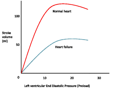
The pathophysiology of HF is complex and includes structural, neurohumoral, cellular, and molecular mechanisms activation to maintain physiologic functioning (maladaptation, myocyte hypertrophy, myocyte death/apoptosis/regeneration, and remodeling).[20] The performance of LV function and stroke volume is under the control of preload (venous return and ventricular end-diastolic volume), myocardial contractility, and afterload (the impedance during ejection from the aorta and wall stress). The Frank-Starling curve explains the relationship between stroke volume/cardiac output and left ventricle end-diastolic pressure (LVEDP) or pulmonary capillary wedge pressure (PCWP) in which there is a steep and positive relationship between increased cardiac filling pressures and increased stroke volume/cardiac output. This relationship is right-shifted, representing the decreased contractility, and higher pressure is required to achieve the same cardiac output and flattened in advanced disease, which means that augmentation in venous return and LVEDP fails to increase the stroke volume. (Figure 1) 

HFpEF has the same pathophysiologic processes as HFrEF but in response to increased ventricular stiffness and altered relaxation than CO in HFrEF. This stiffness and altered relaxation cause concentric LVH (instead of eccentric LVH as in HFrEF) and shift the pressure-volume curve to the left.

HF (HFrEF, HFpEF, and HFmrEF) causes activation of neurohumoral systems to maintain perfusion of vital organs: sympathetic nervous systems (SNS), renin-angiotensin-aldosterone system (RAAS), antidiuretic hormone, and other vasoactive substances (brain natriuretic peptide (BNP), nitric oxide, and endothelin). HF causes decreased carotid baroreceptor response, which in turn increases sympathetic nervous activity (SNS) and leads to increased cardiac contractility and heart rate, vasoconstriction, and increased afterload. Activation of RAAS in response to low renal perfusion from HF causes salt/water retention and increases preload. RASS activation increases angiotensin II, which leads to vasoconstriction and more salt and water retention, which further stress the ventricular wall and cause dilatation (remodeling) and worsening ventricular function, and further HF. Those compensatory mechanisms cause negative remodeling of the heart (inflammation, apoptosis, hypertrophy, and fibrosis) and worsening left ventricular function.

History and Physical

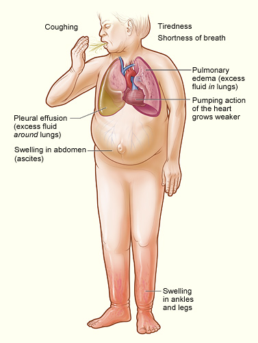
A thorough history and physical exam should be obtained and performed in all patients with suspected HF, as the entire basis of the diagnosis is on clinical symptoms and signs. It also should include assessment of risk factors and possible etiologies of the HF. Symptoms of HF are similar regardless of the EF. Symptoms are more severe with exertion and either secondary to fluid accumulation (dyspnea, orthopnea, edema, and abdominal discomfort from hepatic congestion and ascites in the setting of right heart failure) or due to decreased cardiac output (fatigue, anorexia, and weakness). Other less typical symptoms include nocturnal cough, loss of appetite, wheezing, palpitations, depression, syncope, bendopnea (shortness of breath while bending forward), and dizziness. In advanced HF, patients may have resting sinus tachycardia, diaphoresis, narrow pulse pressure (less than 25 mmHg due to decreased cardiac output), and peripheral vasoconstriction (cool and pale extremities due to decreased perfusion). Volume overload manifests as peripheral edema (extremities edema, ascites, scrotal edema, and hepatosplenomegaly), elevated jugular venous pressure (JVP), and pulmonary congestion (rales on the exam and pleural effusions). Displaced apical impulse (laterally past the midclavicular line, which is a sign of LV enlargement), parasternal lift (right ventricular enlargement), and an S3 gallop. At each clinic visit, symptoms and signs of HF require assessment to monitor response to therapy and stability over time. It is also important to check vital signs and assess volume status during each clinic visit. 

Evaluation

  • Complete blood count to rule out anemia as a possible cause of patients' symptoms. Besides, anemia is associated with higher HF severity, and intravenous iron replacement is important to improve functional status and quality of life if ferritin is under 100 ng/ml or 100 to 300 ng/ml if transferrin saturation is below 20%.[21]
  • Serum electrolytes (including calcium and magnesium levels) and kidney functions (blood urea nitrogen and serum creatinine): serum creatinine and blood urea are prognostic factors in hospitalized patients with HF, and hyponatremia plays a prognostic factor role in chronic patients with HF. Besides, some HF medications can cause electrolyte abnormalities and kidney dysfunction (like spironolactone, angiotensin-converting enzyme inhibitor (ACEi), and furosemide). 
  • Measurement of B-type Natriuretic peptide (BNP) or N-terminal pro-B-type natriuretic peptide (NT-proBNP) is helpful to support the clinical diagnosis of HF in the ambulatory setting and to support the diagnosis of acutely decompensated HF in hospitalized/ER patients. 
  • Other laboratory workups include glucose, fasting lipid profile, liver function tests, and thyroid-stimulating hormone. 
  • EKG is necessary for all patients with suspected HF, and it helps rule out HF if completely normal, with a sensitivity of 89% but low specificity.[22] Abnormal EKG increases the likelihood of HF diagnosis.[23] It also can provide information on etiology (e.g., history of previous myocardial infarction makes CAD a possible cause of HF, arrhythmia as a potential cause of tachycardia-mediated cardiomyopathy HF, LV hypertrophy indicates hypertension-induced HF, widened QRS complex/left bundle branch block may suggest idiopathic dilated cardiomyopathy, and heart blocks as seen in patients with cardiac sarcoidosis) and provide indications for therapy (anticoagulation if atrial fibrillation, pacemaker in some bradycardia, and cardiac resynchronization therapy (CRT) if broadened QRS). 
  • Chest X-ray is also useful and may show pleural effusions secondary to volume overload, cardiomegaly, and Kerley B-lines (interstitial edema).
  • Transthoracic echocardiogram (TTE) is the most useful test that helps establish the diagnosis of HF and classify it as HFrEF, HFmrEF, or HFpEF. Abnormal parameters in HFrEF that can be measured by echocardiogram include increased end-diastolic diameter and volume (LV diameter over 60 mm or 32 mm/m with LV volume exceeding 97 mL/m) and end-systolic diameter and volume (LV diameter greater than 45 mm or 25 mm/m with LV volume over 43 mL/m).[24] Echocardiogram also helps to assess LVEF in HFrEF to guide evidence-based medical and device therapies (implantable cardioverter-defibrillator (ICD) and CRT), evaluate the valves, provide information on ventricular wall thickness, and it is vital in the risk stratification of patients with HF.[25]
  • Genetic testing: some CMP can be genetic, especially DCM, HCM, and autosomal dominant ARVD/C, and screening the family members may be important in family-based management. 
  • Computed tomography (CT) scanning or cardiac magnetic resonance imaging (cMR): neither are routinely indicated in the diagnosis and management of HF, and the role of imaging modalities other than TTE was restricted in the 2106 ESC guidelines. Cardiac CT can help to evaluate the coronary arteries in patients with HF with low to intermediate pretest probability of CAD. They provide information about cardiac function and have a high anatomical resolution of all aspects of the heart and surrounding structures (ventricular mass, chamber size, heart valves, and pericardium and wall motion).[26][27] Cardiac MRI assesses LV volume, LVEF, myocardial perfusion, viability, and fibrosis and helps identify HF etiology (ischemic versus non-ischemic disease, infiltrative disease, and hypertrophic disease) and assess prognosis. CMR can determine viability, help the success of revascularization in patients with low EF, and is the gold standard for evaluating RV function. However, cMR is costly and cannot be performed with implantable defibrillators all the time (some of the defibrillators as MRI-compatible these days). Also, both cardiac CT and cMR have low accuracy in patients with high heart rates. 
  • Left heart catheterization or coronary angiography is indicated in patients with HF and angina symptoms or ischemic changes by ECG or noninvasive testing. Indications also include worsening HF symptoms without a clear cause, when the pretest probability of underlying ICMP is high, before cardiac transplantation or LVAD, and in post-infarction mechanical complications like a ventricular aneurysm. It may be useful in patients with HF without angina but with LV dysfunction. In patients without CAD, CAD should be considered as a potential etiology of HF and impaired LV function and should be excluded whenever possible. It is only necessary if patients are potentially eligible for revascularization.[2]
  • Stress nuclear imaging or echocardiography may be an acceptable option for assessing ischemia in patients presenting with HF who have known CAD and no angina unless they are ineligible for revascularization. 

Diagnosis

The diagnosis of HF is based on clinical signs and symptoms, laboratory workup, and echocardiogram. The Framingham clinical criteria help to diagnose (with low specificity) HF.[28] It includes major and minor criteria, and HF diagnosis requires two major or one major and two minor criteria. Major criteria include orthopnea, pulmonary rales, S3, cardiomegaly on chest X-ray, pulmonary edema on chest X-ray, elevated jugular venous pressure, paroxysmal nocturnal dyspnea, and weight loss over 4.5 kg in five days in response to treatment of presumed HF. Minor criteria include nocturnal cough, dyspnea on ordinary exertion, pleural effusion, hepatomegaly, tachycardia with a heart rate over 120 beats/min, bilateral leg edema, and weight loss under 4.5 kg in five days. However, Framingham's clinical criteria have excellent sensitivity to exclude the diagnosis of HF in the absence of these symptoms/signs, but it has poor specificity to confirm the diagnosis.[28] On the other hand, some patients with HF may not have symptoms. Only 50% of patients with LV dysfunction on an echocardiogram are symptomatic. Symptoms and signs of HF are either too nonspecific or too infrequent, which makes the clinical diagnosis of HF challenging with <25% of accuracy for most of the clinical features.

Measurement of natriuretic peptides (NPs) like BNP/NT-proBNP is also helpful in supporting the diagnosis in ambulatory and inpatient settings. BNP has 70% sensitivity and 99% specificity for HF diagnosis, and NT-proBNP has 99% sensitivity and 85% specificity.[29] NPs have a very high negative predictive value of 0.94 to 0.98 and can exclude the diagnosis of HF (both HFpEF and HFrEF) if the values are below the cut-off. On the other hand, the positive predictive value of NPs is low (0.66 to 0.67) and cannot confirm the diagnosis.[30]

Screening

The cost-effectiveness of routine periodic population screening for asymptomatic reduced LVEF is not a current recommendation. Screening for hemochromatosis or HIV is reasonable in selected patients with HF (Class IIa, LOE: C).[2] Some genetic screening tests are also recommendations for HCM, idiopathic DCM, ARVC, isolated non-compaction CMP, and restrictive CMP.  

Treatment / Management

Treatment of HF starts with the prevention of overt HF by controlling the risk factors. Hypertension is a significant risk factor, and the cause of HF and intensive blood pressure control (systolic BP under 120 mmHg) was more beneficial for the prevention of cardiovascular diseases than standard treatment (systolic BP goal of less than 140 mmHg) in the SPRINT trial.[31] Recent studies have shown that sodium-glucose cotransporter-2 (SGLT-2) inhibitors reduced mortality and HF hospitalization in patients with type 2 diabetes.[32] In patients with STEMI, rapid primary percutaneous coronary intervention (PCI) and administration of ACEI, beta-blocker, mineralocorticoid receptor antagonists (MRAs), and statin can reduce HF hospitalization and mortality.

Treatment of HF is indicated in all patients with LV dysfunction (diastolic or systolic) regardless of symptoms. The goal of treatment is to improve survival and symptoms, reduce the length of stay and HF readmission, decrease morbidity, prevent organ damage from HF, and prevent symptoms in asymptomatic LV dysfunction. Treatment of HF can fall into non-pharmacological therapy, pharmacological therapy, devices and non-surgical interventions, and invasive strategies. 

Nonpharmacological therapy

Non-pharmacological therapy is indicated for all types of HF regardless of EF. It is consistent with behavioral and lifestyle modifications, including dietary and nutritional consultation (sodium restriction to 2 to 3 g/day, fluid restriction to 2 L/day if there is hyponatremia, and caloric supplementations if cardiac cachexia), strict adherence to therapy and diet, daily weight and diuretic dosing adjustment for sudden weight changes, patient education to facilitate self-care/close observation/follow up, aerobic exercise training (can reverse LV remodeling in stable patients), controlling of HBP, DM and lipid disorders, and smoking/alcohol/illicit drug use cessation.

Pharmacologic therapy for HFrEF

Initial pharmacologic therapy includes a combination of diuretics (as needed for volume overload to improve symptoms, but they do not improve survival and routine use of loop diuretics without volume overload signs or symptoms may even increase mortality [33]), an angiotensin system blocker (ACEi, ARB, or ARNI) or hydralazine plus nitrate as an alternative if no tolerance to angiotensin system blockers, and a beta-blocker. Other medications that are options for selected patients are MRA, ivabradine, and digoxin. 

1- ACEi or ARBs (class IA) are the first-line therapies, and the dose should titrate up to the maximum tolerated evidence-based doses. ACEis inhibit the activity of angiotensin-converting enzyme (ACE) and therefore prevent the formation of angiotensin II from angiotensin I, and that causes natriuresis, diuresis, and a reduction in arterial blood pressure and, thereby, afterload. ACEIs have been in use for many years, and multiple clinical trials have shown survival benefits and decreased hospitalization in chronic symptomatic HFrEF, as in the Cooperative North Scandinavian Enalapril Survival Study (CONSENSUS) and the Studies of Left Ventricular Dysfunction (SOLVD).[34][35] ARBs have not consistently been proven to reduce mortality in patients with HFrEF, and their use should be restricted to patients intolerant of an ACEI. According to the current ESC and AHA/ACCF guidelines, every patient with HFrEF should receive an ACEI independent of symptoms and if no contraindications. This group includes captopril, lisinopril, ramipril, enalapril, quinapril, losartan, valsartan, irbesartan, and candesartan. The routine combination of ACEi, ARB, and MRA is not recommended and may cause more symptomatic hypotension and worsening renal function (class III C). 

Angiotensin receptor-neprilysin inhibitor (ARNI) sacubitril/valsartan was approved by FAD in 2015 for HFrEF treatment after the Prospective Comparison of ARNI with ACE-I to Determine Impact on Global Mortality and Morbidity in Heart Failure (PARADIGM-HF) trial in 2014 that found a markedly decreased cardiovascular and all-cause mortality in patients with HFrEF in the sacubitril/valsartan group compared to enalapril.[36] It is indicated in patients with symptomatic HFrEF with EF ≤40%, elevated plasma NP levels (BNP greater than equal to 150 pg/mL or NT-proBNP ≥600 pg/mL or, if they had been hospitalized for HF within the previous 12 months, BNP ≥100 pg/mL or NT-proBNP greater than equal to 400 pg/mL), and an estimated GFR (eGFR) ≥30 mL/min/1.73 m of body surface area as per the ESC 2016 guidelines.[37] Sacubitril/valsartan has some safety issues, and long-term safety is a concern. It can cause more symptomatic hypotension, especially in the elderly, and had more angioedema cases than enalapril in the PARADIGM-HF trial. To minimize the angioedema risk caused by overlapping ACE and neprilysin inhibition, the clinicians should withhold the ACEi for at least 36 hours before initiating the sacubitril/valsartan. In a recent meta-analysis, ARNI improved LVH, LV size, and reverse cardiac remodeling compared with ACEi/ARBs in patients with HFrEF and caused marked changes in the left ventricular mass index and left atrial volume in HFpEF.[38][38] 

2- Beta Blockers (Class IB): one of the pathophysiologies of HFrEF is ongoing sympathetic activation that adversely affects the cardiac myocytes and contractility, so blocking this pathway with beta-blockers reverses this mechanism and helps improve HFrEF. Beta-Blockers improve survival, reduce HF hospitalizations, and increase LVEF in chronic HFrEF.[39] Several studies have shown the benefits of beta-blockers on survival, including the Study of the Effects of Nebivolol Intervention on Outcomes and Re-hospitalization in Seniors with Heart Failure (SENIORS) and Carvedilol Prospective Randomized Cumulative Survival (COPERNICUS), among many other studies.[40] Randomized controlled trials support carvedilol, metoprolol succinate, and bisoprolol. According to the current ESC guidelines, ACEIs and beta-blockers should be started immediately after the diagnosis of HFrEF.[41]   

3- Mineralocorticoid Receptor Antagonists (MRAs): the class IA indication for MRAs as per the 2013 ACCF/AHA guidelines is in patients with NYHA class II-IV who have EF of 35% or less, serum creatinine less than 2 .5 mg/dL, or an estimated glomerular filtration rate over 30 mL/min/1.73 m, and stable serum potassium less than 5.0 mEq/L at the initiation of therapy. MRAs also decrease mortality and hospitalization in chronic HFrEF (NYHA class II-IV). However, a meta-analysis of 1575 patients enrolled in 14 studies reported an improvement in LVEF of 3.2% and a significant improvement in NYHA class among subjects treated with aldosterone antagonists (p<0.001) regardless of baseline LVEF or NYHA class.[42] 

4- Nitrates Plus Hydralazine: they both decrease afterload, and nitrates also reduce the preload. According to the 2013 ACCF/AHA guidelines, it is a Class IA indication that decreases mortality and morbidity in HFrEF among African Americans, with NYHA class III-IV HF receiving optimal medical therapy (OMT) with ACEi and beta-blockers. 

5- Digoxin: it can decrease hospitalization of HFrEF (class IIA, level of evidence B), but it does not improve survival. Some evidence suggests that digoxin increases mortality in women but not in men; its use requires caution in women with symptomatic heart failure.[43]  

6- Aspirin and statin for ischemic HF. 

7- Ivabradine was approved by FAD in 2015 to reduce the risk of HF hospitalization. It is indicated in stable, symptomatic patients with chronic HFrEF with EF under 35%, who are in sinus rhythm and demonstrate a resting heart rate exceeding 70 bpm on the maximum tolerated dose of beta-blocker or have contraindications for beta-blockers.[44]

8- Sodium-glucose cotransporter 2 inhibitor: in patients with HFrEF who still have symptoms and elevated BNP levels on optimal medical therapy (OMT) and device therapy, the recommendation is to add dapagliflozin (versus no additional drug therapy) (grade 1 B) even in patients without DM as shown by the DAPA-HF Trial.[45] It is contraindicated in patients with symptomatic hypotension, SBP under 95 mmHg, eGFR below 30 ml per minute per 1.73 m

9- Continuous intravenous inotrope (dobutamine or milrinone) is reasonable as bridge therapy for cardiac transplantation or Ventricular Assist Devices (VADs) in patients with stage D HF refractory to Guideline Directed Medical Therapy (GDMT) (class IIa, LOE:B) or as a palliative care as per the 2013 ACCF/AHA guidelines. 

  • Medications to avoid in HF

According to the ESC guidelines, some treatments can be harmful in patients with HF and should be avoided[37]

1- Non-steroidal anti-inflammatory drugs (NSAIDs), COX-2 inhibitors: can worsen HF, increase hospitalization, worsen kidney functions, and cause more sodium and water retention.[46]

2- Calcium Channel Blockers (CCB), excluding amlodipine and felodipine: [47] they have negative inotropic effects and may worsen HF and increase hospitalizations.

3- Thiazolidinediones (TZDs): can cause fluid extension and exacerbate an existing HF and increase the risk of HF in patients without HF. The benefit/risk profile of TZDsmerit consideration when treating DM in patients with or without prior HF.[48]

4- Adding an ARB to an ACEi and an MRA possibly will worsen kidney function and increase the risk of hyperkalemia.

5- Dronedarone should not be an option in patients with NYHA class III-IV HFrEF as per ANDROMEDA (antiarrhythmic trial with dronedarone in moderate to severe CHF evaluating morbidity decrease study). Dronedarone also demonstrated worse cardiovascular outcomes in patients with HFpEF.[49]

Other medications that should be avoided or at least used with caution in patients with HF are metformin (increase the risk of potentially lethal lactic acidosis), phosphodiesterase inhibitor (PDE-3 inhibitors increased mortality, PDE-5 inhibitors are harmful in patients with HF who have borderline low BP and/or low volume status), antiarrhythmic agents (especially class I and III) due to negative inotrope activity (amiodarone is the preferred drug for the treatment of arrhythmias in patients with HF.  

  • Devices and non-surgical interventions

These include implantable cardioverter-defibrillator (ICD) and cardiac resynchronization therapy (CRT). Before proceeding with device therapy, patients should be treated with ACEi/ARB plus Beta-blockers for at least three months and then reassess the LVEF. If EF remains less than or equal to 35%, the recommendation is a referral for device therapy. 

ICD

ACC/AHA 2013 guidelines [2] recommend ICD for primary prevention of sudden cardiac death for non-ischemic dilated cardiomyopathy (NIDCM) or ischemic CMP (ICM) at least 40 days post-MI on chronic goal-directed medical therapy (GDMT) with either LVEF ≤ 35% and NYHA class II or III symptoms (I-A) or LVEF ≤ 30% and NYHA class I symptom (I-B) 

ESC 2016 guidelines [1] recommend ICD for primary prevention in symptomatic HF (NYHA II-III) with LVEF ≤ 35% despite ≥ three months of GDMT in ICM (IA) or NIDCM (IB)

CRT

ACC/AHA 2013 guidelines recommend CRT for patients with HF with sinus rhythm, LVEF ≤ 35%, left bundle branch block (LBBB) with QRS duration ≥ 150 ms, and NYHA class III (I-A) or ambulatory IV (I-A) or II (I-B) on GDMT. 

ESC 2016 guidelines recommend CRT for symptomatic HF with sinus rhythm and LVEF ≤ 35% despite GDMT with LBBB with QRSd ≥ 150 ms (I-A) or 130 to 149 ms (I-B). 

  • Invasive therapies

Coronary revascularization (Coronary Artery Bypass Grafting or CABG, angioplasty, and Percutaneous Coronary Intervention or PCI) is indicated for patients with HF (HFpEF, HFrEF, or HFmrEF) on GDMT with angina and suitable coronary anatomy (class IC). 

  • Surgical treatment of HF

Despite the advances in the medical management of HF, there are some circumstances in which surgery is the best treatment option. Surgical approaches to HF treatment include heart transplantation and procedures that reshape the heart, repair the heart, or replace all or part of the heart function. The basis for any decision to surgically treat HF depends on functional status, prognosis, and severity of the underlying HF and comorbidities. It should take place in centers with multidisciplinary medical and surgical teams.  

Heart Transplantation

According to Heart Failure Society of America (HFSA) 2010 heart failure guidelines, it is recommended to evaluate patients for heart transplantation in severe HF, debilitating refractory symptoms, ventricular arrhythmia, or congenital heart disease that remains uncontrolled despite drug, device, or alternative surgical therapy (strength of evidence B).[50] Data from the registry of the International Society of Heart and Lung Transplantation indicates a current 1-year survival of 84.5% and 5-year survival of 72.5%; subsequently, survival decreases linearly by approximately 3.4% per year.[51]

Ventricular Assist Devices (VADs)

The blood is removed from the failing ventricle and diverted into a pump that delivers the blood to either the aorta (in case of LV failure and LVAD) or the pulmonary artery (in case of right ventricle failure and RVAD). Devices for this process include a left ventricular assist device (LVAD), right ventricular assist device (RVAD), or biventricular assist devices (BiVAD), which is inserted percutaneously into the LV and draws blood from LV and expels it into the ascending aorta. These VADs are used as a bridge therapy to heart transplantation in refractory stage D heart failure according to the ACCF/AHA, ESC, and HFSA guidelines, or as a destination therapy if not a candidate for heart transplantation as LVADs as destination therapy, in this case, is superior to medical therapy according to the Randomized Evaluation of Mechanical Assistance for the Treatment of Congestive Heart Failure (REMATCH) trial.[2][37][50][51][52]

Surgical Ventricular Restoration (SVR)

One of the mechanisms of HF pathophysiology is ventricular remodeling and the shape change from an elliptical shape to a spherical shape, especially post transmural MI. Correction of this pathologic remodeling by incising and excluding the nonviable myocardium with either patch or primary reconstruction to decrease the ventricular volume may be hypothetically helpful in treating patients with HF. However, the STICH trial, which is the major study for ventricular reconstruction, found that adding ventricular surgical reconstruction to CABG reduced the left ventricular volume, as compared with CABG alone, but this did not correlate with a greater improvement in symptoms or exercise tolerance or with a reduction in the mortality rate or hospitalization for cardiac causes.[53] SVR is not routinely recommended and is not in the ESC, AHA/ACCF, or HFAS guidelines. 

Treatment of HFpEF and HFmrEF

There are no disease-modifying therapies for HFpEF or HFmrEF that improve outcomes compared to HFrEF agents. Efficacious therapies for HFrEF have failed to demonstrate a benefit for HFpEF, and more research is required to evaluate them in HFmrEF.[1] Spironolactone has been shown to reduce HF hospitalization rates in HFpEF, but no treatment has demonstrated improved survival.[54] Treatment focuses on controlling BP using beta-blockers, ACEi or ARBs is reasonable to control BP in patients with HFpEF (class IIA), diuretics to relieve symptoms of volume overload, and to address the risk factors and comorbidities. There are no studies to determine the impact of revascularization on symptoms or outcomes, specifically in patients with HFpEF. It might be reasonable to consider revascularization in patients for whom ischemia appears to contribute to HF symptoms.

Treatment of Right Heart Failure

The most frequent causes of RV failure are LV failure and primary pulmonary diseases, and treating the underlying cause is essential in RV failure treatment. ACEi/ARB and beta-blockers efficacy in isolated RV failure is not known, but they are beneficial if RV failure is secondary to LV failure. If severe unstable RV failure, the use of inotrope may be a consideration. The prognosis in patients with RV failure depends on the etiology. Volume overload, pulmonary stenosis, and Eisenmenger syndrome are associated with a better prognosis. Decreased exercise tolerance predicts poor survival. 

Treatment of Acute Decompensated Heart Failure (ADHF) 

Diagnosis of ADHF is based on symptoms and signs with the help of BNP, NT-proBNP concentration if the clinical diagnosis is uncertain. Hospitalization is necessary if severely decompensated HF (hypotension, worsening renal function, or altered mentation), dyspnea at rest, hemodynamically significant arrhythmia (including new-onset rapid atrial fibrillation), or acute coronary syndromes. Successful inpatient therapy for ADHF involves a comprehensive care plan. Etiology and precipitating factors require identification, chronic oral therapy should be optimized, treatment to relieve symptoms should be applied (loop intravenous diuretics or ultrafiltration if diuretic resistance, and vasodilators to decrease preload and afterload), the patient that may benefit from revascularization or device therapies should be identified, education about dietary sodium restriction/self-assessment of volume status and principal cardiac medications should be provided. The use of non-invasive positive pressure ventilation may be useful in severely dyspneic patients with evidence of pulmonary edema. The cornerstone of ADHF is volume removal. Loop diuretics (furosemide, torsemide, or bumetanide) are the preferred initial diuretics, and if inadequate diuresis, a second diuretic can be added (e.g., a thiazide). Vasodilators (nitroprusside, nesiritide, or nitroglycerin) are also recommended as an adjuvant to diuresis to relieve symptoms but should be avoided if systolic BP is less than 90 mmHg or in cases of significant aortic or mitral stenosis.[1] ACEi/ARBs and Beta-blockers should be continued during the HF exacerbation in HFrEF if no contraindication (in patients with significant worsening renal function, the clinician can temporarily discontinue ACEi/ARBs and/or MRA until renal function improves. Most patients will tolerate continuing the beta-blockers well, but this can be held if marked volume overload or marginal/low CO. Medical treatment should be optimized. If the patient is not on ACEi/ARB/ARNI or beta-blockers, those should be started when feasible while inpatient.

Differential Diagnosis

The diagnosis of HF is mostly clinical, as mentioned above, but many other medical conditions can cause the same signs and symptoms. There is a broad differential diagnosis of HF, which includes but is not limited to bacterial pneumonia, chronic obstructive pulmonary disease (COPD), liver cirrhosis, acute kidney injury, idiopathic pulmonary fibrosis, nephrotic syndrome, pulmonary embolism respiratory failure, primary pulmonary hypertension, anemia, and venous insufficiency.

  • Acute kidney injury
  • Bacterial pneumonia
  • Acute respiratory distress syndrome
  • Cardiogenic pulmonary edema
  • Interstitial (non-idiopathic) pulmonary fibrosis
  • Chronic obstructive pulmonary disease
  • Cirrhosis
  • Goodpasture syndrome
  • Community-acquired pneumonia
  • Idiopathic pulmonary fibrosis
  • Myocardial infarction
  • Pulmonary embolism
  • Nephrotic syndrome
  • Neurogenic pulmonary edema
  • Pneumothorax
  • Respiratory failure
  • Viral pneumonia
  • Venous insufficiency

Staging

Heart failure divides into four stages, according to the 2013 ACCF/AHA Guidelines [55]

Stage A- At high risk for HF but no structural heart disease or symptoms of HF

Stage B- Asymptomatic LV dysfunction: structural heart disease but no symptoms or signs of HF 

Stage C- Overt HF: structural heart disease with symptoms of HF 

Stage D- Refractory HF. 

Symptoms of HF are only in stages C and D.

Classifications

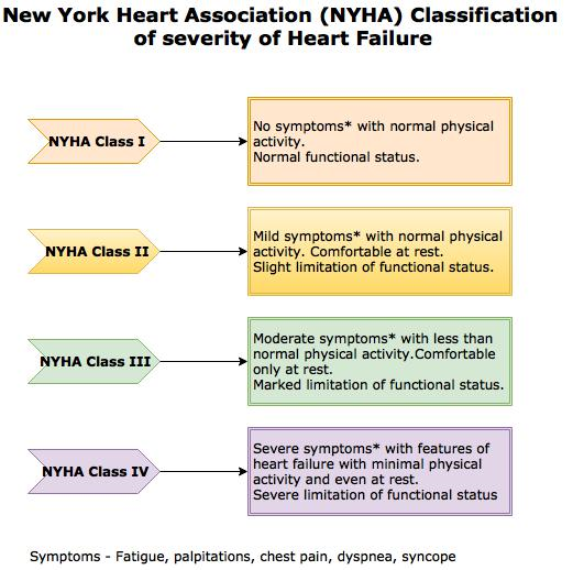
New York Heart Association ( NYHA) classifies HF based on HF symptoms and functional limitations into four classes[56]:

Class I: asymptomatic LV dysfunction with no limitations on physical activity or symptoms. 

Class II: mild symptoms with slight limitation of physical activity. Ordinary activities lead to symptoms.

Class III: moderate symptoms with marked limitation of physical activity. Less than ordinary activities lead to symptoms.

Class IV: severe symptoms at rest.

Prognosis

Changes in EF over time are more prognostic of HF than baseline EF. Patients who progress from HFmrEF to HFrEF have a worse prognosis than those who progress to HFpEF or remain stable in HFmrEF. The mortality rate is higher in HFrEF than HFmrEF and HFpEF, according to the OPTIMIZE-HF trail [57], showed a mortality rate of 3.9% for HFrEF, 3% for HFmrEF, and 2.9% for HFpEF. The mortality rate is also higher in symptomatic patients. There are some predictors of poor prognosis and increased mortality in hospitalized patients, which include systolic blood pressure less than 115 mmHg, serum creatinine greater than 2.7 mg/dL, serum urea over15 mmol/L, NT-pro-BNP exceeding 986 pg/mL, and LVEF under 45%.[58] Poor prognostic factors in chronic heart failure include S3 gallop, DM, hyponatremia, decreased LVEF, high NYHA functional class, reduced cardiac index, and increased pulmonary artery capillary wedge pressure.[59]

Complications

HF may cause multiple complications, including but not limited to:

  • Arrhythmias: Atrial fibrillation (Afib) can be a cause or a consequence of HF and may present in 10% to 50% of chronic HF patients, and those patients with HF and Afib have a poor prognosis. Malignant ventricular arrhythmias (like sustained monomorphic ventricular tachycardia, sustained polymorphic ventricular tachycardia, and torsades de pointes) are common in end-stage heart failure, especially if precipitating or factors are present like electrolyte disturbance, prolonged QT interval, and digoxin toxicity. Bradyarrhythmias may also happen.[60]
  • Thromboembolism: HF is a cause of stroke in 9% of patients.[61] Between 10 and 24% of patients with stroke have HF.[62][63] There is a high relative risk for deep venous thrombosis (DVT) and pulmonary embolism (PE) in patients with HF, especially those who were under 60 years of age.[64]
  • Gastrointestinal: liver shock (ischemic hepatitis), liver cirrhosis, and cardiac cachexia due to decreases intestinal blood flow in patients with HF.[65] 
  • Renal: renal function may get worse in both acute and chronic HF, and that predicts poor prognosis, and even a small transient rise in creatinine will be clinically relevant.[66]
  • Respiratory: pulmonary congestion, respiratory muscle weakness, and rarely pulmonary hypertension 

Deterrence and Patient Education

Emphasizing diet and medical compliance to patients with HF is important as one of the most common causes of HF readmission is the failure to comply either with diet or medications. A single-session intervention could be beneficial as a randomized control trial of 605 patients with HF found that the incidence of all-cause hospitalization or mortality was not significantly reduced in patients receiving multisession self-care training compared to those receiving a single-session intervention.[67]

Enhancing Healthcare Team Outcomes

Heart failure is a leading cause of hospitalization and represents a significant clinical and economic burden. The long-term goal of treatment is to avoid exacerbation of HF and decrease hospital readmission rates. It needs an interprofessional approach involving patients, physicians, nurses, pharmacists, families, and caretakers. Those strategies include early identification of high-risk patients, patient education, improving medication and dietary compliance, assuring close follow-up, introducing end-of-care issues, and tele-home monitoring if available. Primary care and emergency department providers often are the first to make this diagnosis. Referral to cardiologists is often appropriate. Cardiology, medical/surgical, and critical care nurses administer treatment, provide education, monitor patients, and communicate with the rest of the team so that everyone on the healthcare team operates from the same data set. The managing clinician would do well to consult with a board-certified cardiology pharmacist when initiating pharmaceutical care in HF cases. Pharmacists also review medicines, check the dosages, detect drug-drug interactions, and stress to patients and their families the importance of compliance. In end-stage cases, hospice care and hospice nurses can work with the patient and their family to provide comfort care. These interprofessional collaborations will optimize patient outcomes in HF cases. [Level 5]


(Click Image to Enlarge)
The illustration shows the major signs and symptoms of heart failure.
Contributed by National Heart, Lung, and Blood Institute, National Institutes of Health (NIH)

(Click Image to Enlarge)
NYHA Classification - Heart failure
Contributed by the New York Health Association (NYHA)

(Click Image to Enlarge)
Frank-Starling Curve
Contributed by Said Hajouli
Details

Author

Said Hajouli

Editor:

Dipesh Ludhwani

Updated:

12/23/2022 5:42:58 PM

References


[1]

Ponikowski P,Voors AA,Anker SD,Bueno H,Cleland JGF,Coats AJS,Falk V,González-Juanatey JR,Harjola VP,Jankowska EA,Jessup M,Linde C,Nihoyannopoulos P,Parissis JT,Pieske B,Riley JP,Rosano GMC,Ruilope LM,Ruschitzka F,Rutten FH,van der Meer P, 2016 ESC Guidelines for the diagnosis and treatment of acute and chronic heart failure: The Task Force for the diagnosis and treatment of acute and chronic heart failure of the European Society of Cardiology (ESC)Developed with the special contribution of the Heart Failure Association (HFA) of the ESC. European heart journal. 2016 Jul 14;     [PubMed PMID: 27206819]


[2]

Yancy CW,Jessup M,Bozkurt B,Butler J,Casey DE Jr,Drazner MH,Fonarow GC,Geraci SA,Horwich T,Januzzi JL,Johnson MR,Kasper EK,Levy WC,Masoudi FA,McBride PE,McMurray JJ,Mitchell JE,Peterson PN,Riegel B,Sam F,Stevenson LW,Tang WH,Tsai EJ,Wilkoff BL, 2013 ACCF/AHA guideline for the management of heart failure: a report of the American College of Cardiology Foundation/American Heart Association Task Force on practice guidelines. Circulation. 2013 Oct 15;     [PubMed PMID: 23741058]

Level 3 (low-level) evidence

[3]

Yusuf S,Rangarajan S,Teo K,Islam S,Li W,Liu L,Bo J,Lou Q,Lu F,Liu T,Yu L,Zhang S,Mony P,Swaminathan S,Mohan V,Gupta R,Kumar R,Vijayakumar K,Lear S,Anand S,Wielgosz A,Diaz R,Avezum A,Lopez-Jaramillo P,Lanas F,Yusoff K,Ismail N,Iqbal R,Rahman O,Rosengren A,Yusufali A,Kelishadi R,Kruger A,Puoane T,Szuba A,Chifamba J,Oguz A,McQueen M,McKee M,Dagenais G, Cardiovascular risk and events in 17 low-, middle-, and high-income countries. The New England journal of medicine. 2014 Aug 28     [PubMed PMID: 25162888]


[4]

Savarese G,Lund LH, Global Public Health Burden of Heart Failure. Cardiac failure review. 2017 Apr     [PubMed PMID: 28785469]


[5]

Purek L,Laule-Kilian K,Christ A,Klima T,Pfisterer ME,Perruchoud AP,Mueller C, Coronary artery disease and outcome in acute congestive heart failure. Heart (British Cardiac Society). 2006 May     [PubMed PMID: 16159982]


[6]

Phillips HR,O'Connor CM,Rogers J, Revascularization for heart failure. American heart journal. 2007 Apr     [PubMed PMID: 17394905]


[7]

A Randomized Trial of Intensive versus Standard Blood-Pressure Control. The New England journal of medicine. 2017 Dec 21     [PubMed PMID: 29262284]

Level 1 (high-level) evidence

[8]

Mozaffarian D,Benjamin EJ,Go AS,Arnett DK,Blaha MJ,Cushman M,Das SR,de Ferranti S,Després JP,Fullerton HJ,Howard VJ,Huffman MD,Isasi CR,Jiménez MC,Judd SE,Kissela BM,Lichtman JH,Lisabeth LD,Liu S,Mackey RH,Magid DJ,McGuire DK,Mohler ER 3rd,Moy CS,Muntner P,Mussolino ME,Nasir K,Neumar RW,Nichol G,Palaniappan L,Pandey DK,Reeves MJ,Rodriguez CJ,Rosamond W,Sorlie PD,Stein J,Towfighi A,Turan TN,Virani SS,Woo D,Yeh RW,Turner MB, Executive Summary: Heart Disease and Stroke Statistics--2016 Update: A Report From the American Heart Association. Circulation. 2016 Jan 26;     [PubMed PMID: 26811276]


[9]

Benjamin EJ,Blaha MJ,Chiuve SE,Cushman M,Das SR,Deo R,de Ferranti SD,Floyd J,Fornage M,Gillespie C,Isasi CR,Jiménez MC,Jordan LC,Judd SE,Lackland D,Lichtman JH,Lisabeth L,Liu S,Longenecker CT,Mackey RH,Matsushita K,Mozaffarian D,Mussolino ME,Nasir K,Neumar RW,Palaniappan L,Pandey DK,Thiagarajan RR,Reeves MJ,Ritchey M,Rodriguez CJ,Roth GA,Rosamond WD,Sasson C,Towfighi A,Tsao CW,Turner MB,Virani SS,Voeks JH,Willey JZ,Wilkins JT,Wu JH,Alger HM,Wong SS,Muntner P, Heart Disease and Stroke Statistics-2017 Update: A Report From the American Heart Association. Circulation. 2017 Mar 7;     [PubMed PMID: 28122885]


[10]

Ponikowski P,Anker SD,AlHabib KF,Cowie MR,Force TL,Hu S,Jaarsma T,Krum H,Rastogi V,Rohde LE,Samal UC,Shimokawa H,Budi Siswanto B,Sliwa K,Filippatos G, Heart failure: preventing disease and death worldwide. ESC heart failure. 2014 Sep;     [PubMed PMID: 28834669]


[11]

Mozaffarian D,Benjamin EJ,Go AS,Arnett DK,Blaha MJ,Cushman M,Das SR,de Ferranti S,Després JP,Fullerton HJ,Howard VJ,Huffman MD,Isasi CR,Jiménez MC,Judd SE,Kissela BM,Lichtman JH,Lisabeth LD,Liu S,Mackey RH,Magid DJ,McGuire DK,Mohler ER 3rd,Moy CS,Muntner P,Mussolino ME,Nasir K,Neumar RW,Nichol G,Palaniappan L,Pandey DK,Reeves MJ,Rodriguez CJ,Rosamond W,Sorlie PD,Stein J,Towfighi A,Turan TN,Virani SS,Woo D,Yeh RW,Turner MB, Heart Disease and Stroke Statistics-2016 Update: A Report From the American Heart Association. Circulation. 2016 Jan 26     [PubMed PMID: 26673558]


[12]

Ho KK,Pinsky JL,Kannel WB,Levy D, The epidemiology of heart failure: the Framingham Study. Journal of the American College of Cardiology. 1993 Oct;     [PubMed PMID: 8376698]


[13]

Huffman MD,Berry JD,Ning H,Dyer AR,Garside DB,Cai X,Daviglus ML,Lloyd-Jones DM, Lifetime risk for heart failure among white and black Americans: cardiovascular lifetime risk pooling project. Journal of the American College of Cardiology. 2013 Apr 9     [PubMed PMID: 23500287]


[14]

Najafi F,Jamrozik K,Dobson AJ, Understanding the 'epidemic of heart failure': a systematic review of trends in determinants of heart failure. European journal of heart failure. 2009 May     [PubMed PMID: 19251729]

Level 3 (low-level) evidence

[15]

Tsuji K,Sakata Y,Nochioka K,Miura M,Yamauchi T,Onose T,Abe R,Oikawa T,Kasahara S,Sato M,Shiroto T,Takahashi J,Miyata S,Shimokawa H, Characterization of heart failure patients with mid-range left ventricular ejection fraction-a report from the CHART-2 Study. European journal of heart failure. 2017 Oct;     [PubMed PMID: 28370829]


[16]

Bhatia RS,Tu JV,Lee DS,Austin PC,Fang J,Haouzi A,Gong Y,Liu PP, Outcome of heart failure with preserved ejection fraction in a population-based study. The New England journal of medicine. 2006 Jul 20;     [PubMed PMID: 16855266]


[17]

Bursi F,Weston SA,Redfield MM,Jacobsen SJ,Pakhomov S,Nkomo VT,Meverden RA,Roger VL, Systolic and diastolic heart failure in the community. JAMA. 2006 Nov 8;     [PubMed PMID: 17090767]


[18]

Owan TE,Hodge DO,Herges RM,Jacobsen SJ,Roger VL,Redfield MM, Trends in prevalence and outcome of heart failure with preserved ejection fraction. The New England journal of medicine. 2006 Jul 20;     [PubMed PMID: 16855265]


[19]

Andersson C,Vasan RS, Epidemiology of heart failure with preserved ejection fraction. Heart failure clinics. 2014 Jul     [PubMed PMID: 24975902]


[20]

Tanai E,Frantz S, Pathophysiology of Heart Failure. Comprehensive Physiology. 2015 Dec 15     [PubMed PMID: 26756631]


[21]

Ponikowski P,van Veldhuisen DJ,Comin-Colet J,Ertl G,Komajda M,Mareev V,McDonagh T,Parkhomenko A,Tavazzi L,Levesque V,Mori C,Roubert B,Filippatos G,Ruschitzka F,Anker SD, Beneficial effects of long-term intravenous iron therapy with ferric carboxymaltose in patients with symptomatic heart failure and iron deficiency†. European heart journal. 2015 Mar 14;     [PubMed PMID: 25176939]


[22]

Mant J,Doust J,Roalfe A,Barton P,Cowie MR,Glasziou P,Mant D,McManus RJ,Holder R,Deeks J,Fletcher K,Qume M,Sohanpal S,Sanders S,Hobbs FD, Systematic review and individual patient data meta-analysis of diagnosis of heart failure, with modelling of implications of different diagnostic strategies in primary care. Health technology assessment (Winchester, England). 2009 Jul;     [PubMed PMID: 19586584]

Level 1 (high-level) evidence

[23]

Kelder JC,Cramer MJ,van Wijngaarden J,van Tooren R,Mosterd A,Moons KG,Lammers JW,Cowie MR,Grobbee DE,Hoes AW, The diagnostic value of physical examination and additional testing in primary care patients with suspected heart failure. Circulation. 2011 Dec 20;     [PubMed PMID: 22104551]


[24]

Gimelli A,Lancellotti P,Badano LP,Lombardi M,Gerber B,Plein S,Neglia D,Edvardsen T,Kitsiou A,Scholte AJ,Schröder S,Cosyns B,Gargiulo P,Zamorano JL,Perrone-Filardi P, Non-invasive cardiac imaging evaluation of patients with chronic systolic heart failure: a report from the European Association of Cardiovascular Imaging (EACVI). European heart journal. 2014 Dec 21     [PubMed PMID: 25416326]


[25]

Agha SA,Kalogeropoulos AP,Shih J,Georgiopoulou VV,Giamouzis G,Anarado P,Mangalat D,Hussain I,Book W,Laskar S,Smith AL,Martin R,Butler J, Echocardiography and risk prediction in advanced heart failure: incremental value over clinical markers. Journal of cardiac failure. 2009 Sep;     [PubMed PMID: 19700135]


[26]

Paterson I,Mielniczuk LM,O'Meara E,So A,White JA, Imaging heart failure: current and future applications. The Canadian journal of cardiology. 2013 Mar     [PubMed PMID: 23439018]


[27]

Butler J, The emerging role of multi-detector computed tomography in heart failure. Journal of cardiac failure. 2007 Apr     [PubMed PMID: 17448420]


[28]

Jimeno Sainz A,Gil V,Merino J,García M,Jordán A,Guerrero L, [Validity of Framingham criteria as a clinical test for systolic heart failure]. Revista clinica espanola. 2006 Nov;     [PubMed PMID: 17203567]


[29]

Dassanayaka S,Jones SP, Recent Developments in Heart Failure. Circulation research. 2015 Sep 11;     [PubMed PMID: 26358111]


[30]

Roberts E,Ludman AJ,Dworzynski K,Al-Mohammad A,Cowie MR,McMurray JJ,Mant J, The diagnostic accuracy of the natriuretic peptides in heart failure: systematic review and diagnostic meta-analysis in the acute care setting. BMJ (Clinical research ed.). 2015 Mar 4     [PubMed PMID: 25740799]

Level 1 (high-level) evidence

[31]

Wright JT Jr,Williamson JD,Whelton PK,Snyder JK,Sink KM,Rocco MV,Reboussin DM,Rahman M,Oparil S,Lewis CE,Kimmel PL,Johnson KC,Goff DC Jr,Fine LJ,Cutler JA,Cushman WC,Cheung AK,Ambrosius WT, A Randomized Trial of Intensive versus Standard Blood-Pressure Control. The New England journal of medicine. 2015 Nov 26     [PubMed PMID: 26551272]

Level 1 (high-level) evidence

[32]

Pradhan A,Vohra S,Vishwakarma P,Sethi R, Review on sodium-glucose cotransporter 2 inhibitor (SGLT2i) in diabetes mellitus and heart failure. Journal of family medicine and primary care. 2019 Jun     [PubMed PMID: 31334145]


[33]

Dini FL,Ghio S,Klersy C,Rossi A,Simioniuc A,Scelsi L,Genta FT,Cicoira M,Tavazzi L,Temporelli PL, Effects on survival of loop diuretic dosing in ambulatory patients with chronic heart failure using a propensity score analysis. International journal of clinical practice. 2013 Jul     [PubMed PMID: 23758444]


[34]

Effects of enalapril on mortality in severe congestive heart failure. Results of the Cooperative North Scandinavian Enalapril Survival Study (CONSENSUS). The New England journal of medicine. 1987 Jun 4     [PubMed PMID: 2883575]

Level 3 (low-level) evidence

[35]

Yusuf S,Pitt B,Davis CE,Hood WB,Cohn JN, Effect of enalapril on survival in patients with reduced left ventricular ejection fractions and congestive heart failure. The New England journal of medicine. 1991 Aug 1     [PubMed PMID: 2057034]


[36]

McMurray JJ,Packer M,Desai AS,Gong J,Lefkowitz MP,Rizkala AR,Rouleau JL,Shi VC,Solomon SD,Swedberg K,Zile MR, Angiotensin-neprilysin inhibition versus enalapril in heart failure. The New England journal of medicine. 2014 Sep 11     [PubMed PMID: 25176015]


[37]

Ponikowski P,Voors AA,Anker SD,Bueno H,Cleland JG,Coats AJ,Falk V,González-Juanatey JR,Harjola VP,Jankowska EA,Jessup M,Linde C,Nihoyannopoulos P,Parissis JT,Pieske B,Riley JP,Rosano GM,Ruilope LM,Ruschitzka F,Rutten FH,van der Meer P, 2016 ESC Guidelines for the diagnosis and treatment of acute and chronic heart failure: The Task Force for the diagnosis and treatment of acute and chronic heart failure of the European Society of Cardiology (ESC). Developed with the special contribution of the Heart Failure Association (HFA) of the ESC. European journal of heart failure. 2016 Aug     [PubMed PMID: 27207191]


[38]

Wang Y,Zhou R,Lu C,Chen Q,Xu T,Li D, Effects of the Angiotensin-Receptor Neprilysin Inhibitor on Cardiac Reverse Remodeling: Meta-Analysis. Journal of the American Heart Association. 2019 Jul 2     [PubMed PMID: 31240976]

Level 1 (high-level) evidence

[39]

Chatterjee S,Biondi-Zoccai G,Abbate A,D'Ascenzo F,Castagno D,Van Tassell B,Mukherjee D,Lichstein E, Benefits of β blockers in patients with heart failure and reduced ejection fraction: network meta-analysis. BMJ (Clinical research ed.). 2013 Jan 16     [PubMed PMID: 23325883]

Level 1 (high-level) evidence

[40]

Flather MD,Shibata MC,Coats AJ,Van Veldhuisen DJ,Parkhomenko A,Borbola J,Cohen-Solal A,Dumitrascu D,Ferrari R,Lechat P,Soler-Soler J,Tavazzi L,Spinarova L,Toman J,Böhm M,Anker SD,Thompson SG,Poole-Wilson PA, Randomized trial to determine the effect of nebivolol on mortality and cardiovascular hospital admission in elderly patients with heart failure (SENIORS). European heart journal. 2005 Feb     [PubMed PMID: 15642700]

Level 1 (high-level) evidence

[41]

Packer M,Fowler MB,Roecker EB,Coats AJ,Katus HA,Krum H,Mohacsi P,Rouleau JL,Tendera M,Staiger C,Holcslaw TL,Amann-Zalan I,DeMets DL, Effect of carvedilol on the morbidity of patients with severe chronic heart failure: results of the carvedilol prospective randomized cumulative survival (COPERNICUS) study. Circulation. 2002 Oct 22     [PubMed PMID: 12390947]

Level 1 (high-level) evidence

[42]

Phelan D,Thavendiranathan P,Collier P,Marwick TH, Aldosterone antagonists improve ejection fraction and functional capacity independently of functional class: a meta-analysis of randomised controlled trials. Heart (British Cardiac Society). 2012 Dec     [PubMed PMID: 22791658]

Level 1 (high-level) evidence

[43]

Rathore SS,Wang Y,Krumholz HM, Sex-based differences in the effect of digoxin for the treatment of heart failure. The New England journal of medicine. 2002 Oct 31;     [PubMed PMID: 12409542]


[44]

Borer JS,Böhm M,Ford I,Komajda M,Tavazzi L,Sendon JL,Alings M,Lopez-de-Sa E,Swedberg K, Effect of ivabradine on recurrent hospitalization for worsening heart failure in patients with chronic systolic heart failure: the SHIFT Study. European heart journal. 2012 Nov     [PubMed PMID: 22927555]


[45]

McMurray JJV,Solomon SD,Inzucchi SE,Køber L,Kosiborod MN,Martinez FA,Ponikowski P,Sabatine MS,Anand IS,Bělohlávek J,Böhm M,Chiang CE,Chopra VK,de Boer RA,Desai AS,Diez M,Drozdz J,Dukát A,Ge J,Howlett JG,Katova T,Kitakaze M,Ljungman CEA,Merkely B,Nicolau JC,O'Meara E,Petrie MC,Vinh PN,Schou M,Tereshchenko S,Verma S,Held C,DeMets DL,Docherty KF,Jhund PS,Bengtsson O,Sjöstrand M,Langkilde AM, Dapagliflozin in Patients with Heart Failure and Reduced Ejection Fraction. The New England journal of medicine. 2019 Nov 21     [PubMed PMID: 31535829]


[46]

Rotunno R,Oppo I,Saetta G,Aveta P,Bruno S, NSAIDs and heart failure: A dangerous relationship. Monaldi archives for chest disease = Archivio Monaldi per le malattie del torace. 2018 Jun 7     [PubMed PMID: 29877658]


[47]

Girouard C,Grégoire JP,Poirier P,Moisan J, Effect of contraindicated drugs for heart failure on hospitalization among seniors with heart failure: A nested case-control study. Medicine. 2017 Mar     [PubMed PMID: 28248890]

Level 2 (mid-level) evidence

[48]

Hernandez AV,Usmani A,Rajamanickam A,Moheet A, Thiazolidinediones and risk of heart failure in patients with or at high risk of type 2 diabetes mellitus: a meta-analysis and meta-regression analysis of placebo-controlled randomized clinical trials. American journal of cardiovascular drugs : drugs, devices, and other interventions. 2011     [PubMed PMID: 21294599]

Level 1 (high-level) evidence

[49]

Connolly SJ,Camm AJ,Halperin JL,Joyner C,Alings M,Amerena J,Atar D,Avezum Á,Blomström P,Borggrefe M,Budaj A,Chen SA,Ching CK,Commerford P,Dans A,Davy JM,Delacrétaz E,Di Pasquale G,Diaz R,Dorian P,Flaker G,Golitsyn S,Gonzalez-Hermosillo A,Granger CB,Heidbüchel H,Kautzner J,Kim JS,Lanas F,Lewis BS,Merino JL,Morillo C,Murin J,Narasimhan C,Paolasso E,Parkhomenko A,Peters NS,Sim KH,Stiles MK,Tanomsup S,Toivonen L,Tomcsányi J,Torp-Pedersen C,Tse HF,Vardas P,Vinereanu D,Xavier D,Zhu J,Zhu JR,Baret-Cormel L,Weinling E,Staiger C,Yusuf S,Chrolavicius S,Afzal R,Hohnloser SH, Dronedarone in high-risk permanent atrial fibrillation. The New England journal of medicine. 2011 Dec 15     [PubMed PMID: 22082198]


[50]

Lindenfeld J,Albert NM,Boehmer JP,Collins SP,Ezekowitz JA,Givertz MM,Katz SD,Klapholz M,Moser DK,Rogers JG,Starling RC,Stevenson WG,Tang WH,Teerlink JR,Walsh MN, HFSA 2010 Comprehensive Heart Failure Practice Guideline. Journal of cardiac failure. 2010 Jun     [PubMed PMID: 20610207]

Level 1 (high-level) evidence

[51]

Lund LH,Edwards LB,Kucheryavaya AY,Benden C,Christie JD,Dipchand AI,Dobbels F,Goldfarb SB,Levvey BJ,Meiser B,Yusen RD,Stehlik J, The registry of the International Society for Heart and Lung Transplantation: thirty-first official adult heart transplant report--2014; focus theme: retransplantation. The Journal of heart and lung transplantation : the official publication of the International Society for Heart Transplantation. 2014 Oct     [PubMed PMID: 25242124]


[52]

Lietz K,Long JW,Kfoury AG,Slaughter MS,Silver MA,Milano CA,Rogers JG,Naka Y,Mancini D,Miller LW, Outcomes of left ventricular assist device implantation as destination therapy in the post-REMATCH era: implications for patient selection. Circulation. 2007 Jul 31     [PubMed PMID: 17638928]


[53]

Jones RH,Velazquez EJ,Michler RE,Sopko G,Oh JK,O'Connor CM,Hill JA,Menicanti L,Sadowski Z,Desvigne-Nickens P,Rouleau JL,Lee KL, Coronary bypass surgery with or without surgical ventricular reconstruction. The New England journal of medicine. 2009 Apr 23     [PubMed PMID: 19329820]


[54]

Pitt B,Pfeffer MA,Assmann SF,Boineau R,Anand IS,Claggett B,Clausell N,Desai AS,Diaz R,Fleg JL,Gordeev I,Harty B,Heitner JF,Kenwood CT,Lewis EF,O'Meara E,Probstfield JL,Shaburishvili T,Shah SJ,Solomon SD,Sweitzer NK,Yang S,McKinlay SM, Spironolactone for heart failure with preserved ejection fraction. The New England journal of medicine. 2014 Apr 10;     [PubMed PMID: 24716680]


[55]

Yancy CW,Jessup M,Bozkurt B,Butler J,Casey DE Jr,Drazner MH,Fonarow GC,Geraci SA,Horwich T,Januzzi JL,Johnson MR,Kasper EK,Levy WC,Masoudi FA,McBride PE,McMurray JJ,Mitchell JE,Peterson PN,Riegel B,Sam F,Stevenson LW,Tang WH,Tsai EJ,Wilkoff BL, 2013 ACCF/AHA guideline for the management of heart failure: a report of the American College of Cardiology Foundation/American Heart Association Task Force on Practice Guidelines. Journal of the American College of Cardiology. 2013 Oct 15;     [PubMed PMID: 23747642]

Level 3 (low-level) evidence

[56]

Bennett JA,Riegel B,Bittner V,Nichols J, Validity and reliability of the NYHA classes for measuring research outcomes in patients with cardiac disease. Heart     [PubMed PMID: 12122390]


[57]

Abraham WT,Fonarow GC,Albert NM,Stough WG,Gheorghiade M,Greenberg BH,O'Connor CM,Sun JL,Yancy CW,Young JB, Predictors of in-hospital mortality in patients hospitalized for heart failure: insights from the Organized Program to Initiate Lifesaving Treatment in Hospitalized Patients with Heart Failure (OPTIMIZE-HF). Journal of the American College of Cardiology. 2008 Jul 29;     [PubMed PMID: 18652942]


[58]

Ouwerkerk W,Voors AA,Zwinderman AH, Factors influencing the predictive power of models for predicting mortality and/or heart failure hospitalization in patients with heart failure. JACC. Heart failure. 2014 Oct;     [PubMed PMID: 25194294]


[59]

Pocock SJ,Wang D,Pfeffer MA,Yusuf S,McMurray JJ,Swedberg KB,Ostergren J,Michelson EL,Pieper KS,Granger CB, Predictors of mortality and morbidity in patients with chronic heart failure. European heart journal. 2006 Jan;     [PubMed PMID: 16219658]


[60]

Xiao YF, Cardiac arrhythmia and heart failure: From bench to bedside. Journal of geriatric cardiology : JGC. 2011 Sep     [PubMed PMID: 22783298]


[61]

Haeusler KG,Laufs U,Endres M, Chronic heart failure and ischemic stroke. Stroke. 2011 Oct     [PubMed PMID: 21903953]


[62]

Divani AA,Vazquez G,Asadollahi M,Qureshi AI,Pullicino P, Nationwide frequency and association of heart failure on stroke outcomes in the United States. Journal of cardiac failure. 2009 Feb     [PubMed PMID: 19181288]


[63]

Kim W,Kim EJ, Heart Failure as a Risk Factor for Stroke. Journal of stroke. 2018 Jan     [PubMed PMID: 29402070]


[64]

Beemath A,Stein PD,Skaf E,Al Sibae MR,Alesh I, Risk of venous thromboembolism in patients hospitalized with heart failure. The American journal of cardiology. 2006 Sep 15     [PubMed PMID: 16950187]


[65]

Sandek A,Swidsinski A,Schroedl W,Watson A,Valentova M,Herrmann R,Scherbakov N,Cramer L,Rauchhaus M,Grosse-Herrenthey A,Krueger M,von Haehling S,Doehner W,Anker SD,Bauditz J, Intestinal blood flow in patients with chronic heart failure: a link with bacterial growth, gastrointestinal symptoms, and cachexia. Journal of the American College of Cardiology. 2014 Sep 16     [PubMed PMID: 25212642]


[66]

Udani SM,Koyner JL, The effects of heart failure on renal function. Cardiology clinics. 2010 Aug     [PubMed PMID: 20621250]


[67]

DeWalt DA,Schillinger D,Ruo B,Bibbins-Domingo K,Baker DW,Holmes GM,Weinberger M,Macabasco-O'Connell A,Broucksou K,Hawk V,Grady KL,Erman B,Sueta CA,Chang PP,Cene CW,Wu JR,Jones CD,Pignone M, Multisite randomized trial of a single-session versus multisession literacy-sensitive self-care intervention for patients with heart failure. Circulation. 2012 Jun 12     [PubMed PMID: 22572916]

Level 1 (high-level) evidence