Continuing Education Activity
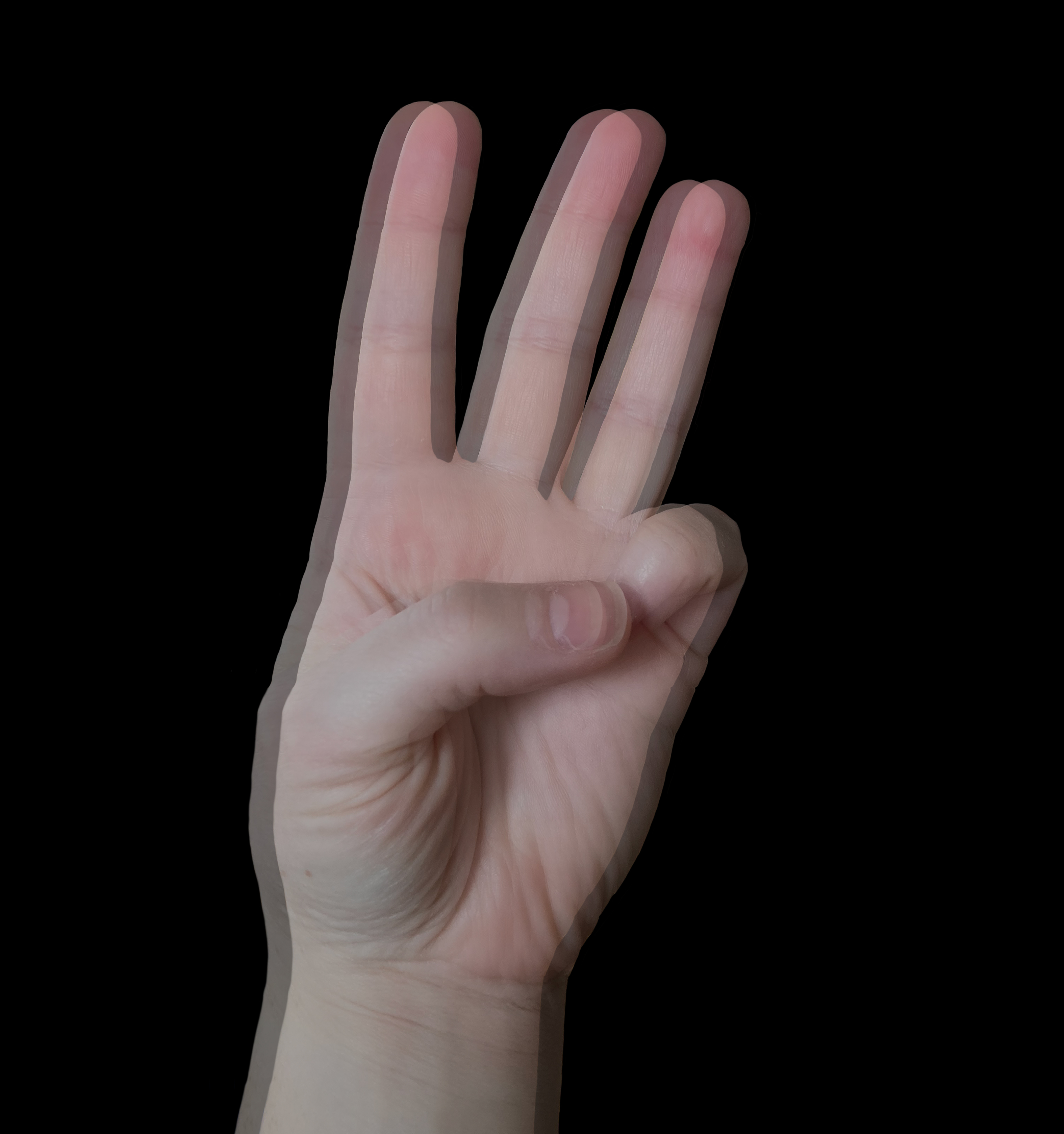
Diplopia refers to seeing two images and is due either to ocular misalignment, in which case it disappears when either eye is occluded or to an optical problem, in which case it is termed monocular diplopia and does not disappear with monocular viewing. Patients with ocular misalignment can harbor serious pathology and should be evaluated in a systematic and thorough manner in order to uncover all potentially serious cases. This activity will present an approach to diplopic patients that will assist providers in evaluating the condition, making a diagnosis early, and improving outcomes.
Objectives:
- Review of the approach to the patient with diplopia.
- Describe the evaluation process for a patient with diplopia.
- Summarize the treatment of diplopia.
- Outline the evaluation and management of a patient with diplopia and the role of interprofessional team members in collaborating to provide well-coordinated care and enhance patient outcomes.
Introduction
Diplopia refers to seeing two images and is due either to ocular misalignment in which case it disappears when either eye is occluded or to an optical problem in which case it is termed monocular diplopia and does not disappear with monocular viewing.
Patients with ocular misalignment can harbor serious pathology and should be evaluated in a systematic and thorough manner in order to uncover all potentially serious cases. This article will present an outline of the approach to diplopic patients.
Etiology
The etiology of diplopia is either eye misalignment if diplopia is binocular or an optical phenomenon if it is monocular. Eye misalignment can occur due to a variety of causes, and in order to understand it, one has to follow the anatomical algorithm of what needs to happen in order for the eyes to be aligned together. Both eyes have to receive equal innervation of all of their extraocular muscles in order to be in the so-called primary position when the innervation to antagonist extraocular muscles in each eye is equal. Innervation to extraocular muscles is provided by the 3rd, 4th, and 6th cranial nerves; thus, all three have to be working well on each side in order to maintain both eyes in the primary position and aligned with each other. The nuclei of all three of these cranial nerves originate in the brainstem (3rd and 4th in the midbrain and 6th in the pons). Their fascicles then traverse the brainstem exiting it ventrally in the case of the 3rd and 6th nerve and dorsally in the case of the 4th nerve. The nerves then travel variable distances in the subarachnoid space, where they are susceptible to a variety of pathologies affecting cerebrospinal fluid (inflammatory conditions, infections, and malignancies). Eventually, all oculomotor nerves (3rd, 4th, and 6th) emerge from the subarachnoid space and converge in the space filled with venous blood located laterally to each side of the pituitary gland, which is termed cavernous sinus. The cavernous sinus is surrounded by a lot of structures, and pathologies affecting these neighboring structures (sphenoid sinus, pituitary gland, nasopharynx, intracavernous carotid arteries, etc.) can affect any of the oculomotor nerves and thus cause diplopia. As all three oculomotor nerves are located in close proximity to each other in the cavernous sinus, when encountering a patient with more than one oculomotor nerve palsies, one should think of cavernous sinus as the most likely location of the causative lesion. From the cavernous sinuses, all three oculomotor nerves travel in close proximity to each other to enter the superior orbital fissure, where they are also located close to the 2nd cranial nerve (optic nerve); thus, lesions in the superior orbital fissure produce decrease vision and often forward displacement of the globe (proptosis) in addition to multiple oculomotor nerve palsies. From the superior orbital fissure, each nerve travels to the extraocular muscle it innervates through the orbit; thus, orbital pathology can produce diplopia as well. Eventually, the nerve will join the muscle that it innervates, and pathology at the neuromuscular junction can also cause ocular misalignment and thus binocular diplopia. Finally, extraocular muscles themselves need to be intact in order for the eyes to be aligned with each other; thus, myopathies can also produce diplopia.
Monocular diplopia is almost always an ophthalmological problem and stems most commonly from the cataractous changes in the crystalline lens, abnormalities in the corneal surface (keratoconus or uncorrected astigmatism), and exceedingly rarely lesions affecting occipital cortex can produce monocular diplopia as well (termed "cortical polyopia") in which case they are almost always accompanied by homonymous visual field defects. Finally, when no organic etiology for monocular diplopia can be found, and diplopia does not disappear when looking through a pinhole, one has to assume that it is functional in nature.
Epidemiology
Diplopia is a common complaint in both the ambulatory setting as well as in the emergency department, with one study reporting almost 805000 ambulatory and 50000 emergency department visits in the United States yearly with the chief complaint of diplopia[1]. Diplopia, particularly when acute in onset, is very unsettling to the patient and will prompt most to visit an emergency department. While we are always concerned about the potential sinister and even life-threatening causes of diplopia, only 16% of patients with diplopia were found to have potentially life-threatening etiologies in one study.[2]
Pathophysiology
Binocular diplopia occurs because the image falls outside of the fovea in one eye, thus triggering the perception of two separate images. If eye misalignment is horizontal, diplopia is horizontal, and if the eye misalignment is vertical, it will be vertical.
History and Physical
When evaluating a diplopic patient, one has first to determine whether diplopia is monocular or binocular. While this has already been mentioned above, it is of paramount importance as skipping this step will lead to unnecessary investigations and anxiety for the patient.
The next most important step is to search for accompanying “brainstem” symptoms. While isolated brainstem strokes are uncommon, diplopia can be the main complaint in patients with strokes involving diencephalon or brainstem, which involve either the nuclei or fascicles of the IIIrd, IVth, or VIth cranial nerves, medial longitudinal fasciculus, or vertibulo-ocular pathways producing the so-called skew deviation[3].
Any patient with an acute onset of binocular diplopia who has any accompanying symptoms that can be caused by brainstem dysfunction (vertigo, dizziness, dysarthria, crossed motor or sensory symptoms, ataxia, imbalance, etc.) should be immediately referred to an emergency department for in order have an urgent MRI of the brain with attention to the brainstem performed. MRI should include diffusion-weighted as well as susceptibility-weighted images in order to detect subtle ischemic and/or hemorrhagic lesions.
All diplopic patients should be asked about the fatiguability and variability of their symptoms as well as the presence of symptoms of increased intracranial pressure, and all patients over the age of 50 should be asked about the symptoms of giant cell arteritis.
Next, a careful examination should be performed, first testing ocular motility in order to try to identify obvious deficits and determine whether they map out to the dysfunction of the 3rd or 6th cranial nerves. Motility testing should be performed by slowly moving a target in all directions of gaze and should be checked in each eye separately. After motility testing, ocular alignment should be checked by performing alternate cover testing. If a vertical deviation is uncovered, testing of misalignment in ipsi- and contralateral gaze as well as ipsi- and contralateral head tilt positions should be performed to determine if the deviation fits the pattern of the so-called 3-step test where the hypertropia increases in contralateral gaze and ipsilateral head tilt. A positive 3-step test indicates 4th nerve paresis[3].
If ocular motility is full and eye misalignment remains the same in all positions of gaze, it is termed comitant and is almost always secondary to decompensated congenital strabismus. It does not require further investigations or testing other than a referral for ophthalmological evaluation. After ocular motility and alignment have been examined and recorded (we prefer recording ocular motility in percentages, i.e., "there was only 70% of expected supraduction"), pupillary examination, the position of the lids, and movement of the lid during the motility testing, presence of proptosis, and orbicularis strength should all be examined and recorded. Dilated fundus examination looking for the presence of optic nerve head edema and any signs of venous stasis retinopathy should be performed in all diplopic patients as well.
Evaluation
After performing careful testing of extraocular motility in each eye separately followed by ocular alignment testing (which should be performed by doing alternate cover testing and abnormalities recorded in each cardinal position of gaze utilizing measurements in prism diopters), an examiner has to combine all findings to figure out whether they can be due to an isolated palsy of the 3rd, 4th or 6th cranial nerves. The management of each one of these entities is different and is discussed below. Identifying complete third nerve palsy and sixth nerve palsy is relatively straightforward, but diagnosing partial third nerve paresis can be tricky. The examiner has to be very familiar with the 3-step text described above in order to diagnose 4th nerve palsy.
If there is more than one cranial nerve palsy, urgent neuroimaging (MRI of the brain and orbits with contrast enhancement) is mandatory, looking for the lesions in the area of the cavernous sinuses, superior orbital fissure, and extraocular muscles. If fatiguability, variability, decreased orbicularis strength or any other symptoms of ocular or generalized myasthenia gravis are present, acetylcholine receptor antibody testing should be performed, although its sensitivity is only about 50 to 70%[4]. If the diagnosis is strongly suspected despite the negative acetylcholine receptor antibody titers, a single fiber electromyogram of the orbicularis muscle should be performed as in the hands of an experienced operator it has a sensitivity of about 95% in diagnosing abnormalities specific to ocular myasthenia[5].
All patients with the third nerve palsy (regardless of whether it is complete or partial, pupillary sparing or pupillary involving) should undergo an emergency CT angiography of the brain in order to rule out an aneurysmal compressive lesion of the third cranial nerve in its subarachnoid compartment[6]. CTA is widely available and is very sensitive for detecting any aneurysms larger than 2-3 mm in diameter (it is generally agreed upon that an aneurysm has to be at least 4 mm in diameter in order to produce compression of the third nerve). The main limitation of the CT angiogram in detecting all relevant third nerve palsies is the experience and knowledge of the interpreting radiologist[7]. If there is pupillary involvement and the CTA has been interpreted as normal, a clinician should personally communicate their clinical findings to the radiologist and ensure that they have carefully reviewed the junction between the internal carotid and posterior communicating arteries as this is the most common location of an intracranial aneurysm that can produce third nerve palsy. If the CTA is normal and the patient is over the age of 50 with vasculopathic risk factors, it is reasonable to assume that the etiology of the third nerve palsy is likely microischemic in nature and observe them for 2-3 months when the resolution or significant improvement of the nerve palsy is expected. If the patient is under the age of 50 or does not have any microvascular ischemic risk factors and the CTA is negative, urgent MRI with contrast utilizing steady-state imaging sequences should be performed in order to follow the entire course of the third nerve and rule out any pathology affecting it along its course[6].
For patients with the 4th nerve palsy, if the hyper deviation in the affected eye is greater in upgaze than in downgaze, it can be safely assumed that it is likely either decompensated congenital, post-traumatic, or microischemic in nature and should either be palliated with the prisms in the spectacles or observed for spontaneous improvement[8]. If the deviation is worse in downgaze, an MRI of the brain with contrast and steady-state imaging should be performed, evaluating the course of the fourth cranial nerve.
Patients with acute onset of 6th nerve palsy who have vascular risk factors who are older than 50 years of age should be observed if it can be assured that cranial nerve palsy is isolated and imaged only if their motility deficit does not improve after 2-3 months. Patients without microvascular risk factors and all patients under 50 years of age with the 6th nerve palsy should have an MRI with contrast and steady-state imaging acquisition performed in order to rule out demyelinating or compressive etiology anywhere along the course of the nerve.[3]
Treatment / Management
Treatment options for patients with diplopia are limited. Patients with monocular diplopia should be evaluated for possible cataract surgery, and their refractive errors should be corrected. Patients with binocular diplopia who are symptomatic should ideally first try to palliate the diplopia with prisms in their spectacles. Prisms are generally effective only for patients with small (~up to 10-12 prism diopters) misalignments. For patients with larger misalignments, occlusion of either eye with a patch or a tape applied to one lens in the spectacles can be an effective way to eliminate diplopia. Patients with stable misalignments should be assessed by a strabismologist for potential surgical correction of the eye misalignment. Injection of Botulinum toxin into the antagonist of the paretic muscle has also been suggested but can be difficult as it is hard to predict the reaction of each patient to a specific dose of Botulinum toxin, and its effect will eventually wear off[9]. Monovision (placing a contact lens in one eye in order to make this eye hyperopic or myopic and thus disrupting binocular viewing) has also been tried for patients with diplopia who have small misalignments and could be an effective treatment option for some[10].
Differential Diagnosis
The differential diagnosis for patients with binocular diplopia is vast. The anatomical approach in formulating the differential diagnosis will again be used.
Any lesions affecting the brainstem can affect the nuclei or the fascicles of the 3rd, 4th, and 6th cranial nerves, involve the vestibulo-ocular pathways or medial longitudinal fascicule.
Any lesions affecting the horizontal or vertical gaze centers (located in the pons and midbrain, respectively) can rarely cause diplopia.
Lesions of the medial longitudinal fasciculus, which connects the sixth cranial nerve to the medial rectus subnucleus on the other side, can also produce diplopia. These lesions tend to be demyelinating in patients under the age of 50 and ischemic in those older than 50.
Pathology affecting the CSF (malignant cell circulating in it, inflammatory processes disturbing normal CSF composition, increased protein, etc.) can also produce oculomotor palsies.
Guillain-Barre syndrome can cause diplopia when it preferentially affects the oculomotor nerves (3rd, 4th, and 6th) in the so-called Miller-Fisher variant where the levels of GQ1b antibodies are often elevated.
Thiamine deficiency (Wernicke encephalopathy) can produce diplopia as well; thus, intravenous thiamine should be administered in all diplopic patients who are confused, have nystagmus, or ataxia.
A variety of lesions can affect the cavernous sinus, often producing multiple cranial nerve palsies. These include inflammatory lesions from idiopathic cavernous sinus syndrome, sarcoidosis, IgG4 disease or granulomatosis with polyangiitis; vascular lesions from the expanding aneurysms of the intra-cavernous carotid artery, carotid-cavernous fistulas; mass lesions due to expanding pituitary apoplexy, neoplastic or inflammatory lesions in the sphenoid sinus; infections lesions due to cavernous sinus thrombosis, or direct spread of infection from the face; spread of malignancy from the nasopharynx or sphenoid sinus, as well as distant metastasis.
A multitude of lesions affecting superior orbital fissures can cause diplopia by affecting one or several cranial nerves that travel through it.
Orbital disease (such as thyroid eye disease, any tumors or vascular lesions of the orbit) can also cause diplopia.
Finally, disorders of neuromuscular junctions and myopathies affecting extraocular muscles can also produce diplopia.
Prognosis
The prognosis of diplopia is very variable and depends entirely on the underlying condition that is causing it.
Complications
The main complication from diplopia itself is the discomfort experienced by the patient and the inability to drive for many patients. Of course, individual etiologies that can cause diplopia have their own complications associated with them.
Deterrence and Patient Education
Patients should be educated about the nature of diplopia and that treating binocular diplopia can be challenging. They should be explained what the thinking process of the examiner is and how they will approach the diagnostic and treatment process based on their clinical findings.
Enhancing Healthcare Team Outcomes
Patients with diplopia may initially present to the emergency department physician, primary care provider, and nurse practitioner. However, because there are many causes of diplopia and the workup is beyond the scope of the primary provider, these patients should be referred to an ophthalmologist and preferably to a neuro-ophthalmologist for definitive diagnosis and treatment planning. The prognosis depends on the cause. Emergency department and ophthalmology nurses are involved in the initial evaluation and coordination of evaluation. This interprofessional team can coordinate care to improve outcomes. [Level 5]

