Blunt Head Trauma

Earn CME/CE in your profession:


Continuing Education Activity

Traumatic Brain Injury is a significant cause of morbidity and mortality in the United States, with an annual occurrence of more than 1.5 million. Falls are the most common cause of traumatic brain injury, and motor vehicle-related incidents are the second leading cause. Blunt head trauma is classified as mild, moderate, or severe traumatic brain injury. This activity describes the etiology, classification, pathophysiology, evaluation, and management of blunt head trauma and highlights the role of interprofessional teams in improving outcomes for such patients.

Objectives:

  • Describe the etiology, epidemiology, pathophysiology, and classification of patients with blunt head trauma.
  • Outline the evaluation and resuscitation of patients with blunt head trauma.
  • Review the conservative and surgical treatment options available for patients with blunt head trauma.
  • Explain interprofessional team strategies for enhancing care coordination to facilitate rapid diagnosis and targeted management of patients with blunt head trauma.

Introduction

Traumatic Brain Injury (TBI) is a significant cause of morbidity and mortality in the United States, with an annual occurrence of more than 1.5 million. Patients with moderate and severe TBI comprise about 20% of TBI, and those with moderate TBI have a mortality of about 15% while those with severe TBI have associated mortality approaching 40%. The majority (approximately 80%) of patients with TBI have mild TBI which is associated with a less than 0.5% mortality, but about 25% experience extended post-concussive symptoms including a headache, dizziness, difficulty concentrating, and depression. [1][2][3]

Etiology

Falls are the most common cause of TBI, and motor vehicle-related incidents are the second leading cause of TBI. Motor vehicle-related TBI includes automobile, motorcycle, and bicycle accidents and pedestrians struck by those vehicles. Sports, recreation, and work-related injuries are the third leading cause of TBI, and assaults are the fourth leading cause of TBI. Blast injuries are the leading cause of TBI in active duty military personnel in war zones.[4][5][6]

Epidemiology

TBI is the most common cause of death in people younger than the age of 25. The majority of fatal TBI is due to motor vehicle-related incidents, falls, and assaults. Mortality due to motor vehicle accidents is greatest in the young-adult age group attributed to alcohol use and excessive speed. Mortality due to falls is greatest in patients over age 65, which is also the age group with the highest mortality in any TBI. Neurosurgical intervention such as craniotomy, elevation of skull fracture, intracranial pressure (ICP) monitor, or ventriculostomy is required in about 40% of patients with severe TBI and about 10% of patients with moderate TBI.  [7][8]

Pathophysiology

Most patients with moderate to severe TBI have a combination of intracranial injuries. The majority of patients with moderate to severe TBI have related diffuse axonal injury to some degree. The diffuse axonal injury typically is caused by a rapid rotational or deceleration force that causes stretching and tearing of neurons, leading to focal areas of hemorrhage and edema that are not always detected on the initial computed tomogram (CT) scan. Subarachnoid hemorrhage (SAH) is the most common CT finding in TBI and is caused by tears in the pial vessels. Subdural and epidural hematomas are the most frequent type of mass lesion identified in TBI. Cerebral contusions occur in about a third of patients with moderate to severe TBI, caused by direct impact or acceleration-deceleration forces that cause the brain to strike the frontal or temporal regions of the skull. Intracerebral bleeding or hematoma, caused by coalescence of contusions or a tear in a parenchymal vessel, occurring in up to a third of patients with moderate to severe TBI.  [9][10]

History and Physical

The majority of patients with TBI have a straightforward clinical presentation, but it is also important to solicit the mechanism of injury, current anticoagulation use, symptoms of the head or neck pain, post-traumatic seizure, and any history of repeat head injury or past central nervous system surgeries.

The initial resuscitation should proceed in a step-wise fashion to identify all injuries and optimize cerebral perfusion by maintaining hemodynamic stabilization and oxygenation. The initial survey also should include a brief, focused neurological examination with attention to the Glasgow Coma Scale (GCS), pupillary examination, and motor function.

After addressing any airway or circulatory deficits, a thorough head-to-toe physical examination must be performed with vigilance for occult injuries and careful attention to detect any of the following warning signs[11]:

  • Inspection for cranial nerve deficits, periorbital or postauricular ecchymoses[12], cerebrospinal fluid rhinorrhea or otorrhea, hemotympanum (signs of a base of skull fracture)
  • Fundoscopic examination for retinal hemorrhage (a potential sign of abuse in children) and papilledema (a sign of increased ICP)
  • Optic nerve sheath diameter of greater than 5 mm on ultrasound has been shown to correlate well with increased intracranial pressure in patients with TBI
  • Palpation of the scalp for hematoma, crepitus, laceration, and bony deformity (markers of skull fractures)
  • Auscultation for carotid bruits, painful Horner syndrome or facial/neck hyperesthesia (markers of carotid or vertebral dissection)
  • Evaluation for cervical spine tenderness, paresthesias, incontinence, extremity weakness, priapism (signs of spinal cord injury)
  • Examination of motor and sensory extremities (for signs of spinal cord injury)

Evaluation

Non-contrast cranial CT is the imaging modality of choice for patients with TBI. CT findings associated with a poor outcome in TBI include midline shift, subarachnoid hemorrhage into the verticals, and compression of the basal cisterns. Magnetic Resonance Imaging scan may be indicated when the clinical picture remains unclear after a CT to identify more subtle lesions.[13][14]

Treatment / Management

Airway adjuncts are indicated in patients not able to maintain an open airway or maintain more than 90% oxygen saturation with supplementary oxygen. Oxygenation parameters should be monitored using continuous pulse oximetry with a target of more than 90% oxygen saturation. Ventilation should be monitored with continuous capnography with an end-tidal CO2 target of 35 mmHg to 40 mmHg. Placement of a definitive airway is recommended in the patient with a Glasgow Coma Scale (GCS) score of less than 9.  [15]

Systemic hypotension negatively impacts the outcome in the setting of TBI, and current studies have demonstrated improved outcomes in patients with systolic blood pressure (BP) greater than or equal to 120 mmHg. Isotonic crystalloids should be used to prevent and correct hypotension; colloidal solutions have not been shown to improve outcomes.

Serial neurological examinations allow for early identification of patients with elevated ICP, and subsequent implementation of primary bedside interventions to improve venous outflow and reduce metabolic demands. Initial bedside approaches to increase ICP include elevating the head of the patient's bed 30 degrees, ascertaining that the cervical collar is not impeding venous outflow, and appropriate analgesics and sedation.

Routine hyperventilation should be avoided during the first 24 hours, and should only be used as a temporizing measure in the setting of impending herniation. Hyperosmolar therapy such as mannitol or hypertonic saline can further reduce intracerebral volume. ICP monitoring is indicated in patients with TBI when they have a GCS score of less than 9, an abnormal CT, and the approach to refractory elevated intracranial pressure includes high-dose barbiturates and possibly a decompressive hemicraniectomy.[16]

Differential Diagnosis

  • Cerebral aneurysms
  • Confusional states and acute memory disorders
  • Emergent management of subarachnoid haemorrhage
  • Epileptic and epileptiform encephalopathies
  • Frontal lobe syndrome
  • Generalized tonic-clonic seizures
  • Hydrocephalus
  • Prion related diseases
  • Subdural empyema
  • Temporal lobe epilepsy

Complications

  • Intracranial hemorrhage
  • Brain edema
  • Elevated ICP
  • Vasospasm
  • Seizures
  • Infection
  • Hydrocephalus
  • Memory loss
  • Hyponatremia

Pearls and Other Issues

Patients with pre-existing anticoagulant or antiplatelet use have a poorer outcome with TBI. Acute, rapid reversal of anticoagulant agents should be considered early. Correction agents for patients on antiplatelet or anticoagulant agents with TBI include Vitamin K, Protamine Sulfate, Prothrombin Complex Concentrate (PCC), Fresh Frozen Plasma (FFP), Activated factor VIIa (rFVIIa). The identification of an up-to-date medication list is extremely important to choose the appropriate correction agent.

Enhancing Healthcare Team Outcomes

Blunt head trauma is a common presentation to the emergency room. The key to the treatment of blunt head trauma is prevention. Most cases are preventable, and hence the education of the public is essential. At the time of injury, the patient and family should be educated on using safety devices during sports and other high-risk activities. The caregiver of the elderly should be educated on the prevention of falls in the home and parents should be educated on child safety during driving and playground activities. Those who have a neurological deficit may benefit from physiotherapy and occupational therapy. The pharmacist should educate the patient on abstaining from alcohol and the use of sedative drugs. In addition, the patient should be warned about seizures and the need to take anticonvulsants.[17][18] (Level V)

Outcomes

The outcome after blunt head trauma depends on the type of injury, the severity of the injury, patient age, the presence of any neurological deficit, comorbidity and any secondary injury. The GCS at 24 hours is a strong predictor of cognitive recovery at 24 months after the injury. The mortality rates do vary from 4-40% at five years after blunt head trauma. Younger people fare much better than older individuals. Individuals with a neurological deficit and those taking oral anticoagulants at the time of injury often have a poor outcome. The pupillary function before and after the injury is also predictive of outcome. Those with bilateral unresponsive pupils tend to have a grave prognosis and a higher risk of remaining in a vegetative state. Finally, studies show that diabetics also tend to fare worse with blunt head trauma compared to nondiabetics.[19][20] (Level V)


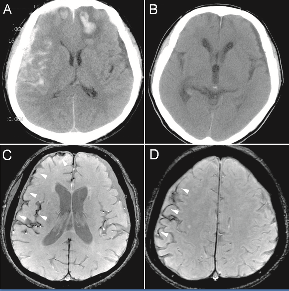
(Click Image to Enlarge)
Original description: Figure 2
Original description: Figure 2. CT and enhanced gradient echo T2 star-weighted angiography (ESWAN) images of the brain of a 54-year-old man who experienced a traumatic brain injury. An axial head CT image displays right frontotemporal SAH (Fisher grade 4) with bilateral frontal contusions and intracerebral hematoma (A). A follow-up CT image 26 weeks after the brain injury indicates that the hemorrhages were completely resolved and the lateral ventricles were mildly enlarged (B). A follow-up MRI (1.5T) image was obtained 26 weeks following the head injury (C,D). The axial ESWAN image displays a rim of hypointensity (arrowheads), with hemosiderin deposits forming along the cerebral convexity (C, D).
Contributed by Hongwei Zhao, Jin Wang, Zhonglie Lu, Qingjie Wu, Haijuan Lv, Hu Liu, Xiangyang Gong
Details

Editor:

Bracken Burns

Updated:

8/8/2023 12:28:43 AM

References


[1]

Lamb LC, DiFiori M, Comey C, Feeney J. Cost Analysis of Direct Oral Anticoagulants Compared with Warfarin in Patients with Blunt Traumatic Intracranial Hemorrhages. The American surgeon. 2018 Jun 1:84(6):1010-1014     [PubMed PMID: 29981640]


[2]

Daher P, Teixeira PG, Coopwood TB, Brown LH, Ali S, Aydelotte JD, Ford BJ, Hensely AS, Brown CV. Mild to Moderate to Severe: What Drives the Severity of ARDS in Trauma Patients? The American surgeon. 2018 Jun 1:84(6):808-812     [PubMed PMID: 29981606]


[3]

Harvell BJ, Helmer SD, Ward JG, Ablah E, Grundmeyer R, Haan JM. Head CT Guidelines Following Concussion among the Youngest Trauma Patients: Can We Limit Radiation Exposure Following Traumatic Brain Injury? Kansas journal of medicine. 2018 May:11(2):1-17     [PubMed PMID: 29796153]


[4]

Abolfotouh MA, Hussein MA, Abolfotouh SM, Al-Marzoug A, Al-Teriqi S, Al-Suwailem A, Hijazi RA. Patterns of injuries and predictors of inhospital mortality in trauma patients in Saudi Arabia. Open access emergency medicine : OAEM. 2018:10():89-99. doi: 10.2147/OAEM.S166026. Epub 2018 Jul 31     [PubMed PMID: 30104908]

Level 2 (mid-level) evidence

[5]

Joyce T, Gossman W, Huecker MR. Pediatric Abusive Head Trauma. StatPearls. 2023 Jan:():     [PubMed PMID: 29763011]


[6]

Thierauf-Emberger A, Lickert A, Pollak S. A moving human body causes fatal blunt trauma: an unusual traffic accident. International journal of legal medicine. 2019 Mar:133(2):547-551. doi: 10.1007/s00414-018-1855-z. Epub 2018 Jun 6     [PubMed PMID: 29876635]


[7]

George E, Khandelwal A, Potter C, Sodickson A, Mukundan S, Nunez D, Khurana B. Blunt traumatic vascular injuries of the head and neck in the ED. Emergency radiology. 2019 Feb:26(1):75-85. doi: 10.1007/s10140-018-1630-y. Epub 2018 Aug 10     [PubMed PMID: 30097750]


[8]

Joachim M, Tuizer M, Araidy S, Abu El-Naaj I. Pediatric maxillofacial trauma: Epidemiologic study between the years 2012 and 2015 in an Israeli medical center. Dental traumatology : official publication of International Association for Dental Traumatology. 2018 Apr 27:():. doi: 10.1111/edt.12406. Epub 2018 Apr 27     [PubMed PMID: 29701325]


[9]

Leetch AN, Wilson B. Pediatric Major Head Injury: Not a Minor Problem. Emergency medicine clinics of North America. 2018 May:36(2):459-472. doi: 10.1016/j.emc.2017.12.012. Epub 2018 Feb 10     [PubMed PMID: 29622334]


[10]

Essien EO, Fioretti K, Scalea TM, Stein DM. Physiologic Features of Brain Death. The American surgeon. 2017 Aug 1:83(8):850-854     [PubMed PMID: 28822390]


[11]

Clark A, M Das J, Weisbrod LJ, Mesfin FB. Trauma Neurological Exam. StatPearls. 2023 Jan:():     [PubMed PMID: 29939692]


[12]

M Das J, Munakomi S. Raccoon Sign. StatPearls. 2023 Jan:():     [PubMed PMID: 31194384]


[13]

Masterson Creber RM, Dayan PS, Kuppermann N, Ballard DW, Tzimenatos L, Alessandrini E, Mistry RD, Hoffman J, Vinson DR, Bakken S, Pediatric Emergency Care Applied Research Network (PECARN) and the Clinical Research on Emergency Services and Treatments (CREST) Network. Applying the RE-AIM Framework for the Evaluation of a Clinical Decision Support Tool for Pediatric Head Trauma: A Mixed-Methods Study. Applied clinical informatics. 2018 Jul:9(3):693-703. doi: 10.1055/s-0038-1669460. Epub 2018 Sep 5     [PubMed PMID: 30184559]


[14]

McGrew PR, Chestovich PJ, Fisher JD, Kuhls DA, Fraser DR, Patel PP, Katona CW, Saquib S, Fildes JJ. Implementation of a CT scan practice guideline for pediatric trauma patients reduces unnecessary scans without impacting outcomes. The journal of trauma and acute care surgery. 2018 Sep:85(3):451-458. doi: 10.1097/TA.0000000000001974. Epub     [PubMed PMID: 29787555]

Level 1 (high-level) evidence

[15]

Bukur M, Teurel C, Catino J, Kurek S. The Price of Always Saying Yes: A Cost Analysis of Secondary Overtriage to an Urban Level I Trauma Center. The American surgeon. 2018 Aug 1:84(8):1368-1375     [PubMed PMID: 30185318]


[16]

Rybkin I, Kim M, Amin A, Tobias M. Development of Delayed Posttraumatic Acute Subdural Hematoma. World neurosurgery. 2018 Sep:117():353-356. doi: 10.1016/j.wneu.2018.06.135. Epub 2018 Jun 26     [PubMed PMID: 29959076]


[17]

McLoughlin RJ, Green J, Nazarey PP, Hirsh MP, Cleary M, Aidlen JT. The risk of snow sport injury in pediatric patients. The American journal of emergency medicine. 2019 Mar:37(3):439-443. doi: 10.1016/j.ajem.2018.06.006. Epub 2018 Jun 2     [PubMed PMID: 29884589]


[18]

Khor D,Wu J,Hong Q,Benjamin E,Xiao S,Inaba K,Demetriades D, Early Seizure Prophylaxis in Traumatic Brain Injuries Revisited: A Prospective Observational Study. World journal of surgery. 2018 Jun     [PubMed PMID: 29159600]

Level 2 (mid-level) evidence

[19]

Watanabe T, Kawai Y, Iwamura A, Maegawa N, Fukushima H, Okuchi K. Outcomes after Traumatic Brain Injury with Concomitant Severe Extracranial Injuries. Neurologia medico-chirurgica. 2018 Sep 15:58(9):393-399. doi: 10.2176/nmc.oa.2018-0116. Epub 2018 Aug 11     [PubMed PMID: 30101808]


[20]

Aiolfi A, Khor D, Cho J, Benjamin E, Inaba K, Demetriades D. Intracranial pressure monitoring in severe blunt head trauma: does the type of monitoring device matter? Journal of neurosurgery. 2018 Mar:128(3):828-833. doi: 10.3171/2016.11.JNS162198. Epub 2017 May 26     [PubMed PMID: 28548592]