Hepatic Cyst

Earn CME/CE in your profession:


Continuing Education Activity

Hepatic cysts are fluid-filled cavities in the liver generally asymptomatic and found incidentally on imaging studies. They can sometimes be associated with serious complications such as infection, hemorrhage, rupture, or compression of the biliary tree. This activity explains the evaluation and treatment of hepatic cyst and reviews the role of the interprofessional team in managing patients with this condition.

Objectives:

  • Summarize the pathophysiology of hepatic cysts.
  • Identify the use of CT, MRI, and an ultrasound in the evaluation of hepatic cysts.
  • Describe the use of percutaneous aspiration, laparoscopic deroofing, and complete cyst excision in the treatment of symptomatic hepatic cysts.
  • Outline the importance of improving care coordination among the interprofessional team members to enhance the delivery of care and improve outcomes for patients with hepatic cysts.

Introduction

Hepatic cysts are generally found incidentally on imaging studies. They are usually asymptomatic but can sometimes be associated with serious complications. As opposed to smaller cysts, larger cysts are more likely to result in symptoms from complications such as hemorrhage, rupture, infection, or compression of the biliary tree.

Etiology

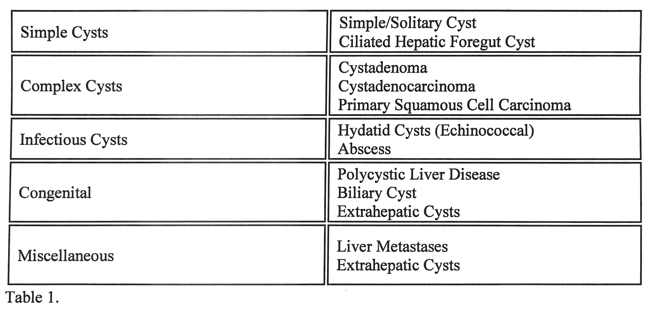
Hepatic cysts can be due to a spectrum of etiologies ranging from benign to malignant and infectious.[1] They are described in detail in Table 1.

Epidemiology

In general, non-infectious hepatics cysts are most prevalent in the female population. Their incidence also tends to increase with age. On the contrary, ciliated hepatic foregut cysts occur more commonly in men. Echinococcal hepatics cysts (also known as hydatid cysts) are more common in their endemic geographical regions which include South America, the Middle East, and the eastern Mediterranean, some sub-Saharan African countries, western China and the former Soviet Union.[2] Biliary cysts can present in infants due to congenital abnormalities.[3]

Pathophysiology

Simple hepatic cysts are the most common type of hepatic cysts. They contain a clear fluid, and their size can vary from a few millimeters to tens of centimeters in diameter. Primary squamous cell carcinoma can arise in hepatic cysts. Liver metastases, which originate from organs such as the ovaries, kidneys, colon, or pancreas, may appear as hepatic cysts. Polycystic liver disease is associated with polycystic kidney disease and leads to many hepatic cysts.[4] Echinococcal cysts are associated with a parasitic infection (by tapeworm Echinococcus) which is transmitted by the fecal (from dogs) oral (to humans) route through environmental contamination of water or vegetables.[5] Biliary cysts arise from the biliary ductal system and can be congenital in origin.

Histopathology

Usually, histological examination is not necessary to make a diagnosis of a hepatic cyst as the diagnosis is often made on radiologic and clinical findings. Regardless, there are differences in terms of histopathology when it comes to different hepatic cysts.

Simple cysts are comprised of a thin outer layer, dense fibrous tissue, and an inner epithelial lining. This epithelial lining is made up of a single layer of cuboidal or columnar cells. Simple cysts lack cellular atypia or mesenchymal stroma. They are fluid-filled, and most are less than 3 cm in size. The fluid within the cyst is sterile and cytologically negative. A distinct histological feature of an echinococcal cyst is that it is associated with calcifications. Ciliated hepatic foregut cysts consist are comprised of a ciliated pseudostratified columnar epithelium, a subepithelial connective tissue layer, smooth muscle, and a fibrous capsule.

History and Physical

When hepatic cysts do become large enough to cause symptoms, they can cause abdominal pain and gastrointestinal symptoms such as nausea and anorexia. Significantly large cysts may even be palpable on abdominal examination. Echinococcal cysts can cause elevated body temperature, bloody sputum, and pruritis.[6] Biliary cysts may present as ta riad of pain, jaundice, and abdominal mass.[7]

Evaluation

As mentioned, hepatic cysts are usually incidentally found on radiological imaging.[8] While an ultrasound in the most practical test, computed tomography (CT) and magnetic resonance imaging (MRI) can also be utilized in diagnosis. Ultrasound can generally differentiate a simple cyst from a complicated one. Simple hepatic cysts will appear as a unilocular space filled with fluids. Walls are thin and there is posterior acoustic enhancement. These cysts do not contain any septations or papillary projections which are findings often seen in complicated hepatic cysts. On CT and MRI, simple hepatic cysts do not enhance even if contrast is used. Complicated hepatic cysts may show up as hypoechoic lesions with irregular and thick walls with echoes representing debris or wall nodularity. Echinococcal cysts can often appear like simple cysts on radiological imaging. When present with daughter cysts, ultrasound can detect characteristic internal septations. Other characteristic findings on imaging include hydatid sand (hooklets and scolexes from the protoscolices), inner cyst wall infoldings, and separation of the hydatid membrane from the wall of the cyst. In addition to imaging, serological testing is generally used to diagnosis of echinococcal cysts.

Treatment / Management

Incidentally found asymptomatic simple hepatics cysts do not require any treatment. However, cysts larger than 4 cm in diameter should be monitored. Follow-up ultrasounds can be performed to monitor the size and stability of the cysts. They are done at 3- and 12-month intervals. If the cyst is stable for 2 to 3 years, there is no need for further ultrasound follow-up.[9]

Regarding symptomatic hepatic cysts, there are three common procedures which can either decrease the cyst size or even remove the cyst altogether. These procedures include percutaneous aspiration, laparoscopic deroofing, and complete cyst excision.[10]

Percutaneous aspiration can be performed on its own or alongside sclerosing agents such as alcohol or minocycline hydrochloride. On its own, percutaneous aspiration does not prevent recurrence. With the help of a sclerosing agent, the recurrence rate is decreased significantly. This method is safe and minimally invasive. Therefore, this approach is generally considered first-line treatment. If percutaneous aspiration is not a viable option or has not provided the patient with the desired outcome, deroofing can be performed laparoscopically. There is a much lower recurrence rate for this method with the downside of higher morbidity. Complete cyst excision or hepatectomy is the last line of treatment options.[11] These options are more radical and should only be performed if necessary.

Differential Diagnosis

Diagnosis is made via clinical setting and with radiographic findings. However, differential diagnoses should include other possibilities such as cholelithiasis, gastroesophageal reflux disease, peptic ulcer disease, gastric dysmotility, hepatic abscess, hemangioma, and/or hamartoma. These conditions may elicit similar symptoms as compared to symptomatic hepatic cysts such as abdominal pain. Imaging modalities used to diagnose hepatic cysts can help to exclude some of these conditions. If inconclusive, esophagogastroduodenoscopy may be performed to assess the possibility of gastroesophageal reflux disease or peptic ulcer disease. Motility and transit studies can rule out gastric dysmotility.

Prognosis

Hepatic cysts large enough to produce symptoms can compress the biliary tree. This opens the possibility of rupture. Rupture into the peritoneal cavity or bile duct can happen spontaneously and may lead to infection.[12] There are specific complications related to certain cysts. For example, hydatid cysts have the potential to induce anaphylactic shock which can sometimes be fatal. Moreover, mucinous cysts can transform into a malignancy which generally has a poor prognosis. Similarly, a metastatic disease that presents with hepatic cysts is also associated with poor outcomes.

Enhancing Healthcare Team Outcomes

Diagnosis and management of hepatic cysts often require an interprofessional approach involving various sub-specialties: radiology, internal medicine, hepatology, infectious diseases, and surgery. It is imperative for the involved parties to coordinate patient care together to provide the best healthcare possible and improve patient outcomes.


(Click Image to Enlarge)
Hepatic Cyst Etiology
Contributed by Michelle Bernshteyn, MD
Details

Editor:

Umair Masood

Updated:

6/12/2023 8:10:56 PM

References


[1]

Mavilia MG, Pakala T, Molina M, Wu GY. Differentiating Cystic Liver Lesions: A Review of Imaging Modalities, Diagnosis and Management. Journal of clinical and translational hepatology. 2018 Jun 28:6(2):208-216. doi: 10.14218/JCTH.2017.00069. Epub 2018 Jan 5     [PubMed PMID: 29951366]


[2]

Lembarki G, El Benna N. Echinococcal Cysts in the Liver. The New England journal of medicine. 2018 Jul 12:379(2):181. doi: 10.1056/NEJMicm1702005. Epub     [PubMed PMID: 29996075]


[3]

Recinos A, Zahouani T, Guillen J, Rajegowda B. Congenital Hepatic Cyst. Clinical medicine insights. Pediatrics. 2017:11():1179556517702853. doi: 10.1177/1179556517702853. Epub 2017 Apr 10     [PubMed PMID: 28469521]


[4]

Aussilhou B, Dokmak S, Dondero F, Joly D, Durand F, Soubrane O, Belghiti J. Treatment of polycystic liver disease. Update on the management. Journal of visceral surgery. 2018 Dec:155(6):471-481. doi: 10.1016/j.jviscsurg.2018.07.004. Epub 2018 Aug 23     [PubMed PMID: 30145049]


[5]

Paulsen JD Jr, Elgert P, Yee-Chang M, Wei XJ, Shi Y. Cytomorphologic features of echinococcal cysts. Diagnostic cytopathology. 2017 Aug:45(8):731-734. doi: 10.1002/dc.23733. Epub 2017 Apr 24     [PubMed PMID: 28440023]


[6]

Türkoğlu E, Demirtürk N, Tünay H, Akıcı M, Öz G, Baskin Embleton D. Evaluation of Patients with Cystic Echinococcosis. Turkiye parazitolojii dergisi. 2017 Mar:41(1):28-33. doi: 10.5152/tpd.2017.4953. Epub     [PubMed PMID: 28483731]


[7]

Martínez Ortiz CA, Jiménez-López M, Serrano Franco S. Biliary cysts in adults. 26 years experience at a single center. Annals of medicine and surgery (2012). 2016 Nov:11():29-31. doi: 10.1016/j.amsu.2016.08.016. Epub 2016 Aug 31     [PubMed PMID: 27656283]


[8]

Hasan A, Alrayashi W, Waisel D, Boretsky K. A Case Report of Incidental Hepatic Cysts Found on Ultrasound Imaging: Implications for the Anesthesiologist. A&A practice. 2018 Jan 15:10(2):33-35. doi: 10.1213/XAA.0000000000000627. Epub     [PubMed PMID: 28937424]

Level 3 (low-level) evidence

[9]

Lee DM, Kwon OS, Choi YI, Shin SK, Jang SJ, Seo H, Lee JJ, Choi DJ, Kim YS, Kim JH. [Spontaneously Resolving of Huge Simple Hepatic Cyst]. The Korean journal of gastroenterology = Taehan Sohwagi Hakhoe chi. 2018 Aug 25:72(2):86-89. doi: 10.4166/kjg.2018.72.2.86. Epub     [PubMed PMID: 30145861]


[10]

Sumer F, Kayaalp C, Polat Y, Ertugrul I, Karagul S. Transgastric removal of a polycystic liver disease using mini-laparoscopic excision. Interventional medicine & applied science. 2016 Jun 1:8(2):89-92. doi: 10.1556/1646.8.2016.2.3. Epub     [PubMed PMID: 28386465]


[11]

Smerieri N, Fiorentini G, Ratti F, Cipriani F, Belli A, Aldrighetti L. Laparoscopic left hepatectomy for mucinous cystic neoplasm of the liver. Surgical endoscopy. 2018 Feb:32(2):1068-1069. doi: 10.1007/s00464-017-5736-1. Epub 2017 Jul 21     [PubMed PMID: 28733729]


[12]

Yang C, Yang H, Deng S, Zhang Y. Intraperitoneal Rupture of Hepatic Hydatid Cysts. Journal of gastrointestinal surgery : official journal of the Society for Surgery of the Alimentary Tract. 2019 Jan:23(1):173-175. doi: 10.1007/s11605-018-3778-x. Epub 2018 Apr 20     [PubMed PMID: 29679343]