Lateral Wall Myocardial Infarction

Earn CME/CE in your profession:


Continuing Education Activity

Isolated lateral wall myocardial infarction (LMI), similar to other acute myocardial infarctions (MI), is caused by acute atherosclerotic plaque rupture with subsequent thrombus formation in the left circumflex (LCx) coronary artery or one of its branches. More commonly the left anterior descending (LAD) coronary artery is involved in the ensuing anterolateral MI. In patients with recent drug-eluting stents, medication non-compliance can cause stent restenosis resulting in acute MI. This activity reviews the medical team's role in evaluating acute lateral wall myocardial infarction and in treating this condition.

Objectives:

  • Describe the causes of a lateral wall myocardial infarction.
  • Review the electrocardiogram (ECG) findings in a lateral wall myocardial infarction.
  • Outline the treatment options of a lateral wall myocardial infarction.
  • Summarize the interprofessional coordination of management of a patient with a lateral wall myocardial infarction.

Introduction

The collaborative expert consensus of the European Society of Cardiology (ESC)/American College of Cardiology (ACC)/American Heart Association (AHA) and World Heart Federation (WHF) released the fourth universal definition of Myocardial Infarction (MI) in August 2018. Diagnosis of type I MI focuses on the detection of a rise and/or fall of cardiac Troponin (cTn) with at least one value above the 99th percentile and with the presence of at least one of the following factors:

  1. Symptoms of acute MI
  2. New signs of ischemia on electrocardiogram (ECG)
  3. The occurrence of pathological Q waves
  4. New wall motion abnormality or loss of viable myocardium as evident on imaging
  5. Coronary thrombus identification on angiography[1] 

MI can further be classified based on presenting EKG findings and the location of ischemia. The left ventricular lateral wall is supplied by branches of the left anterior descending artery (LAD) and left circumflex artery (LCx). Lateral and posterior walls together form the left ventricular free wall which is a common site for free-wall rupture (FWR) post-MI. Isolated lateral wall involvement is sporadic and is usually seen as part of multi-territorial infarction such as anterolateral, posterolateral, and inferolateral MI. Occlusion of the obtuse marginal branch of the LCx or diagonal branch of LAD can cause isolated lateral myocardial infarction (LMI).

Etiology

Isolated LMI, similar to other acute MI, is caused by acute atherosclerotic plaque rupture with subsequent thrombus formation in LCx or one of its branches. More commonly, LAD is involved in the ensuing anterolateral MI. In patients with recent drug-eluting stents, medication non-compliance can cause stent restenosis resulting in acute MI.  On rare occasions, patients can present with myocardial infarction with non-obstructive coronary arteries (MINOCA). The prevalence of MINOCA is estimated at around 6% based on a meta-analysis done in 2015.[2] Some of the causes of MINOCA include:

  • Coronary artery spasm
  • Spontaneous coronary artery dissection (SCAD)
  • Coronary artery embolism (CAE): Atrial myxoma, atrial fibrillation, infective endocarditis, rheumatic valve disease, mitral stenosis
  • Autoimmune conditions such as Takayasu arteritis
  • Myocardial bridging

Epidemiology

Recent trends suggest that the incidence of ST-elevation myocardial infarction (STEMI) has declined and incidence of non-ST-elevated MI (NSTEMI) has increased.[3] Around 25% to 40% of patients with MI present as STEMI and of those, 23% have diabetes mellitus. A study based on atherosclerosis risk in communities (ARIC), reported an overall LMI incidence of 13.4% amongst patients presenting with STEMI.[4] Patients with traditional cardiovascular risk factors such as age, family history, obesity, smoking, hypertension, dyslipidemia, diabetes, and alcohol use are predisposed to suffer from LMI.

Pathophysiology

Atherosclerotic plaques with large lipid-rich core and covered by a thin, fibrous cap are extremely unstable and susceptible to rupture. Such plaques may not necessarily cause obstruction. Metalloproteases and cytokines released by T-lymphocytes and macrophages present at the site of plaque weaken the fibrous tissue rendering it vulnerable to erosion.[5] Sudden acute plaque rupture exposes sub-endothelial collagen which serves as a nidus for future platelet adhesion and activation. Thromboxane A2, 5-hydroxytryptamine, platelet-activating factor and ADP released by platelets promote platelet aggregation and vessel vasoconstriction. Activation of coagulation cascade results in fibrin formation and stabilization of occlusive thrombus. Prolonged ischemia leads to necrosis and infarction. The sub-endocardial region of the ventricular wall is most prone to ischemia however with time it can involve the entire wall thickness. Such ischemic events spanning the entire myocardial wall thickness present as STEMI. Infarcted tissue undergoes a series of changes from necrosis to granulation tissue and subsequent scar formation. During this interval from the appearance of granulation tissue to scar formation (between 4 to 14 days), infarcted myocardium is extremely prone to rupture.

History and Physical

Most MI, regardless of location, present with a similar array of symptoms. Some of the common presenting symptoms include left-sided chest pain radiating to arm/neck, shortness of breath, nausea, vomiting, palpitation, diaphoresis, and fatigue. People with a history of previous MI can relate current symptoms to previous episodes and tend to seek care sooner. Silent MI refers to a group of patients presenting with no acute signs or symptoms. The prevalence of silent MI is reported as high as 23%.[6]

All patients suspected of having LMI should have a thorough history and physical exam performed. Details regarding the nature of chest pain including onset, aggravating/relieving factors and radiation can help make an important clinical decision. Patients should be examined for signs of excessive lipid accumulation such as xanthoma and xanthelasma.[7] Physical findings such as diaphoresis, extra heart sounds, heart murmurs, and elevated jugular venous pressure can guide toward the diagnosis of LMI.

Evaluation

Patients suspected of having acute LMI should have a prompt evaluation with an electrocardiogram (ECG) and measurements of serial cardiac troponins.[8] Recognizing distinct ST-T involvement patterns can aid in early diagnosis of MI.

ECG Findings

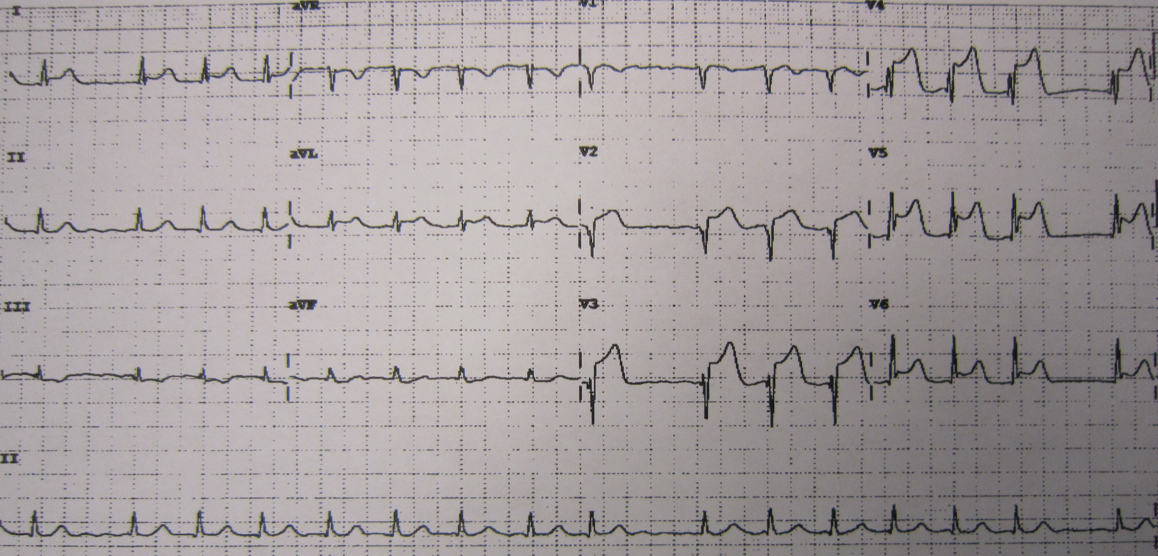
  • ST-Elevated LMI: ST elevation in lead I, aVL, V5, and V6; Reciprocal ST depression in inferior lead III and aVF
  • High lateral STEMI: High lateral STEMI can present as ST-elevation involving lead I and aVL. Subtle ST elevation in V5, V6 and reciprocal changes in lead III and avF may be present. This is usually caused by occlusion of the first diagonal branch of LAD and is sometimes referred to as the South African flag sign.
  • Old LMI presents with deep and broad Q waves I leads I and aVL
  • Inferolateral STEMI presents with ST-elevation involving lateral (I, aVL, V5, V6) and inferior leads (II, III, aVF): This is usually seen with occlusion of the proximal LCx artery.
  • Anterolateral STEMI presents with ST-elevation involving lateral (I, aVL, V5, V6) and anterior leads (V1, V2, V3): This is highly indicative of proximal LAD occlusion.

In patients with normal troponin and non-diagnostic ECG, a quick evaluation with an echocardiogram can be done. Echocardiogram has high sensitivity and low specificity when diagnosing MI.[9] Severe ischemia produces regional wall motion abnormalities (RWMA) which can be visualized on echocardiogram. However, it is difficult to differentiate acute ischemia from old infarct based on RWMA. Patients with normal echocardiogram but having moderate pre-test probability should be evaluated with a stress test.[10] Coronary angiogram remains the most definitive and diagnostic test of choice.

Treatment / Management

LMI presenting as STEMI should be treated immediately. Early reperfusion has shown benefits with improved clinical outcomes.[11] Percutaneous intervention (PCI) has demonstrated superior results when compared to fibrinolytic therapy.[12] ACC/AHA guidelines for STEMI management recommend early PCI with preferable door to balloon time of fewer than 90 minutes at PCI capable facility and less than 120 minutes at non-PCI capable facility.[13] Antiplatelet therapy with aspirin and either P2Y12 inhibitor (Clopidogrel, Ticagrelor, Prasugrel)  or Glycoprotein IIb/IIIa inhibitor is recommended before and after PCI. Multiple studies have demonstrated mortality benefits with beta-blockers (Metoprolol, Carvedilol, Bisoprolol) and high-intensity high potency statins (Rosuvastatin).[14] Patients presenting with cardiogenic shock or mitral valve rupture post-MI may need a mechanical supportive device such as an intra-aortic balloon pump (IABP) after PCI.

Additionally, symptomatic management includes the usage of Morphine, Oxygen (in patients with hypoxemia), and nitrates (nitroglycerin).

LMI patients presenting as NSTEMI should receive initial medical therapy with oxygen (if hypoxic, Saturation of oxygen <94%), beta-blockers (Carvedilol, Metoprolol, Bisoprolol), and a statin (Rosuvastatin or Atorvastatin). Antithrombotic therapy including antiplatelet medications (using aspirin and a P2Y12 inhibitor such as Ticagrelor, Clopidogrel or Prasugrel) and anticoagulants such as unfractionated heparin should be initiated as soon as possible. NSTEMI patients have also shown better outcomes with early reperfusion strategy.[15] Contrary to STEMI fibrinolysis has no role in the management of NSTEMI.[16]

All patients with LMI should be placed on life-long therapy with aspirin, beta-blockers, and high intensity-high potency statins.

Differential Diagnosis

Patients presenting with chest pain will have the following in the differential diagnosis:

  • Acute pericarditis
  • Acute gastritis
  • Peptic ulcer disease
  • Aortic dissection
  • Esophagitis
  • Cardiac tamponade
  • Costochondritis
  • Myocarditis
  • Pneumonia
  • Pneumothorax
  • Prinzmetal’s angina
  • Pulmonary embolism
  • Cocaine-induced vasospasm

Prognosis

LMI has overall favorable outcomes. Long-term outcomes in patients with STEMI and NSTEMI have improved over the last decade.[17] Hreybe et al. reported marginally elevated risk of ventricular fibrillation in patients presenting with an anterior or lateral MI.[18]

Prognosis tends to be worse in patients presenting with life-threatening complications such as arrhythmias, sudden cardiac arrest, free wall/papillary muscle rupture and cardiogenic shock. Risk stratification using TIMI scores can help identify patients with increased in-hospital mortality.[19] Thirty-day mortality rates for STEMI are 13% with medical therapy alone, 6-7% with optimal fibrinolytic therapy, and 3% to 5% with primary percutaneous coronary intervention when performed within 2 hours of hospital arrival. There is an even lower risk of death of only 2% in patients with NSTEMI after 30 days, as compared to STEMI.

Complications

  1. Left ventricular free wall rupture
  2. Pericarditis (Fibrinous)
  3. Dressler Syndrome
  4. Ventricular aneurysm
  5. Acute mitral regurgitation
  6. Left ventricular thrombus
  7. Congestive heart failure
  8. Ventricular fibrillation
  9. Ventricular tachycardia
  10. Sudden cardiac arrest
  11. Cardiogenic shock
  12. PCI related complications

Deterrence and Patient Education

Patient education is the cornerstone of preventing future MI events. Interventions such as smoking cessation, weight reduction, medication adherence, and dietary modification have all been well established in improving overall cardiovascular mortality.[20][21] Targeting strict blood pressure, glycemic, and lipid goals can help decrease the risk of complications after MI. Patients should be encouraged to participate in cardiac rehabilitation programs to improve overall cardiac function and achieve better heart rate control.

Enhancing Healthcare Team Outcomes

An interprofessional approach has been shown to improve health care outcomes in patients admitted with MI.[22][23] Adherence to guideline-directed therapy is equally important. Establishing a cardiac alert response team for emergencies can help provide timely dedicated care.


(Click Image to Enlarge)
An ECG showing pardee waves indicating acute myocardial infarction in the inferior leads II, III and aVF with reciprocal changes in the anterolateral leads
An ECG showing pardee waves indicating acute myocardial infarction in the inferior leads II, III and aVF with reciprocal changes in the anterolateral leads.
Contributed by Wikimedia Commons, Glenlarson (Public Domain-Self)

(Click Image to Enlarge)
Inferior wall MI
Inferior wall MI
Contributed by Steven Mountfort

(Click Image to Enlarge)
ECG- Left Bundle Branch Block and inferior wall MI
Contributed by Steven Mountfort
Details

Editor:

Nagendra Gupta

Updated:

1/28/2023 5:34:19 AM

References


[1]

Thygesen K,Alpert JS,Jaffe AS,Chaitman BR,Bax JJ,Morrow DA,White HD, Fourth universal definition of myocardial infarction (2018). European heart journal. 2018 Aug 25     [PubMed PMID: 30165617]


[2]

Pasupathy S,Air T,Dreyer RP,Tavella R,Beltrame JF, Systematic review of patients presenting with suspected myocardial infarction and nonobstructive coronary arteries. Circulation. 2015 Mar 10     [PubMed PMID: 25587100]

Level 1 (high-level) evidence

[3]

Roger VL,Weston SA,Gerber Y,Killian JM,Dunlay SM,Jaffe AS,Bell MR,Kors J,Yawn BP,Jacobsen SJ, Trends in incidence, severity, and outcome of hospitalized myocardial infarction. Circulation. 2010 Feb 23     [PubMed PMID: 20142444]


[4]

Newman JD,Shimbo D,Baggett C,Liu X,Crow R,Abraham JM,Loehr LR,Wruck LM,Folsom AR,Rosamond WD, Trends in myocardial infarction rates and case fatality by anatomical location in four United States communities, 1987 to 2008 (from the Atherosclerosis Risk in Communities Study). The American journal of cardiology. 2013 Dec 1     [PubMed PMID: 24063834]

Level 3 (low-level) evidence

[5]

Tibaut M,Mekis D,Petrovic D, Pathophysiology of Myocardial Infarction and Acute Management Strategies. Cardiovascular     [PubMed PMID: 27993119]


[6]

Arenja N,Mueller C,Ehl NF,Brinkert M,Roost K,Reichlin T,Sou SM,Hochgruber T,Osswald S,Zellweger MJ, Prevalence, extent, and independent predictors of silent myocardial infarction. The American journal of medicine. 2013 Jun     [PubMed PMID: 23597799]


[7]

Esmat S,Abdel-Halim MR,Fawzy MM,Nassef S,Esmat S,Ramzy T,El Fouly ES, Are normolipidaemic patients with xanthelasma prone to atherosclerosis? Clinical and experimental dermatology. 2015 Jun     [PubMed PMID: 25683563]


[8]

Bose A,Jain V,Kawthekar G,Chhabra C,Hemvani N,Chitnis DS, The Importance of Serial Time Point Quantitative Assessment of Cardiac Troponin I in the Diagnosis of Acute Myocardial Damage. Indian journal of critical care medicine : peer-reviewed, official publication of Indian Society of Critical Care Medicine. 2018 Sep     [PubMed PMID: 30294127]


[9]

Sabia P,Afrookteh A,Touchstone DA,Keller MW,Esquivel L,Kaul S, Value of regional wall motion abnormality in the emergency room diagnosis of acute myocardial infarction. A prospective study using two-dimensional echocardiography. Circulation. 1991 Sep     [PubMed PMID: 1884510]


[10]

Fihn SD,Gardin JM,Abrams J,Berra K,Blankenship JC,Dallas AP,Douglas PS,Foody JM,Gerber TC,Hinderliter AL,King SB 3rd,Kligfield PD,Krumholz HM,Kwong RY,Lim MJ,Linderbaum JA,Mack MJ,Munger MA,Prager RL,Sabik JF,Shaw LJ,Sikkema JD,Smith CR Jr,Smith SC Jr,Spertus JA,Williams SV, 2012 ACCF/AHA/ACP/AATS/PCNA/SCAI/STS guideline for the diagnosis and management of patients with stable ischemic heart disease: executive summary: a report of the American College of Cardiology Foundation/American Heart Association task force on practice guidelines, and the American College of Physicians, American Association for Thoracic Surgery, Preventive Cardiovascular Nurses Association, Society for Cardiovascular Angiography and Interventions, and Society of Thoracic Surgeons. Circulation. 2012 Dec 18     [PubMed PMID: 23166210]

Level 1 (high-level) evidence

[11]

Anderson JL,Karagounis LA,Califf RM, Metaanalysis of five reported studies on the relation of early coronary patency grades with mortality and outcomes after acute myocardial infarction. The American journal of cardiology. 1996 Jul 1     [PubMed PMID: 8712096]


[12]

Keeley EC,Boura JA,Grines CL, Primary angioplasty versus intravenous thrombolytic therapy for acute myocardial infarction: a quantitative review of 23 randomised trials. Lancet (London, England). 2003 Jan 4     [PubMed PMID: 12517460]

Level 1 (high-level) evidence

[13]

O'Gara PT,Kushner FG,Ascheim DD,Casey DE Jr,Chung MK,de Lemos JA,Ettinger SM,Fang JC,Fesmire FM,Franklin BA,Granger CB,Krumholz HM,Linderbaum JA,Morrow DA,Newby LK,Ornato JP,Ou N,Radford MJ,Tamis-Holland JE,Tommaso CL,Tracy CM,Woo YJ,Zhao DX,Anderson JL,Jacobs AK,Halperin JL,Albert NM,Brindis RG,Creager MA,DeMets D,Guyton RA,Hochman JS,Kovacs RJ,Kushner FG,Ohman EM,Stevenson WG,Yancy CW, 2013 ACCF/AHA guideline for the management of ST-elevation myocardial infarction: a report of the American College of Cardiology Foundation/American Heart Association Task Force on Practice Guidelines. Circulation. 2013 Jan 29     [PubMed PMID: 23247304]

Level 3 (low-level) evidence

[14]

O'Gara PT,Kushner FG,Ascheim DD,Casey DE Jr,Chung MK,de Lemos JA,Ettinger SM,Fang JC,Fesmire FM,Franklin BA,Granger CB,Krumholz HM,Linderbaum JA,Morrow DA,Newby LK,Ornato JP,Ou N,Radford MJ,Tamis-Holland JE,Tommaso JE,Tracy CM,Woo YJ,Zhao DX, 2013 ACCF/AHA guideline for the management of ST-elevation myocardial infarction: executive summary: a report of the American College of Cardiology Foundation/American Heart Association Task Force on Practice Guidelines. Circulation. 2013 Jan 29     [PubMed PMID: 23247303]

Level 3 (low-level) evidence

[15]

Shetabi K,Ullah R,Patel R,Wilson T,Siddiqua T,Olet S,Ammar KA,Jahangir A,Allaqaband SQ,Bajwa T,Jan MF, Contemporary practice pattern of revascularization in a large tertiary care referral center in non-ST elevation myocardial infarction: A propensity-matched 10-year experience. Catheterization and cardiovascular interventions : official journal of the Society for Cardiac Angiography     [PubMed PMID: 30265421]


[16]

Amsterdam EA,Wenger NK,Brindis RG,Casey DE Jr,Ganiats TG,Holmes DR Jr,Jaffe AS,Jneid H,Kelly RF,Kontos MC,Levine GN,Liebson PR,Mukherjee D,Peterson ED,Sabatine MS,Smalling RW,Zieman SJ, 2014 AHA/ACC guideline for the management of patients with non-ST-elevation acute coronary syndromes: executive summary: a report of the American College of Cardiology/American Heart Association Task Force on Practice Guidelines. Circulation. 2014 Dec 23     [PubMed PMID: 25249586]

Level 3 (low-level) evidence

[17]

McManus DD,Gore J,Yarzebski J,Spencer F,Lessard D,Goldberg RJ, Recent trends in the incidence, treatment, and outcomes of patients with STEMI and NSTEMI. The American journal of medicine. 2011 Jan     [PubMed PMID: 21187184]


[18]

Hreybe H,Saba S, Location of acute myocardial infarction and associated arrhythmias and outcome. Clinical cardiology. 2009 May     [PubMed PMID: 19452487]


[19]

Morrow DA,Antman EM,Parsons L,de Lemos JA,Cannon CP,Giugliano RP,McCabe CH,Barron HV,Braunwald E, Application of the TIMI risk score for ST-elevation MI in the National Registry of Myocardial Infarction 3. JAMA. 2001 Sep 19     [PubMed PMID: 11560541]


[20]

Wing RR,Lang W,Wadden TA,Safford M,Knowler WC,Bertoni AG,Hill JO,Brancati FL,Peters A,Wagenknecht L,Look AHEAD Research Group., Benefits of modest weight loss in improving cardiovascular risk factors in overweight and obese individuals with type 2 diabetes. Diabetes care. 2011 Jul     [PubMed PMID: 21593294]


[21]

Choi YJ,Park JS,Kim U,Lee SH,Son JW,Shin DG,Kim YJ,Jeong MH,Ahn YK,Cho MC,Bae JW,Kim CJ,Cho JM,Han KR,Lee JH, Changes in smoking behavior and adherence to preventive guidelines among smokers after a heart attack. Journal of geriatric cardiology : JGC. 2013 Jun     [PubMed PMID: 23888174]


[22]

Coons JC,Fera T, Multidisciplinary team for enhancing care for patients with acute myocardial infarction or heart failure. American journal of health-system pharmacy : AJHP : official journal of the American Society of Health-System Pharmacists. 2007 Jun     [PubMed PMID: 17563049]


[23]

Fanari Z,Barekatain A,Kerzner R,Hammami S,Weintraub WS,Maheshwari V, Impact of a Multidisciplinary Team Approach Including an Intensivist on the Outcomes of Critically Ill Patients in the Cardiac Care Unit. Mayo Clinic proceedings. 2016 Dec     [PubMed PMID: 28126152]