Continuing Education Activity
Mediastinoscopy is a critical surgical procedure primarily used to diagnose and stage diseases within the mediastinum, including bronchogenic carcinoma and other mediastinal pathologies. This procedure is traditionally regarded as the gold standard for staging mediastinal lymph nodes, providing essential information for the diagnosis and treatment planning of non–small cell lung cancer. The procedure involves general anesthesia, precise dissection, and careful navigation to obtain tissue samples. The procedure continues to evolve with advancements such as video-assisted mediastinoscopy and complementary techniques like endobronchial ultrasound-guided fine-needle aspiration, enhancing surgical visualization and diagnostic accuracy.
Clinicians participating in this course gain a comprehensive understanding of mediastinoscopy, including its indications, procedural techniques, and potential complications. The course covers preoperative and postoperative care, patient selection, and integrating advanced imaging techniques to improve patient outcomes. Additionally, participants learn about the importance of interprofessional communication and care coordination, enhancing their ability to work effectively within a multidisciplinary team. This knowledge provides clinicians with the skills needed to optimize patient safety, improve diagnostic accuracy, and deliver high-quality, patient-centered care in the context of mediastinoscopy.
Objectives:
Identify patients who would benefit from mediastinoscopy based on clinical presentations and diagnostic imaging.
Screen for potential contraindications and risks associated with mediastinoscopy in high-risk patients.
Differentiate between mediastinoscopy and other diagnostic procedures for mediastinal assessment.
Collaborate with multidisciplinary teams to ensure comprehensive patient care and optimal outcomes.
Introduction
Mediastinoscopy is a crucial diagnostic and staging procedure used primarily in managing lung cancer. At the time of diagnosis, approximately 75% of patients with lung cancer present with either locally advanced or metastatic disease, underscoring the importance of precise staging for effective treatment planning. Mediastinoscopy involves making a small incision at the base of the neck to insert a mediastinoscope, which allows for direct visualization and biopsy of the mediastinal lymph nodes. This invasive technique offers an 80% to 95% sensitivity rate for detecting tumor cells in these lymph nodes. However, its specificity rate of 91% to 95% reflects limitations in accessing certain lymph node stations, such as the paraesophageal, aortopulmonary, and lower pulmonary ligament nodes.[1]
Mediastinoscopy can be categorized into 2 types: cervical mediastinoscopy and transthoracic mediastinoscopy. Cervical mediastinoscopy is a more commonly performed procedure that provides access to the pretracheal, paratracheal, and anterior subcarinal lymph nodes. Transthoracic mediastinoscopy, also known as the Chamberlain procedure or anterior mediastinotomy, is a more involved procedure that allows for the dissection of the aortopulmonary lymph nodes.
The history of mediastinal surgery dates back to 1899 when a superior mediastinal abscess was successfully drained. However, it wasn't until the late 1950s, with the introduction of the mediastinoscope by Eric Carlens of Sweden, that the procedure became widely used outside Europe. This innovation allowed for more precise biopsies of the paratracheal and hilar lymph nodes, significantly improving the accuracy of lung cancer staging and, thus, the overall management of thoracic oncologic diseases.[2]
Anatomy and Physiology
Understanding mediastinoscopy requires knowledge of the mediastinum's anatomy. The mediastinum is the central region of the chest, located between the 2 pleural cavities and extending from the thoracic inlet to the diaphragm. Several vital organs, vessels, and nerves, including the heart, great vessels, trachea, esophagus, phrenic and vagus nerves, thymus, and lymph nodes, are contained within the mediastinum.
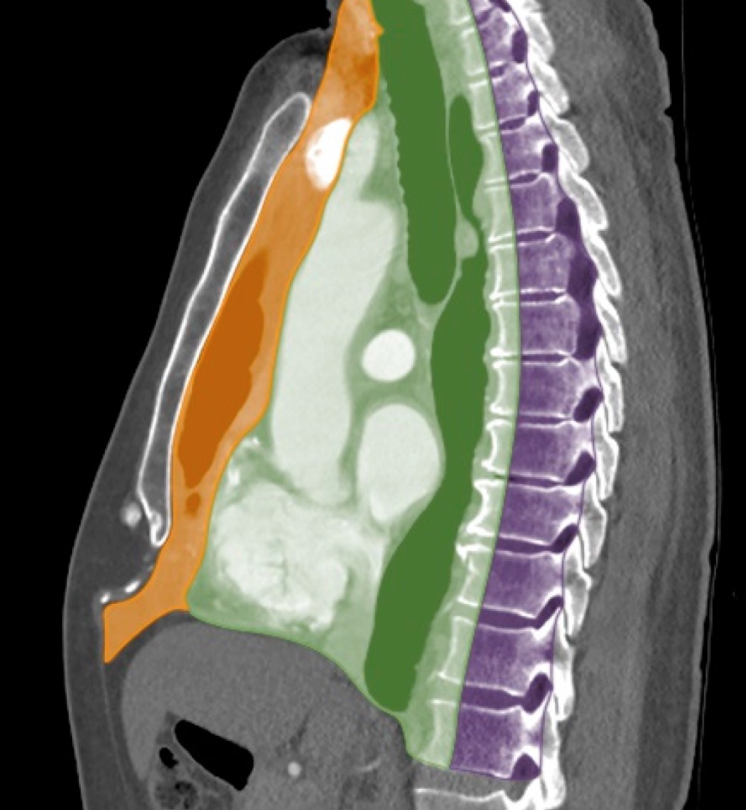
Traditionally, the mediastinum is segmented into 4 compartments based on their relationship to the pericardium: superior, anterior, middle, and posterior. A more contemporary classification, utilizing boundaries defined by computed tomography (CT), identifies the prevascular (anterior), visceral (middle), and paravertebral (posterior) compartments. The prevascular (anterior) compartment includes the thymus, lymph nodes, and the left brachiocephalic vein. The visceral (middle) compartment comprises various vascular structures such as the heart, thoracic aorta, intrapericardial pulmonary arteries, thoracic duct, superior vena cava, trachea, esophagus, and lymph nodes. The paravertebral (posterior) compartment contains the thoracic spine and adjacent soft tissue (see Image. Mediastinum, Computed Tomography Scan).[3][4]
Indications
The primary clinical indications for a mediastinoscopy are:
- Evaluation of lymph node involvement in patients with carcinoma of the lung
- Tissue biopsy of suspected tumors
- Removal of mediastinal masses and enlarged lymph nodes
Mediastinoscopy allows access to mediastinal lymph node groups, proximal hilar lymph nodes, and superior and anterior subcarinal lymph nodes. This procedure is invaluable for diagnosing mediastinal conditions that may present with mediastinal masses, such as tuberculosis, sarcoidosis, histoplasmosis, coccidioidomycosis, and various cancers and tumors (lymphoma, thyroid and parathyroid, esophageal, neurogenic tumors, and germ cell tumors).[3] Additionally, mediastinoscopy is crucial in managing lung cancer by providing tissue to diagnose and stage the disease, essential for determining the appropriate treatment. Notably, there are also vascular conditions that can present with a mediastinal mass like aneurysms and aberrant vessels (eg, persistent left superior vena cava [SVC], anomalous left pulmonary artery) as well as benign conditions such as developmental cysts.[4] Neither requires mediastinoscopy but must be on the differential diagnosis list for any mediastinal mass for which it may be considered.
Contraindications
Contraindications to mediastinoscopy can be classified as either absolute or relative.[5][6] Absolute contraindications include:
- Anterior mediastinal mass
- Inoperable tumor
- Previous recurrent laryngeal nerve injury
- Extremely debilitated patients
- Ascending aortic aneurysm
- Previous mediastinoscopy
- This is a contraindication for a repeat procedure because scar tissue eliminates and distorts the dissection plane.
Relative contraindications include:
- Thoracic inlet obstruction
- SVC syndrome
- Severe tracheal deviation
- History of radiation therapy to the chest
Anything that distorts the anatomy, such as those contraindications listed above, increases the risk of vascular or airway injury, which can be catastrophic in this location.[7]
Equipment
Equipment needed to perform a mediastinoscopy includes:
- A mediastinoscope
- Fiberoptic light cable
- Biopsy forceps
A cardiothoracic setup should be on standby, and an anesthesia machine is needed to perform the procedure if done under general anesthesia.
Personnel
Mediastinoscopy is a complex surgical procedure with several key components and requires a coordinated team effort for successful execution. The following personnel is needed:
- Surgeon
- A cardiothoracic or general surgeon with specialized training in mediastinoscopy is essential. The surgeon must also be capable of performing emergency procedures like thoracotomy and sternotomy if complications arise during the mediastinoscopy.
- Anesthesiologist
- General anesthesia is required for the procedure. An anesthesiologist is responsible for administering anesthesia, monitoring the patient’s vital signs, and managing the airway throughout the surgery.
- Operating room support staff
- The support staff includes surgical nurses and technicians who assist with setting up, sterilizing instruments, and providing intraoperative support.
- Pathologist
- A pathologist is required for the histopathological assessment of the biopsy specimen obtained during the mediastinoscopy. The pathologist examines the tissue to diagnose conditions and assess for malignancies.
- Laboratory personnel
- Lab personnel must analyze molecular markers and perform other diagnostic tests on the biopsy specimens to identify specific characteristics of malignancies, aiding in the precise diagnosis and treatment planning.
Preparation
A preoperative evaluation of a patient undergoing mediastinoscopy is crucial for reducing morbidity and mortality. Particular attention should be given to respiratory symptoms such as wheezing, dyspnea, and orthopnea. These symptoms should be assessed to see if they worsen with exercise or when lying flat, as this may indicate airway obstruction caused by a mediastinal mass.
The preoperative workup should include the following:
- Chest radiograph:
- Obtain posteroanterior and lateral views to assess the overall condition of the chest.
- CT scan:
- Perform a chest and neck CT scan to provide detailed images of the mediastinum and surrounding structures. A review of CT imaging can be constructive in evaluating any compression or deviation of the great vessels.
- Pulmonary function tests:
- Conduct pulmonary function tests with flow-volume loops to evaluate respiratory function.
- Patients with suspected airway obstruction due to a mediastinal mass on physical examination should undergo pulmonary function tests (PFTs) with flow-volume loops. The tests should be performed in both the upright and supine positions. If the PFTs show obstruction while supine during inhalation, the patient's mass is extrathoracic; if the obstruction occurs during expiration, the patient's mass is intrathoracic.
- Additional studies:
- If tracheal deviation is suspected, specific studies such as neck films or tomograms should be conducted to assess the location and extent of the mass and the degree of airway compromise.
By thoroughly evaluating these factors, healthcare providers can better prepare for the mediastinoscopy and manage any potential complications that may arise during the procedure. An intravenous line must be established before proceeding to the operating room. In patients with a large mediastinal mass, SVC syndrome may be present. Obstruction of the superior vena cava causes facial swelling, neck distension, and collateral venous drainage along the thoracic wall; this is commonly associated with dyspnea and dysphagia. Diagnostically, it can be seen on imaging, and a positive Pemberton sign also characterizes it. A positive Pemberton sign occurs when a patient lifts both arms over their head in the upright position, causing marked facial plethora to occur, indicating profound compression of the jugular veins. These patients need intravenous access in the lower extremities because of the resistance to recirculating upper extremity drainage.[2][8][9][10]
Mediastinoscopy can be performed under local or general anesthesia. General anesthesia is preferred at most institutions if the patient shows no preoperative signs or symptoms of airway obstruction.[2][5] The general anesthetic technique typically involves a combination of inhalational and intravenous agents.
After induction and intubation, muscle relaxants ensure a stable operative field, minimizing the risk of sudden movements that could injure adjacent organs with the mediastinoscope. If the preoperative workup indicates airway compromise, the patient may undergo an awake fiberoptic intubation technique, allowing spontaneous breathing during intubation. A standard single-lumen endotracheal tube is used unless contraindications require 1-lung ventilation. In such cases, a large single-lumen tube (7.5 mm or above) is used to facilitate the passage of a bronchial blocker or a double-lumen endotracheal tube.
Following intubation, another large-bore intravenous line and an arterial catheter establish additional vascular access. The arterial catheter allows for continuous beat-to-beat blood pressure monitoring, which is crucial for detecting vascular compression or massive bleeding during the procedure and for regular checks of arterial blood gases. Constant palpation of the right radial or carotid pulse, placement of a right radial arterial line, or continuous plethysmographic monitoring via a pulse oximeter is recommended to detect innominate artery compression by the mediastinoscope. Once the patient is sedated and intubated, the anterior chest wall is prepped for a median sternotomy in case a vascular injury occurs.
In certain situations, mediastinoscopy is indicated despite an anterior mediastinal mass. Surgeons, anesthesiologists, and operating room staff must be keenly aware of the potential complications associated with such masses. Key considerations include managing the airway, as patients may be unable to lie flat due to airway obstruction by the mass, complicating endotracheal intubation. Even with successful intubation and proper airway placement, the airway may still be compromised distally by the mass after muscle paralysis is induced. Therefore, avoiding muscle relaxants in high-risk patients, intubating them while maintaining spontaneous respiration, and using a fiberoptic bronchoscope or direct/indirect laryngoscopy following mask induction is recommended.
An otorhinolaryngology (ENT) surgeon skilled in rigid bronchoscopy should be present to manage potential obstructions that cannot be bypassed with flexible bronchoscopy. Additionally, changing the patient's position from supine to either lateral or prone may temporarily relieve airway obstruction caused by the mass. In cases involving a large anterior mediastinal mass with a risk of cardiovascular collapse upon induction of anesthesia, it is crucial to have cardiopulmonary bypass readily available as a precautionary measure. Taking these precautions and being prepared for potential complications can minimize the risks associated with mediastinoscopy in the presence of an anterior mediastinal mass, ensuring safer outcomes for the patient.
Technique or Treatment
Mediastinoscopy is usually performed in the operating room under general anesthesia. Unless an anterior mediastinal mass causes airway compromise when lying flat, the patient is typically placed supine for the procedure. In patients without such a mass, the procedure begins with intravenous induction. Following intubation, the patient's head is extended and turned to the side. A 3-cm incision is made 2 cm above the suprasternal notch between the anterior borders of the sternocleidomastoid muscles.
The superficial fascia of the neck, deep cervical fascia, and pretracheal fascia are dissected to reach the trachea. Blunt dissection continues down to the anterior trachea, allowing access into the superior mediastinum. The tract is developed through blunt dissection, extending between the anterior trachea and the posterior aspect of the great vessels. A mediastinoscope is then inserted and advanced along this tract, enabling the surgeon to sample lymph nodes and masses within the superior mediastinum.[2]
Complications
While mediastinoscopy is generally safe, the morbidity associated with the procedure is reported to be between 1.5% and 3%, with an overall mortality rate of 0.09%.[11] The procedure is associated with complications that range from minor issues to severe, life-threatening events. The main complications include:
- Bleeding
- Minor
- This can occur at the incision site or during tissue biopsy.
- This is usually controlled with pressure or minor interventions.
- Major
- Injury to major vessels such as the innominate artery, aorta, or pulmonary arteries can lead to significant bleeding, requiring immediate intervention, potentially including sternotomy for control and repair.
- Infection
- Wound Infection
- Infection at the incision site can occur, necessitating antibiotic treatment and possibly surgical drainage.
- Mediastinitis
- Although rare, infection within the mediastinum can be a serious complication requiring aggressive antibiotic treatment and possibly surgical intervention.
- Pneumothorax
- Accidentally entering the pleural space can cause a pneumothorax (collapsed lung), which might require chest tube placement to reexpand the lung.
- Tracheal or esophageal injury
- Blunt or sharp dissection can result in perforation of the trachea or esophagus, leading to air leaks, mediastinal infections, and the need for surgical repair.
- Recurrent laryngeal nerve injury
- Recurrent laryngeal nerve damage can result in vocal cord paralysis, hoarseness, or respiratory difficulties, which may be temporary or permanent.
- Vascular compression
- The mediastinoscope can compress major vessels, such as the innominate artery, reducing blood flow to the brain and potentially causing neurological symptoms and stroke. Continuous monitoring of arterial pressure and pulse oximetry is essential to detect this complication early.
- Air embolism
- Entry of air into the vascular system during the procedure can lead to air embolism, a rare but potentially fatal complication.
- Chylothorax
- Injury to the thoracic duct can lead to chyle leakage into the chest cavity, resulting in chylothorax, which requires dietary modifications, drainage, and sometimes surgical repair.
- Anesthesia-related complications
- As with any procedure requiring general anesthesia, there is a risk of complications such as adverse reactions to anesthetic agents, respiratory depression, and cardiovascular instability.
Given these potential complications, mediastinoscopy should be performed by experienced surgeons in a well-equipped operating room, with appropriate preoperative planning and intraoperative monitoring to minimize risks.
Clinical Significance
Mediastinoscopy, traditionally regarded as the gold standard for staging mediastinal lymph nodes, remains a crucial clinical procedure for diagnosing disease pathologies in the mediastinum, most notably bronchogenic carcinoma. Along with endobronchial ultrasound-guided (EBUS) fine-needle aspiration (FNA), it plays a vital role in staging non–small cell lung cancer. Patients are typically recommended to undergo an EBUS biopsy before induction therapy and then mediastinoscopy following therapy. Video-assisted mediastinoscopy is increasingly used to enhance surgical visualization and allow the use of multiple instruments simultaneously. Despite the rise of EBUS-FNA, which has shown high sensitivity and specificity for diagnosing nodal metastases, mediastinoscopy is essential, especially when combined with additional techniques such as intranodal forceps biopsy and esophageal ultrasonography.[12]
Patient education and coordinated care are paramount in mediastinoscopy cases, particularly given the close association with lung cancer diagnoses. Ensuring that patients, clinicians, and care staff are well-informed about the procedure is critical for optimizing patient outcomes. The number of both mediastinoscopy and EBUS procedures is expected to increase in the future, and a single institution study has shown that mediastinoscopy is marginally less expensive than EBUS biopsy.[13][14]
Enhancing Healthcare Team Outcomes
Effectively managing mediastinoscopy involves a multidisciplinary team approach to enhance patient-centered care, outcomes, patient safety, and team performance. Physicians, advanced practitioners, nurses, pharmacists, and other health professionals must possess a high skill level and strategic planning. Surgeons require specialized training in mediastinoscopy techniques, while anesthesiologists must be adept at managing airways in complex situations, especially when dealing with anterior mediastinal masses. Advanced practitioners and nurses play crucial roles in preoperative and postoperative care, ensuring patients are well-prepared for the procedure and recover safely. Pharmacists contribute by managing medications, ensuring proper anesthesia, and preventing drug interactions.
Interprofessional communication and care coordination are vital to the success of mediastinoscopy. Regular multidisciplinary meetings and case discussions help align the team's strategy and address potential complications proactively. Clear communication among team members ensures that everyone knows their roles and responsibilities, facilitating a smooth workflow. Educating patients and their families about what to expect before, during, and after the procedure enhances patient-centered care. By working collaboratively and maintaining open lines of communication, the healthcare team can optimize patient outcomes, minimize risks, and improve overall team performance.