Neurofibromatosis Type 1

Earn CME/CE in your profession:


Continuing Education Activity

Neurofibromatosis-1 (NF-1), or Von Recklinghausen disease, is one of the inheritable neurocutaneous disorders manifested by developmental changes in the nervous system, bones, and skin. It is an autosomal dominant disorder. Additionally, it is the most common amongst all the hamartoma neoplastic syndromes, including tuberous sclerosis, Gardner, and Cowden syndromes. Other types of neurofibromatosis are Neurofibromatosis type 2 (NF2) and schwannomatosis. Cafe-au-lait macules and neurofibromas are the distinguishing features of NF1. NF2 can have similar cutaneous manifestations as that of NF1. But the hallmark manifestations of NF2 include schwannoma, meningioma, and ependymoma. This activity describes the clinical presentation and management of NF-1 and highlights the interprofessional team's role in managing patients with this disease.

Objectives:

  • Discuss the pathophysiology of neurofibromatosis-1.
  • Describe the appropriate examination and workup of neurofibromatosis-1.
  • Review the management of patients with neurofibromatosis-1.
  • Summarize how interprofessional coordinated activity and information sharing can improve patient care and outcomes.

Introduction

Neurofibromatosis type 1 (NF-1) or Von Recklinghausen disease is one of the inheritable neurocutaneous disorders that also harbinger the risk for bone abnormalities, vasculopathy, and cognitive impairment.[1] It is an autosomal dominant disorder.[2] Moreover, it is the most common hamartoma neoplastic syndrome, such as tuberous sclerosis, Gardner, and Cowden syndromes. Other types of neurofibromatosis are Neurofibromatosis type 2 (NF2) and schwannomatosis.[3] Cafe-au-lait macules and neurofibromas are the distinguishing features of NF1. NF2 can have similar cutaneous manifestations as that of NF1. But the hallmark manifestations of NF2 include schwannoma, meningioma, and ependymoma.[4]

Etiology

NF-1 is an autosomal dominant disorder. The gene for NF-1 is located on chromosome 17 and encodes a gene product called neurofibromin.[5][6] Neurofibromin is widely expressed in a variety of tissues. Neurofibromin, a GTPase-activating protein, normally inhibits the rat sarcoma (RAS) pathway.[7]

The mutation or deletion of this NF-1 gene results in the phenotypic and genotypic manifestations of the disorder. Penetrance is complete, as it is an autosomal dominant disorder. All generations are involved with no skipping. Expression of the disease differs from one affected family to another and between individuals of the same family.[8]

Epidemiology

NF-1 is an autosomal-dominant genetic disorder that affects all the offspring of an affected individual. The incidence of NF-1 is noted to be 1 in 2600 to 3000 individuals. There is no predilection for the male or female gender.[9] Fifty percent of the presentations are familial, while the rest of the cases are sporadic. Paternally derived chromosomes are the source of sporadic mutations.[10] The risk of these sporadic mutations is high with advancing paternal age. The incidence of the so-called segmental NF-1, a form that predominantly has skin manifestations, is 1 in 36,000 to 40,000 individuals.[11]

Pathophysiology

Mutations of the NF-1 gene result in a lack of expression of neurofibromin, thereby promoting tumorigenesis. Neurofibromas develop when both NF-1 alleles get mutated. Neurofibromas embody Schwann cells, perineural cells, mast cells, and fibroblasts.[12] Neurofibromas affect the skin stem from peripheral nerves and their supporting structures, including neurilemmal cells. The fibroblasts in these neurofibromas are derived from factor XIIIa connective tissue cells, which are HLA-DR-positive in peripheral nerves. Dermal fibroblasts secrete increased amounts of hepatocyte growth factor and stem cell factor, contributing to increased melanin deposition in the epidermis.[13] Café-au-lait spots sometimes contain giant pigment granules found in epidermal cells and melanocytes.

There is a high genotype-phenotype correlation observed in NF1.[7] Cutaneous neurofibromas (cNF) involve dermal nerve terminals, whereas plexiform neurofibromas (pNF) characteristically affect the nerve plexuses and fascicles.[7]

Neurofibroma originates from the Schwann cell (SC) lineage.[7]

Neural crest stem cells (NCSCs) form

1. Boundary cap (BC) cells at the motor exit point (MEP) and the dorsal root entry zone (DREZ) that express:

  • Krox20, form SC components of the dorsal and ventral nerve roots and helps in early myelination of the PNS
  • Prss56 forms the SC component in the nerve roots, hypodermis, and dermis.

2. Schwann cell precursors (SCPs) that

  • Have specific glial genes and molecular markers, such as myelin protein 0 (P0), cadherin-19
  • Depending on axon-associated signals, for transition to myelinated or unmyelinated states.[7]

Neurofibromin normally suppresses guanosine triphosphate (GTP)-bound Kirsten rat sarcoma virus (KRAS)

The loss of this function results in the activation of the following:

  • Mitogen-activated protein kinases (MAPKs) and extracellular signal-regulated kinases 1 and 2 (ERK1 and ERK2), and
  • Phosphoinositide 3 kinases (PI3K)-mammalian target of rapamycin (mTOR) pathway causing transcription and cellular proliferation.[14]

Biallelic inactivation of NF1 in SOX10 cells of the SC lineage form neurofibroma.[7]

The pNF arises from the embryonic SC lineage, whereas cNF comes from the mature cell type in the SC lineage.

Loss of contact between SCs and axons and high Ras-Raf-ERK-dependent downregulation of semaphorin 4F (Sema4F) trigger increased proliferation. SC hyperplasia cause increased local innervation with the sprouting of nerve terminals within the upper dermis secondary to the lack of perineurium.[7]

The patterns of mutation involved in the pathogenesis include:

  • Frameshift mutations
  • Nonsense mutations
  • Missense mutations
  • Splice-site mutations, and
  • Large deletions.[15]

The Syn domain is most commonly affected (80%) in the pathogenesis of NF1.[15]

Histopathology

Histopathology of the lesions commonly seen in NF-1 is discussed below:

  • Neurofibroma: These are well-circumscribed spindle cell proliferation, but they rarely are encapsulated. They have a mucinous background and many mast cells. Spindle cells have a wavy appearance. As neurofibromas result from a proliferation of all supporting elements of neural fibers, they may have Schwann cells, perineural cells, and blood vessels. Axon stains show the random distribution of individual axons in neurofibroma. Collagen fibers are interspersed among the spindle cells. Schwann cell is considered the main tumor cell, where both NF-1 alleles are lacking.[16]
  • Plexiform neurofibroma: They are thought to be the pathognomonic sign of the disease. Histologically, they show numerous elongated nerve bundles with an abundant myxoid matrix. They are often mixed with diffuse cutaneous neurofibromas involving the dermis and subcutaneous fat.[17]
  • Cafe-au-lait macules: Histologically, there is an increase in the melanin content of basal keratinocytes and melanocytes. There is also an increase in the number of melanocytes, along with an increase in the amount of melanin. Giant melanosomes or macro melanosomes have been reported.[13]

History and Physical

The course of NF-1 varies considerably in various patients, but most have a benign course of the disease without developing major complications. The variability in presentation appears to be at least partially genetically determined and is unrelated to the unaffected allele. If the clinical presentation is limited to one part of the body, it is called segmental NF-1 (neurofibromatosis type 5) due to mosaic NF-1 gene mutation.[18] If the presentation involves the whole body, it is called generalized NF-1. Cafe-au-lait macules, inguinal and/or axillary freckling, Lisch nodules, and neurofibromas are the initial clinical manifestations of NF-1.[19] Osseous dysplasia and optic glioma usually appear around 1 to 3 years of age. Tumors, both benign and malignant, occur at an increased rate throughout life.[20] Patients with NF-1 may present with the following:

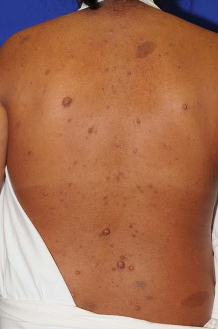
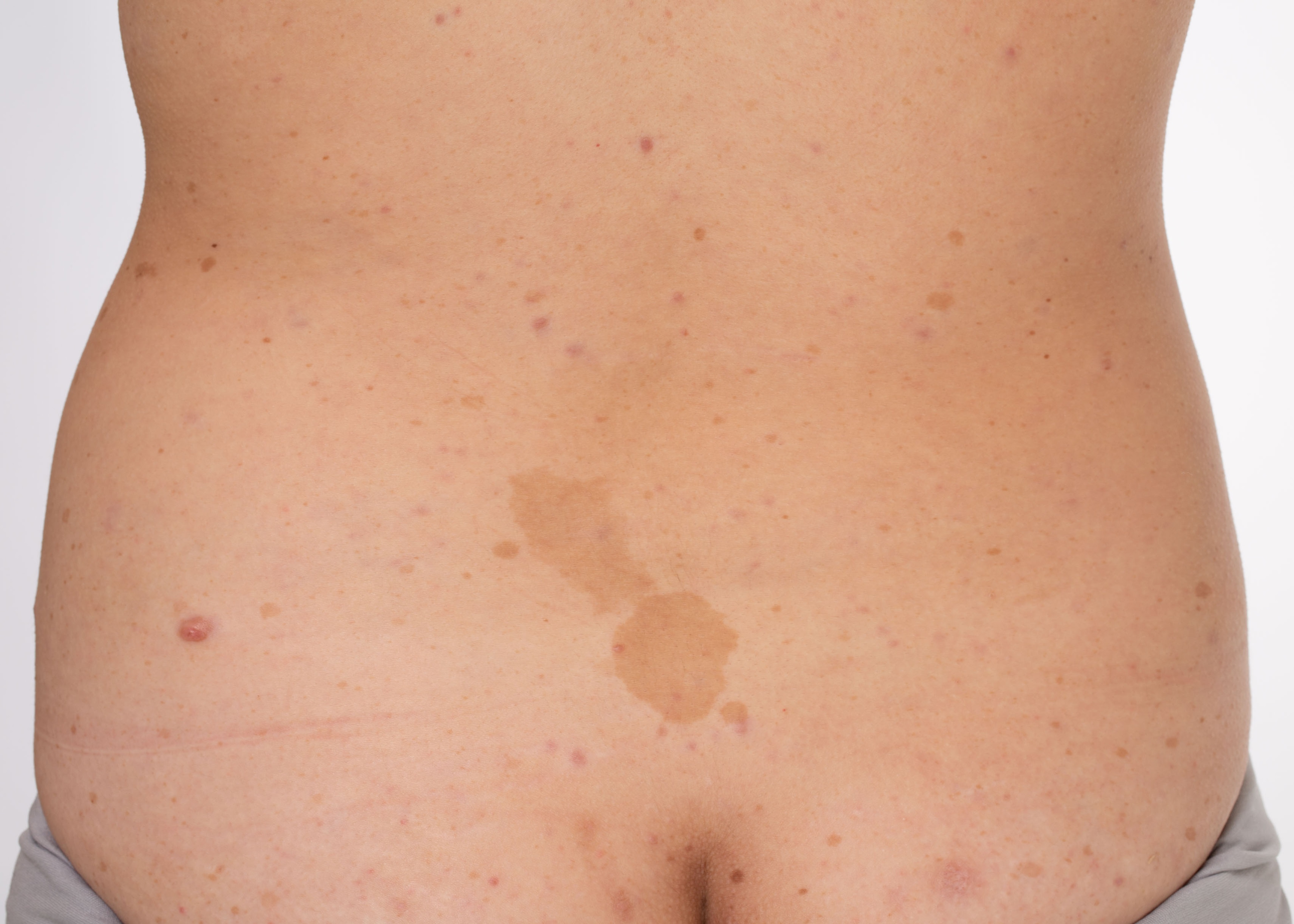
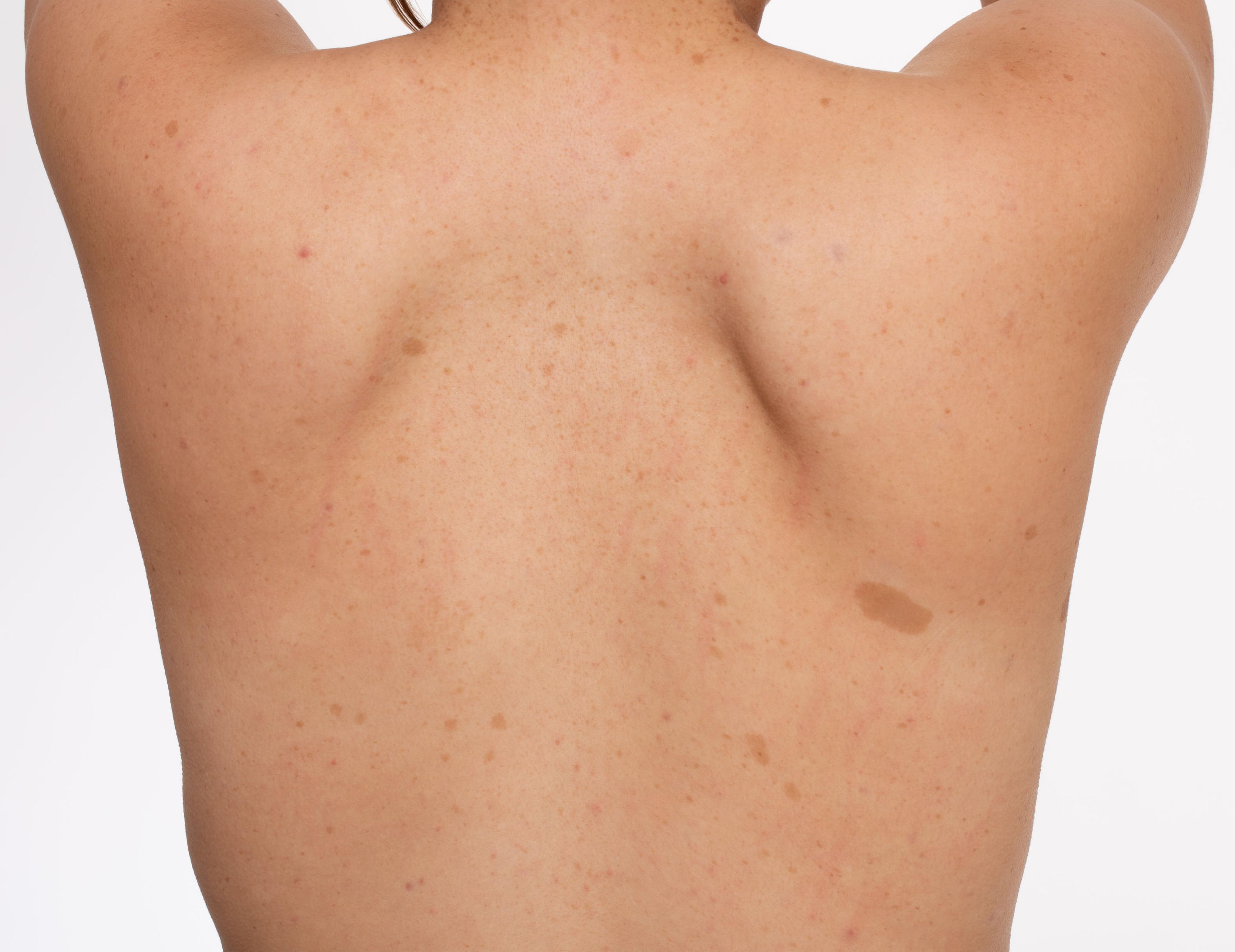
  • Cafe-au-lait macules: Sharply defined light brown patches vary in size from 0.5 cm to 50 cm; the majority are ten cm or less. They are the first feature of the disease and appear in all children affected by the disease. They increase in size and number during the first decade of life. 1 to 3 cafe-au-lait macules are observed in about 15 percent of the normal population. NF-1 is distinguished by having six or more cafe-au-lait macules.[21][22]
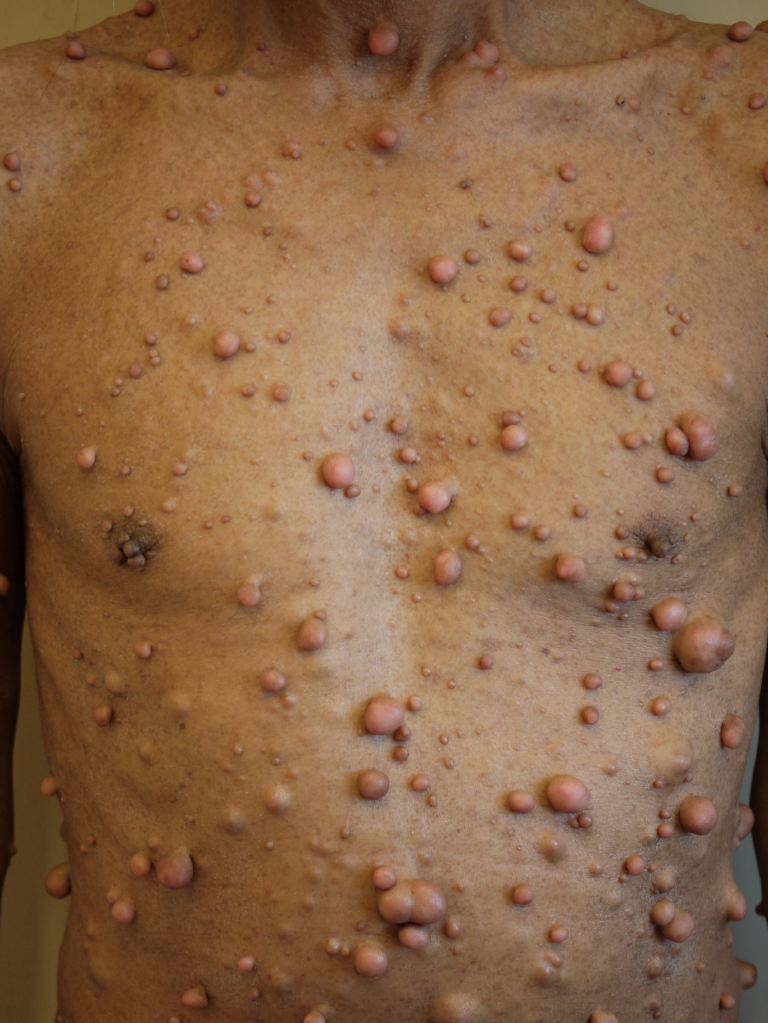
  • Neurofibromas: Neurofibromas are present in the skin (cutaneous), under the skin (nodular), and along with roots of the nerve (plexiform). Pregnancy is associated with an increase in the number and size of tumors, indicating a hormone-responsive state.[23] Cutaneous neurofibromas are soft lilac-pink tumors, mostly sessile and dome-shaped, while some are pedunculated. Mostly found on the trunk and limbs ranging from a few millimeters to several centimeters in diameter. In females, they are prominent on the areola of the breast. They are usually benign and pose a cosmetic concern in adults. The plexiform neurofibroma is a diffuse elongated neuroma along the course of the nerve, frequently involving trigeminal and cervical nerves. They are usually present during the first two years of life. On palpation, they have a characteristic "bag of worms" sensation. Pain and expansion of the lesion suggest malignant transformation and are associated with higher morbidity and mortality.[24] Nodular neurofibromas are firm and rubbery and may cause pain. They do not invade adjacent structures and may become premalignant. 
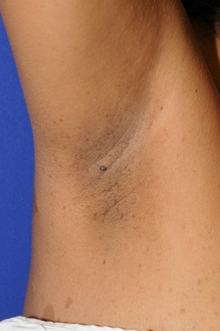
  • Freckling: Axillary and/or inguinal freckling in patients is usually pathognomonic for NF-1, known as Crowe's sign. Freckles are small and grouped when compared to cafe-au-lait macules. It is present in around 70% of individuals and appears later than café-au-lait spots by 3 to 5 years. Other intertriginous areas might also be involved.[21]
  • Lisch nodules: Lisch nodules appear as dome-shaped lesions around the iris on slit-lamp-examination. They are present in over 90% of the affected adults and are also known as pigmented iris hamartomas. They are usually asymptomatic but used to confirm the diagnosis in the affected individual and the parent.[25]
  • Skeletal abnormalities: Kyphoscoliosis occurs in 10 to 25 percent of the affected individuals, and high-level lesions may lead to respiratory difficulties.[26] Pseudoarthrosis of the tibia or radius occurs in about 5% of infants with the male predisposition.[27] Sphenoid wing dysplasia is a characteristic abnormality in NF-1, manifesting as an asymmetry of the face. Nonossifying fibromas, short stature, osteopenia, and osteoporosis are other noted osseous abnormalities.
  • Neurological abnormalities: Learning disability, cognitive dysfunction, developmental delay, autism spectrum disorder, attention deficit hyperactivity disorder, macrocephaly, and dural ectasia are reported.[28][29] The most common neurological manifestations in a cohort comprising 128 patients included macrocephaly (40%) and attention-deficit/hyperactivity disorder (30%), respectively.[30] In a study comprising 1234 patients, the frequency of vascular abnormalities was 3.5%, with stroke and cerebral arterial stenosis being the most common.[31]
  • Malignancies: Neurological tumors consist of optic nerve glioma, astrocytoma, brainstem glioma, and schwannomas. Intracranial tumors can cause headaches and seizures.[32][33] Other malignancies reported to have been associated with the disease are Wilms tumor, rhabdomyosarcoma, malignant peripheral nerve sheath tumors, leukemia, gastrointestinal stromal tumors, pheochromocytoma, retinoblastoma, breast cancer, and malignant melanoma.[34][35] Optic nerve glioma is present in about 15 percent of under-6-year-old children with NF-1.[36] Many children with NF-1 and optic nerve glioma are noted to have normal vision. Puberty may be premature or delayed when optic chiasm is involved due to its effect on the hypothalamus.[37] 
  • Other presentations: Hypertension, congenital heart disease, irritable bowel syndrome, and constipation have been reported with NF-1.[38][39] Papillomatous neurofibromas of the hard palate, tongue, etc., are present in only 5% to 10% of cases.

Lisch nodules are the commonest ophthalmic manifestation of NF1. Freckling is found in approximately 80-90% of the patients. Neurofibroma is seen in almost all NF1 patients over 30 years of age. The plexiform neurofibroma is seen in 25 to 30% of the cases.[40]

Nevus anemicus (NA) and Juvenile Xanthogranulomas (JXG) are also significantly associated with patients with NF1 in the early years of their lives.[41]

A whole body MRI (WBMRI) during the first evaluation revealed  

  • Focal areas of signal intensity (approx 70%)-with cerebellum involvement in 72% of the cases
  • Optic pathway glioma (approx 16%)
  • Bony dysplasia- sphenoid wing involvement in half the cases, followed by tibia and fibula in 40%
  • Spinal dura ectasia , and
  • Plexifirm neurofibroma (PN).[15]

The most frequent NF1-related manifestation in the same study included:

  • Café-au-lait macules (CALMs)(100%)
  • Macrocephaly (55%),
  • Axillary freckling (45%),
  • Lisch nodules (32%), and
  • Cutaneous neurofibromas (18%).[15]

Developmental delay, PN, and NF1 are more common in familial cases.[15]

Evaluation

Diagnosis is based on the National Institute of Health (NIH) and the Revised diagnostic criteria.[42]

National institute of health (NIH) criteria were published in 1987.[43] The diagnosis of NF-1 is mainly clinical, based upon agreed clinical national institutes of health (NIH) criteria which require two or more of the following conditions to be fulfilled:

  • Café-au-lait macules: six or more with the greatest dimension of 5 to 15 mm under normal light
  • Neurofibromas: two or more of any type or one plexiform neurofibroma
  • Freckling: axillary or inguinal
  • Optic gliomas
  • Lisch nodules: two or more
  • Bony lesions: sphenoid dysplasia, pseudoarthrosis, and others
  • Family history: a first-degree relative with the disease.[44]

NIH criteria are both highly sensitive and specific except for young children.[19] Focused symptom assessments of pain, weakness, visual concerns, and headaches should be done along with developmental assessments. Physical exams should be emphasized on the skin, eye, skeletal and nervous systems. Slit-lamp examination of the eye is recommended.

The Revised diagnostic criteria published in 2021 includes the following:

A: A patient with no family history should have two or more of the following:

  • Six or more café-au-lait macules over 5 mm ( before puberty) and over 15 mm (after puberty)
  • Freckling in the axillary or inguinal region
  • Two or more neurofibromas of any type or one plexiform neurofibroma
  • Optic pathway glioma
  • Two or more iris Lisch nodules choroidal abnormalities
  • A distinctive osseous lesion such as sphenoid dysplasia, bowing of the tibia, or pseudarthrosis of a long bone
  • A  pathogenic NF1 variant

B: A child with a positive family history can have only one or more criteria in A.[43]

At least one of the two pigmentary findings (café-au-lait macules or freckling) should be bilateral.

Sphenoid wing dysplasia is not a separate criterion in the case of an ipsilateral orbital plexiform neurofibroma.

Mosaic NF1 should be suspected if the pigmentary lesions are only unilateral. Looking for Lisch nodules/choroidal anomalies or confirmation through genetic testing is the recommended approach in these cohorts.[43]

Neuroradiological findings include neurofibromatosis, bright spots, and a high volume of the brain (megalencephaly).[45] Bright spots are high signal intensity areas identified in T2 weighted magnetic resonance imaging of the brain, often found in the brainstem, cerebellum, and basal ganglia.[46] They are not necessary for the diagnosis of NF-1. Both bright spots and megalencephaly have been associated with variable cognitive dysfunction. 

Other manifestations of NF1 include:

  • Focal areas of signal intensity (FASI)
  • Glioma
  • Interstitial lung disease
  • Gastrointestinal neoplasm
  • Scoliosis.[4]

Genetic testing is not mandatory for the diagnosis. It can be utilized for establishing the diagnosis in questionable presentations, screening of family members, and prenatal screening. Ninety-five percent of mutations can be detected by genetic testing in individuals with NF-1 clinical diagnosis.[47] A negative test does not exclude the disease, as mosaicism may be possible, as in segmental NF-1. Affected individuals should receive multidisciplinary care from geneticists, pediatricians, pediatric neurologists, ophthalmologists, psychiatrists, and psychologists to identify the disorder and related complications, provide the treatment, and need psychosocial support and guidance.

A free-text search algorithm, NF1 ICD-9, and ICD-10 codes are also available for the diagnosis and management of NF1.[48]

Treatment / Management

There is no definitive treatment for NF-1 as it is a genetic disorder with multiple manifestations.[2][49][50][51] Treatment for various NF-1 manifestations is mostly symptomatic, whereas, for plexiform neurofibromas, surgical removal is the only treatment option.[52]

  • Tumors are treated based on the type, involvement of other structures, and associated complications. Radiation therapy in NF-1 is associated with the emergence of secondary tumors, thereby opting for radiation only in essential cases.[53] Cutaneous and nodular neurofibromas are treated when they develop symptoms like pain, increase in size, weakness, vision loss, or cosmetic concerns. Surgery, laser, and electrodesiccation are some of the treatment modalities. Gabapentin has sometimes been utilized for pruritus treatment along with neuropathic pain management. In particular, plexiform neurofibromas (PN) are associated with high morbidity and malignant transformation compared to cutaneous and nodular neurofibromas. Compression of the airway or spinal cord and disfigurement are common reasons for the surgical removal of plexiform neurofibromas.[54] Selumetinib was approved for use in children three years of age and older with PN that cannot be operated on despite having symptoms or being noted to be making progress. Selumetinib is the first drug approved by US food and drug administration for plexiform NF.[43] It is a mitogen-activated protein kinase inhibitor.[55]

Newer advances in therapy include:

  • Tumor-tropic cell carriers harboring therapeutic gene
  • Oncolytic Virotherapy
  • Suicide gene therapy
  • Tumor Suppressor Gene Therapy
  • Immunomodulatory Gene (IFN-β) Interleukin-12, and
  • Gene Target Therapy.[14]
  • Optic pathway gliomas and other low-grade gliomas should be closely monitored for symptoms, visual acuity, and size. Chemotherapy with vincristine, carboplatin, and vinblastine is the first-line therapy in children.[56] No precise therapy is identified for adults with gliomas. Biopsy or surgery is reserved for the evaluation of high-grade gliomas. Malignant peripheral nerve sheath tumors are treated with surgery and radiation. Rhabdomyosarcomas are treated with chemotherapy, surgery, and radiation. 
  • There is no standard management available for cNF. Surgical removal gives excellent cosmetic results for plexiform NF. CO laser ablation, laser photocoagulation, electro-drying, and radiofrequency ablation are fast and effective but can cause depigmentation, hyperpigmentation, or extensive scarring.[57]
  • Seizures or epilepsy should thoroughly be investigated as a neurosurgical intervention is sometimes very beneficial for the patient.
  • Children with learning or cognitive disabilities and neurologic impairment should work closely with speech, occupational, and physical therapists. 
  • Skeletal abnormalities like dysplasias, pseudoarthrosis, and scoliosis might require orthopedic correction.[27][58] Vitamin D and calcium supplementation are recommended for individuals with osteopenia and/or osteoporosis. 
  • Psychotherapy and counseling can assist with psychosocial functioning and boost morale.[59]
  • Follow-up every 12 months is recommended to assess for different complications of the disease.
  • All infants are screened with cranial MRI to look for neurological abnormalities.[60][61][62]
  • Genetic counseling is very important in disease management. Informing the patients about the different complications of the disease is very important. And it should be made very clear to the patients regarding their children that 50% are likely to be affected, and the condition may be severe. First-degree relatives who have no disease manifestations are unlikely to carry the gene, and the risk for their offspring is small but not absent.

Differential Diagnosis

Legius syndrome is an autosomal dominant disorder like NF-1 but lacks neurofibromas and nervous system involvement. Cafe-au-lait macules, freckling is noted.[63] Loss of function mutations in SPRED1 protein results in Legius syndrome.

Neurofibromatosis type 2-Affected individuals do not exhibit more cafe-au-lait macules, unlike NF-1. There is no evidence of Lisch nodules, cognitive dysfunction, or malignant peripheral nerve sheath tumors. Meningiomas and acoustic schwannomas are highly associated with neurofibromatosis type 2 (NF2). Mutations in the NF2 tumor suppressor gene located on the 22q chromosome cause the disorder.[64]

Noonan syndrome-Individuals may exhibit cafe-au-lait macules along with low-set ears, hypertelorism, webbed neck, short stature, and pulmonary stenosis. Mutations of the genes involved in RAS and mitogen-activated protein kinase signaling pathway result in the syndrome.[65] 

Constitutional mismatch repair deficiency (CMMR-D) syndrome - This is an autosomal recessive disorder manifesting cafe-au-lait macules, freckling, and Lisch nodules. CMMR-D is typically associated with brain tumors, colorectal cancer, and hematological malignancies, unlike optic glioma and rhabdomyosarcoma associated with NF-1. Mutations in 1 of the 4 mismatch repair genes cause the syndrome.[66]

Other differential diagnoses include McCune-Albright syndrome, Silver-Russell syndrome, phosphatase and tensin homolog (PTEN) hamartoma syndrome, Carney syndrome, ataxia-telangiectasia, Klippel-Trenaunay-Weber syndrome, Turner syndrome, tuberous sclerosis.[13]

If pigmentary lesions are only present, the major differential diagnosis includes:

  • Legius syndrome
  • Noonan syndrome, and
  • Constitutional mismatch repair deficiency.[43]

Prognosis

The outlook for patients with NF-1 is guarded and depends on the severity of the disease, the presence of malignancy, and the extent of the deformity. Those with mild disease can have a reasonable life expectancy, but those with moderate to severe disease have a poor quality of life.[67] Cosmetic and functional limitations significantly impact these patients' quality of life (QOL).[68] Women with NF-1-related breast cancer have a poor 5-year survival rate and high mortality.[69] Patients with central nervous system tumors are at increased risk for secondary malignancies, mortality, and vascular complications.[70] 

Malignant peripheral nerve sheath tumors in association with NF-1 have poor overall survival.[34] The molecular marker of SCs in neurofibromas, such as S100 protein and SOX10, are lost following their transition to Malignant peripheral nerve sheath tumors (MPNST).[7]

The cohort of 2467 individuals from the Denmark RAREDIS Database from (1995-2013) was observed to have increased odds for the risk of:

  • Hospitalizations
  • Psychiatric illnesses
  • Poor academic performances
  • Poor relationship
  • Abortions and stillbirths
  • Poor quality of life, and
  • Increased dependency.[71]

Serial evaluation with WBMRI should be assessed based on the clinical and NF1 domains.[15] Disease and radiological progression are higher, with NF1 mutation affecting GRD domains.[15] The primary contributing variable to mortality among patients with NF1 includes brain tumors and MPNSTs.[4]

Both recent and ongoing research trials have primarily been focused on PN only (approximately 45%).[72]

Complications

Multidisciplinary specialists should follow affected individuals as part of an interprofessional team to provide longitudinal care to identify the manifestations early and treat the complications as they arise.[73] Annual surveillance visits for children should include height, weight, head circumference, and blood pressure checks. Eye, skin, skeletal, neurological, developmental, and cognitive assessments are completed during the annual visits. School performance and depression screening are recommended. Any plexiform neurofibroma that grows rapidly, becomes more nodular (deeper involvement), develops more pain, or new neurological deficit should be imaged with magnetic resonance imaging for possible malignant peripheral nerve sheath tumor transformation. Given the breast cancer association with NF-1, women are advised to have annual mammograms from the age of 30 instead of conventional screening at the age of 40.[69] NF-1 individuals with hypertension, headache, diaphoresis, or palpitations should be evaluated for pheochromocytoma by measuring plasma or urine-free fractionated metanephrines.[74]

Vasculopathy incidence in NF-1 is around 0.4 to 6.4 percent, presenting as essential hypertension, renovascular hypertension, hemorrhagic strokes, aneurysms, or arteriovenous fistulae.[75] Magnetic resonance angiogram, renal angiogram, or computed tomography angiogram should assess the vascular lesions and provide the needed medical and surgical therapy to prevent further complications. Toe or fingertip pain is likely due to glomus tumors which may coexist. Severe vascular occlusion of internal carotid arteries, moyamoya disease, and multiple endocrine neoplasias are noted in association with NF-1.[13]

Deterrence and Patient Education

The affected individuals and their families should be made aware of the nature of the disease, its manifestations, its prognosis, and the possible complications that can develop. Education on the need for periodic surveillance and longitudinal multidisciplinary care aimed at physical and mental well-being should be provided. Genetic counseling should be offered prior to conception to provide the required information to the patients and families for making an informed decision. Neurofibromatosis type 1 community-based model and support group have shown significant success in the transition of care among these cohorts.[76] Lifelong multidisciplinary management is of paramount importance in the management of these patients.[4]

Pearls and Other Issues

All genetic disorders are rare, and they cause extreme anguish for families. The main aim is to ensure proper diagnosis of the disease and genetic counseling of the affected families to prevent their children from the disease or its complications.

Enhancing Healthcare Team Outcomes

The presentation of patients with NF-1 is highly variable. Because the disorder affects many organs, it is best managed by an interprofessional team, including clinicians (including physicians, nurse practitioners, and physician assistants) and specialty-trained nurses. Primary care physicians, geneticists, pediatric neurologists, ophthalmologists, psychiatrists, psychologists, and therapists (physical, occupational, and speech) work collectively as an interprofessional team toward health promotion and maintenance. All interprofessional team members must maintain open communication with the rest of the team so that care can be administered in a coordinated and cohesive fashion, leading to optimal patient outcomes. [Level 5]

A multidisciplinary approach is the cornerstone of management.[77] While the disorder is benign, it is vital that healthcare workers closely monitor patients because many organ systems are involved. To improve patient outcomes, it is essential to remember that a few patients may develop neurological tumors. Other malignancies reported to have been associated with the disease are Wilms tumor, rhabdomyosarcoma, leukemia, retinoblastoma, and malignant melanoma. A thorough exam and serial imaging studies are the only way to identify the presence of these lesions. Early diagnosis of malignant transformation and timely treatment of the same govern the prognosis of these patients.[77]


(Click Image to Enlarge)
Neurofibromatosis
Neurofibromatosis
Source: DermNet

(Click Image to Enlarge)
Neurofibromas and cafe au lait macules on the back
Contributed by Drs. Jason C. Sluzevich, and Thoyaja Koritala

(Click Image to Enlarge)
Cafe au lait macules and neurofibroma on the back
Contributed by Drs. Ashley B. Wentworth and Thoyaja Koritala

(Click Image to Enlarge)
Cafe au lait macules on the back
Contributed by Drs. Ashley B. Wentworth and Thoyaja Koritala

(Click Image to Enlarge)
Axillary freckling in Neurofibromatosis type 1
Contributed by Drs. Jason C. Sluzevich and Thoyaja Koritala
Details

Author

Abdullah Adil

Editor:

Achint K. Singh

Updated:

8/13/2023 2:54:08 AM

References


[1]

Solares I,Vinal D,Morales-Conejo M, Diagnostic and follow-up protocol for adult patients with neurofibromatosis type 1 in a Spanish reference unit. Revista clinica espanola. 2022 Oct;     [PubMed PMID: 35688675]


[2]

Karaconji T,Whist E,Jamieson RV,Flaherty MP,Grigg JRB, Neurofibromatosis Type 1: Review and Update on Emerging Therapies. Asia-Pacific journal of ophthalmology (Philadelphia, Pa.). 2018 Nov 2;     [PubMed PMID: 30387339]


[3]

Farschtschi S,Mautner VF,McLean ACL,Schulz A,Friedrich RE,Rosahl SK, The Neurofibromatoses. Deutsches Arzteblatt international. 2020 May 15;     [PubMed PMID: 32657748]


[4]

Wang MX,Dillman JR,Guccione J,Habiba A,Maher M,Kamel S,Panse PM,Jensen CT,Elsayes KM, Neurofibromatosis from Head to Toe: What the Radiologist Needs to Know. Radiographics : a review publication of the Radiological Society of North America, Inc. 2022 Jul-Aug;     [PubMed PMID: 35749292]


[5]

Williams VC,Lucas J,Babcock MA,Gutmann DH,Korf B,Maria BL, Neurofibromatosis type 1 revisited. Pediatrics. 2009 Jan;     [PubMed PMID: 19117870]


[6]

Gutmann DH, Ferner RE, Listernick RH, Korf BR, Wolters PL, Johnson KJ. Neurofibromatosis type 1. Nature reviews. Disease primers. 2017 Feb 23:3():17004. doi: 10.1038/nrdp.2017.4. Epub 2017 Feb 23     [PubMed PMID: 28230061]


[7]

Ge LL,Xing MY,Zhang HB,Wang ZC, Neurofibroma Development in Neurofibromatosis Type 1: Insights from Cellular Origin and Schwann Cell Lineage Development. Cancers. 2022 Sep 17;     [PubMed PMID: 36139671]


[8]

Easton DF,Ponder MA,Huson SM,Ponder BA, An analysis of variation in expression of neurofibromatosis (NF) type 1 (NF1): evidence for modifying genes. American journal of human genetics. 1993 Aug     [PubMed PMID: 8328449]


[9]

Evans DG,Howard E,Giblin C,Clancy T,Spencer H,Huson SM,Lalloo F, Birth incidence and prevalence of tumor-prone syndromes: estimates from a UK family genetic register service. American journal of medical genetics. Part A. 2010 Feb;     [PubMed PMID: 20082463]


[10]

Stephens K,Kayes L,Riccardi VM,Rising M,Sybert VP,Pagon RA, Preferential mutation of the neurofibromatosis type 1 gene in paternally derived chromosomes. Human genetics. 1992 Jan     [PubMed PMID: 1346385]


[11]

Ruggieri M,Huson SM, The clinical and diagnostic implications of mosaicism in the neurofibromatoses. Neurology. 2001 Jun 12     [PubMed PMID: 11409413]


[12]

Theos A,Korf BR,American College of Physicians.,American Physiological Society., Pathophysiology of neurofibromatosis type 1. Annals of internal medicine. 2006 Jun 6     [PubMed PMID: 16754926]


[13]

Shah KN. The diagnostic and clinical significance of café-au-lait macules. Pediatric clinics of North America. 2010 Oct:57(5):1131-53. doi: 10.1016/j.pcl.2010.07.002. Epub     [PubMed PMID: 20888463]


[14]

Thomas S,Bikeyeva V,Abdullah A,Radivojevic A,Abu Jad AA,Ravanavena A,Ravindra C,Igweonu-Nwakile EO,Ali S,Paul S,Yakkali S,Teresa Selvin S,Hamid P, Systematic Review of Pediatric Brain Tumors in Neurofibromatosis Type 1: Status of Gene Therapy. Cureus. 2022 Aug;     [PubMed PMID: 36120213]

Level 1 (high-level) evidence

[15]

Kang E,Kim YM,Choi Y,Lee Y,Kim J,Choi IH,Yoo HW,Yoon HM,Lee BH, Whole-body MRI evaluation in neurofibromatosis type 1 patients younger than 3 years old and the genetic contribution to disease progression. Orphanet journal of rare diseases. 2022 Jan 29;     [PubMed PMID: 35093157]


[16]

Maertens O,Brems H,Vandesompele J,De Raedt T,Heyns I,Rosenbaum T,De Schepper S,De Paepe A,Mortier G,Janssens S,Speleman F,Legius E,Messiaen L, Comprehensive NF1 screening on cultured Schwann cells from neurofibromas. Human mutation. 2006 Oct     [PubMed PMID: 16941471]


[17]

Khajavi M,Khoshsirat S,Ahangarnazari L,Majdinasab N, A brief report of plexiform neurofibroma. Current problems in cancer. 2018 Mar - Apr     [PubMed PMID: 29449010]


[18]

He D,Li Y,Yu Y,Cai G,Ouyang F,Lin Y,Lu H,Chu L, Segmental neurofibromatosis type 1 complicated with multiple intracranial arteriovenous fistulas: A case study. Clinical neurology and neurosurgery. 2018 May;     [PubMed PMID: 29544172]

Level 3 (low-level) evidence

[19]

DeBella K,Szudek J,Friedman JM, Use of the national institutes of health criteria for diagnosis of neurofibromatosis 1 in children. Pediatrics. 2000 Mar     [PubMed PMID: 10699117]


[20]

Seminog OO,Goldacre MJ, Risk of benign tumours of nervous system, and of malignant neoplasms, in people with neurofibromatosis: population-based record-linkage study. British journal of cancer. 2013 Jan 15     [PubMed PMID: 23257896]


[21]

Korf BR, Diagnostic outcome in children with multiple café au lait spots. Pediatrics. 1992 Dec     [PubMed PMID: 1344978]


[22]

Nunley KS,Gao F,Albers AC,Bayliss SJ,Gutmann DH, Predictive value of café au lait macules at initial consultation in the diagnosis of neurofibromatosis type 1. Archives of dermatology. 2009 Aug     [PubMed PMID: 19687418]


[23]

Dugoff L,Sujansky E, Neurofibromatosis type 1 and pregnancy. American journal of medical genetics. 1996 Dec 2     [PubMed PMID: 8957502]


[24]

Prada CE,Rangwala FA,Martin LJ,Lovell AM,Saal HM,Schorry EK,Hopkin RJ, Pediatric plexiform neurofibromas: impact on morbidity and mortality in neurofibromatosis type 1. The Journal of pediatrics. 2012 Mar     [PubMed PMID: 21996156]


[25]

Lewis RA,Riccardi VM, Von Recklinghausen neurofibromatosis. Incidence of iris hamartomata. Ophthalmology. 1981 Apr     [PubMed PMID: 6789269]


[26]

Crawford AH,Herrera-Soto J, Scoliosis associated with neurofibromatosis. The Orthopedic clinics of North America. 2007 Oct     [PubMed PMID: 17945135]


[27]

Stevenson DA,Birch PH,Friedman JM,Viskochil DH,Balestrazzi P,Boni S,Buske A,Korf BR,Niimura M,Pivnick EK,Schorry EK,Short MP,Tenconi R,Tonsgard JH,Carey JC, Descriptive analysis of tibial pseudarthrosis in patients with neurofibromatosis 1. American journal of medical genetics. 1999 Jun 11;     [PubMed PMID: 10360395]


[28]

Lorenzo J,Barton B,Arnold SS,North KN, Developmental trajectories of young children with neurofibromatosis type 1: a longitudinal study from 21 to 40 months of age. The Journal of pediatrics. 2015 Apr     [PubMed PMID: 25598303]


[29]

Hyman SL,Shores A,North KN, The nature and frequency of cognitive deficits in children with neurofibromatosis type 1. Neurology. 2005 Oct 11     [PubMed PMID: 16217056]


[30]

Sánchez Marco SB,López Pisón J,Calvo Escribano C,González Viejo I,Miramar Gallart MD,Samper Villagrasa P, Neurological manifestations of neurofibromatosis type 1: our experience. Neurologia (Barcelona, Spain). 2022 Jun;     [PubMed PMID: 35672119]


[31]

Sheerin UM,Holmes P,London NF1 Research Group.,Childs L,Roy A,Ferner RE, Neurovascular complications in adults with Neurofibromatosis type 1: A national referral center experience. American journal of medical genetics. Part A. 2022 Oct;     [PubMed PMID: 36097643]


[32]

Créange A,Zeller J,Rostaing-Rigattieri S,Brugières P,Degos JD,Revuz J,Wolkenstein P, Neurological complications of neurofibromatosis type 1 in adulthood. Brain : a journal of neurology. 1999 Mar     [PubMed PMID: 10094256]


[33]

Ostendorf AP,Gutmann DH,Weisenberg JL, Epilepsy in individuals with neurofibromatosis type 1. Epilepsia. 2013 Oct     [PubMed PMID: 24032542]


[34]

Ferrari A,Bisogno G,Macaluso A,Casanova M,D'Angelo P,Pierani P,Zanetti I,Alaggio R,Cecchetto G,Carli M, Soft-tissue sarcomas in children and adolescents with neurofibromatosis type 1. Cancer. 2007 Apr 1     [PubMed PMID: 17330850]


[35]

Suarez-Kelly LP,Yu L,Kline D,Schneider EB,Agnese DM,Carson WE, Increased breast cancer risk in women with neurofibromatosis type 1: a meta-analysis and systematic review of the literature. Hereditary cancer in clinical practice. 2019     [PubMed PMID: 30962859]

Level 1 (high-level) evidence

[36]

Lewis RA,Gerson LP,Axelson KA,Riccardi VM,Whitford RP, von Recklinghausen neurofibromatosis. II. Incidence of optic gliomata. Ophthalmology. 1984 Aug;     [PubMed PMID: 6436764]


[37]

Guillamo JS,Créange A,Kalifa C,Grill J,Rodriguez D,Doz F,Barbarot S,Zerah M,Sanson M,Bastuji-Garin S,Wolkenstein P,Réseau NF France., Prognostic factors of CNS tumours in Neurofibromatosis 1 (NF1): a retrospective study of 104 patients. Brain : a journal of neurology. 2003 Jan     [PubMed PMID: 12477702]

Level 2 (mid-level) evidence

[38]

Fossali E,Signorini E,Intermite RC,Casalini E,Lovaria A,Maninetti MM,Rossi LN, Renovascular disease and hypertension in children with neurofibromatosis. Pediatric nephrology (Berlin, Germany). 2000 Aug     [PubMed PMID: 10955932]


[39]

Ejerskov C,Krogh K,Ostergaard JR,Joensson I,Haagerup A, Gastrointestinal Symptoms in Children and Adolescents With Neurofibromatosis Type 1. Journal of pediatric gastroenterology and nutrition. 2018 Jun     [PubMed PMID: 29240009]


[40]

Sethi NK,Chadha C,Goyal S,Kaur M, Neurofibromatosis 1: A family case series. Journal of family medicine and primary care. 2022 May;     [PubMed PMID: 35800488]

Level 2 (mid-level) evidence

[41]

García-Martínez FJ,Duat-Rodríguez,Andrés Esteban E,Torrelo,Noguera Morel L,Hernández-Martín, Cutaneous Manifestations not Considered Diagnostic Criteria for Neurofibromatosis Type 1. A Case-Control Study. Actas dermo-sifiliograficas. 2022 Sep 23;     [PubMed PMID: 36162491]

Level 2 (mid-level) evidence

[42]

Angelova-Toshkina D,Holzapfel J,Huber S,Schimmel M,Wieczorek D,Gnekow AK,Frühwald MC,Kuhlen M, Neurofibromatosis type 1: A comparison of the 1997 NIH and the 2021 revised diagnostic criteria in 75 children and adolescents. Genetics in medicine : official journal of the American College of Medical Genetics. 2022 Sep;     [PubMed PMID: 35713653]


[43]

Legius E,Messiaen L,Wolkenstein P,Pancza P,Avery RA,Berman Y,Blakeley J,Babovic-Vuksanovic D,Cunha KS,Ferner R,Fisher MJ,Friedman JM,Gutmann DH,Kehrer-Sawatzki H,Korf BR,Mautner VF,Peltonen S,Rauen KA,Riccardi V,Schorry E,Stemmer-Rachamimov A,Stevenson DA,Tadini G,Ullrich NJ,Viskochil D,Wimmer K,Yohay K,International Consensus Group on Neurofibromatosis Diagnostic Criteria (I-NF-DC).,Huson SM,Evans DG,Plotkin SR, Revised diagnostic criteria for neurofibromatosis type 1 and Legius syndrome: an international consensus recommendation. Genetics in medicine : official journal of the American College of Medical Genetics. 2021 Aug;     [PubMed PMID: 34012067]

Level 3 (low-level) evidence

[44]

Rosser T, Neurocutaneous Disorders. Continuum (Minneapolis, Minn.). 2018 Feb;     [PubMed PMID: 29432239]


[45]

Cutting LE,Koth CW,Burnette CP,Abrams MT,Kaufmann WE,Denckla MB, Relationship of cognitive functioning, whole brain volumes, and T2-weighted hyperintensities in neurofibromatosis-1. Journal of child neurology. 2000 Mar     [PubMed PMID: 10757470]


[46]

DiPaolo DP,Zimmerman RA,Rorke LB,Zackai EH,Bilaniuk LT,Yachnis AT, Neurofibromatosis type 1: pathologic substrate of high-signal-intensity foci in the brain. Radiology. 1995 Jun;     [PubMed PMID: 7754001]


[47]

Messiaen LM,Callens T,Mortier G,Beysen D,Vandenbroucke I,Van Roy N,Speleman F,Paepe AD, Exhaustive mutation analysis of the NF1 gene allows identification of 95% of mutations and reveals a high frequency of unusual splicing defects. Human mutation. 2000     [PubMed PMID: 10862084]


[48]

Barnett C,Candido E,Chen B,Pequeno P,Parkin PC,Tu K, Development of algorithms to identify individuals with Neurofibromatosis type 1 within administrative data and electronic medical records in Ontario, Canada. Orphanet journal of rare diseases. 2022 Aug 26;     [PubMed PMID: 36028856]


[49]

Nemmi F,Cignetti F,Assaiante C,Maziero S,Audic F,Péran P,Chaix Y, Discriminating between neurofibromatosis-1 and typically developing children by means of multimodal MRI and multivariate analyses. Human brain mapping. 2019 May 11;     [PubMed PMID: 31077476]


[50]

Guerrini-Rousseau L,Suerink M,Grill J,Legius E,Wimmer K,Brugières L, Patients with High-Grade Gliomas and Café-au-Lait Macules: Is Neurofibromatosis Type 1 the Only Diagnosis? AJNR. American journal of neuroradiology. 2019 May 9;     [PubMed PMID: 31072978]


[51]

Micieli R,Salsberg JM, Visual Dermatology: Mosaic Generalized Neurofibromatosis 1. Journal of cutaneous medicine and surgery. 2019 May/Jun;     [PubMed PMID: 31070102]


[52]

Petrák B,Bendová Š,Lisý J,Kraus J,Zatrapa T,Glombová M,Zámečník J, [Neurofibromatosis von Recklinghausen type 1 (NF1) - clinical picture and molecular-genetics diagnostic]. Ceskoslovenska patologie. 2015;     [PubMed PMID: 25671360]


[53]

Bhatia S,Chen Y,Wong FL,Hageman L,Smith K,Korf B,Cannon A,Leidy DJ,Paz A,Andress JE,Friedman GK,Metrock K,Neglia JP,Arnold M,Turcotte LM,de Blank P,Leisenring W,Armstrong GT,Robison LL,Clapp DW,Shannon K,Nakamura JL,Fisher MJ, Subsequent Neoplasms After a Primary Tumor in Individuals With Neurofibromatosis Type 1. Journal of clinical oncology : official journal of the American Society of Clinical Oncology. 2019 Nov 10     [PubMed PMID: 31532722]


[54]

Canavese F,Krajbich JI, Resection of plexiform neurofibromas in children with neurofibromatosis type 1. Journal of pediatric orthopedics. 2011 Apr-May;     [PubMed PMID: 21415691]


[55]

Dombi E,Baldwin A,Marcus LJ,Fisher MJ,Weiss B,Kim A,Whitcomb P,Martin S,Aschbacher-Smith LE,Rizvi TA,Wu J,Ershler R,Wolters P,Therrien J,Glod J,Belasco JB,Schorry E,Brofferio A,Starosta AJ,Gillespie A,Doyle AL,Ratner N,Widemann BC, Activity of Selumetinib in Neurofibromatosis Type 1-Related Plexiform Neurofibromas. The New England journal of medicine. 2016 Dec 29     [PubMed PMID: 28029918]


[56]

Lassaletta A,Scheinemann K,Zelcer SM,Hukin J,Wilson BA,Jabado N,Carret AS,Lafay-Cousin L,Larouche V,Hawkins CE,Pond GR,Poskitt K,Keene D,Johnston DL,Eisenstat DD,Krishnatry R,Mistry M,Arnoldo A,Ramaswamy V,Huang A,Bartels U,Tabori U,Bouffet E, Phase II Weekly Vinblastine for Chemotherapy-Naïve Children With Progressive Low-Grade Glioma: A Canadian Pediatric Brain Tumor Consortium Study. Journal of clinical oncology : official journal of the American Society of Clinical Oncology. 2016 Oct 10;     [PubMed PMID: 27573663]


[57]

Zhu B,Wei C,Wang W,Gu B,Li Q,Wang Z, [Treatment and progress of cutaneous neurofibroma]. Zhongguo xiu fu chong jian wai ke za zhi = Zhongguo xiufu chongjian waike zazhi = Chinese journal of reparative and reconstructive surgery. 2022 Sep 15;     [PubMed PMID: 36111466]


[58]

Greggi T,Martikos K, Surgical treatment of early onset scoliosis in neurofibromatosis. Studies in health technology and informatics. 2012     [PubMed PMID: 22744522]


[59]

Lemogne C,Bellivier F,Fakra E,Yon L,Limosin F,Consoli SM,Lantieri L,Hivelin M, Psychological and psychiatric aspects of face transplantation: Lessons learned from the long-term follow-up of six patients. Journal of psychosomatic research. 2019 Apr     [PubMed PMID: 30947816]


[60]

Roth J,Ber R,Constantini S, Neurofibromatosis type 1 related hydrocephalus: treatment options and considerations. World neurosurgery. 2019 May 3;     [PubMed PMID: 31059857]


[61]

Armand ML,Taieb C,Bourgeois A,Bourlier M,Bennani M,Bodemer C,Wolkenstein P, Burden of adult neurofibromatosis 1: development and validation of a burden assessment tool. Orphanet journal of rare diseases. 2019 May 3;     [PubMed PMID: 31053133]

Level 1 (high-level) evidence

[62]

Bellampalli SS,Khanna R, Towards a neurobiological understanding of pain in neurofibromatosis type 1: mechanisms and implications for treatment. Pain. 2019 May;     [PubMed PMID: 31009417]

Level 3 (low-level) evidence

[63]

Messiaen L,Yao S,Brems H,Callens T,Sathienkijkanchai A,Denayer E,Spencer E,Arn P,Babovic-Vuksanovic D,Bay C,Bobele G,Cohen BH,Escobar L,Eunpu D,Grebe T,Greenstein R,Hachen R,Irons M,Kronn D,Lemire E,Leppig K,Lim C,McDonald M,Narayanan V,Pearn A,Pedersen R,Powell B,Shapiro LR,Skidmore D,Tegay D,Thiese H,Zackai EH,Vijzelaar R,Taniguchi K,Ayada T,Okamoto F,Yoshimura A,Parret A,Korf B,Legius E, Clinical and mutational spectrum of neurofibromatosis type 1-like syndrome. JAMA. 2009 Nov 18;     [PubMed PMID: 19920235]


[64]

Asthagiri AR,Parry DM,Butman JA,Kim HJ,Tsilou ET,Zhuang Z,Lonser RR, Neurofibromatosis type 2. Lancet (London, England). 2009 Jun 6     [PubMed PMID: 19476995]


[65]

Roberts AE,Allanson JE,Tartaglia M,Gelb BD, Noonan syndrome. Lancet (London, England). 2013 Jan 26     [PubMed PMID: 23312968]


[66]

Wimmer K,Rosenbaum T,Messiaen L, Connections between constitutional mismatch repair deficiency syndrome and neurofibromatosis type 1. Clinical genetics. 2017 Apr     [PubMed PMID: 27779754]


[67]

Taylor LA,Lewis VL Jr, Neurofibromatosis Type 1: Review of Cutaneous and Subcutaneous Tumor Treatment on Quality of Life. Plastic and reconstructive surgery. Global open. 2019 Jan;     [PubMed PMID: 30859021]

Level 2 (mid-level) evidence

[68]

Yoshida Y,Ehara Y,Koga M,Imafuku S, Health-related quality of life in patients with neurofibromatosis 1 in Japan: A questionnaire survey using EQ-5D-5L. The Journal of dermatology. 2022 Jul 3;     [PubMed PMID: 35781730]

Level 2 (mid-level) evidence

[69]

Evans DGR,Kallionpää RA,Clementi M,Trevisson E,Mautner VF,Howell SJ,Lewis L,Zehou O,Peltonen S,Brunello A,Harkness EF,Wolkenstein P,Peltonen J, Breast cancer in neurofibromatosis 1: survival and risk of contralateral breast cancer in a five country cohort study. Genetics in medicine : official journal of the American College of Medical Genetics. 2020 Feb     [PubMed PMID: 31495828]


[70]

de Blank P,Li N,Fisher MJ,Ullrich NJ,Bhatia S,Yasui Y,Sklar CA,Leisenring W,Howell R,Oeffinger K,Hardy K,Okcu MF,Gibson TM,Robison LL,Armstrong GT,Krull KR, Late morbidity and mortality in adult survivors of childhood glioma with neurofibromatosis type 1: report from the Childhood Cancer Survivor Study. Genetics in medicine : official journal of the American College of Medical Genetics. 2020 Nov     [PubMed PMID: 32572180]


[71]

Doser K,Hove H,Østergaard JR,Bidstrup PE,Dalton SO,Handrup MM,Ejerskov C,Krøyer A,Doherty MA,Møllegaard Jepsen JR,Mulvihill JJ,Winther JF,Kenborg L, Cohort profile: life with neurofibromatosis 1 - the Danish NF1 cohort. BMJ open. 2022 Sep 20;     [PubMed PMID: 36127120]


[72]

Acar S,Nieblas-Bedolla E,Armstrong AE,Hirbe AC, A Systematic Review of Recent and Ongoing Clinical Trials in Patients With the Neurofibromatoses. Pediatric neurology. 2022 Sep;     [PubMed PMID: 35759947]

Level 1 (high-level) evidence

[73]

Miller DT,Freedenberg D,Schorry E,Ullrich NJ,Viskochil D,Korf BR,COUNCIL ON GENETICS.,AMERICAN COLLEGE OF MEDICAL GENETICS AND GENOMICS., Health Supervision for Children With Neurofibromatosis Type 1. Pediatrics. 2019 May     [PubMed PMID: 31010905]


[74]

Petr EJ,Else T, Pheochromocytoma and Paraganglioma in Neurofibromatosis type 1: frequent surgeries and cardiovascular crises indicate the need for screening. Clinical diabetes and endocrinology. 2018     [PubMed PMID: 29977594]


[75]

Rea D,Brandsema JF,Armstrong D,Parkin PC,deVeber G,MacGregor D,Logan WJ,Askalan R, Cerebral arteriopathy in children with neurofibromatosis type 1. Pediatrics. 2009 Sep     [PubMed PMID: 19706560]


[76]

Wang ZC,Li HB,Wei CJ,Wang W,Li QF, Community-boosted neurofibromatosis research in China. The Lancet. Neurology. 2022 Sep;     [PubMed PMID: 35963255]


[77]

Purdenko TI,Delva MY,Ostrovskaya LI,Tarianyk KA,Sylenko HY,Pushko OO,Purdenko SV, NEUROFIBROMATOSIS TYPE I AND ITS DIAGNOSTIC CRITERIA: A CLINICAL OBSERVATION. Wiadomosci lekarskie (Warsaw, Poland : 1960). 2022;     [PubMed PMID: 35758466]