Continuing Education Activity
Paget disease is a skeletal growth disorder in which abnormalities such as unusual bone growth can occur in several multifactoral ways. This is often manifested by diffuse pain throughout the musculoskeletal system. It generally manifests with radiologically visible changes. This activity reviews the etiology, presentation, evaluation, and management of Paget disease and reviews the role of the interprofessional team in evaluating, diagnosing, and managing the condition.
Objectives:
- Review the hypothesized etiologies of Paget disease.
- Describe the evaluation of a patient with Paget disease including exam findings, indicated laboratory and diagnostic imaging.
- Summarize the treatment options for Paget disease.
- Explain modalities to improve care coordination among interprofessional team members in order to improve outcomes for patients affected by Paget disease.
Introduction
Paget disease is a skeletal growth disorder in which abnormalities such as unusual bone growth can occur in several multifactoral ways. This is often manifested by diffuse pain throughout the musculoskeletal system.[1][2][3][4]
The condition presents with excess osteoclastic activity followed by a compensatory increase in osteoblastic activity, leading to the formation of disorganized bone, which is less compact, mechanically weaker, highly vascular and more susceptible to fracture. Fortunately, more than 3/4 of patients with Paget disease are asymptomatic. It is the 2nd most common bone disorder in elderly individuals, after osteoporosis. The condition can affect one or multiple bones but the axial skeleton is most often involved (spine, pelvis, and skull). The condition does not spread to other bones but can progress in the preexisting site. A deadly complication of Paget disease is the development of pagetic sarcoma, which is fatal.
The cause of Paget disease remains unknown but there are both genetic and environmental associations. The disorder is most common in Europe, North America, and Australia but rare in Asia and Africa. A number of viruses have been identified in the diseased bone but what their role is in the disease pathology remains a mystery.
Etiology
Some literary sources suggest that the family of paramyxoviruses solely causes Paget. However, many studies have come to determine that the osteoclast generation of a unique cytokine found exclusively in the bone marrow of patients diagnosed with Paget disease may be the primary insult. This cytokine is known as IL-6.
A genetic predisposition to the disorder appears to be strong based on population studies. Besides finding an association between HLA markers, siblings are at high risk for developing the disorder.
Epidemiology
Paget disease is usually seen in individuals older than 50 years. It is common in Caucasians of northern European descent. Paget disease is equally common in males and females. In the US, it is said to affect 1-3 million people but most are asymptomatic. The disorder is slightly more common in white males. The disorder usually presents in the 4-5 decade of life but the diagnosis is often made a decade later.
Pathophysiology
Paget disease occurs when there's an increase in bone resorption that leads to a decrease in bone mass and lytic structures. This process gives rise to osteoblasts from the bone utilizing a sensing system that allows them to increase their activity.
Paget disease pathological process occurs in four stages. Briefly, it begins with osteoclastic activity followed by a hybrid osteoclastic/osteoblastic process. The third stage is where the osteoblastic activity is observed and culminates in the final stage, where malignant degeneration will be seen.
The disease occurs in isolated pockets but is usually progressive. Often there is erythema and warmth over the involved bone due to hypervascularity, which in turn can also lead to high output heart failure.
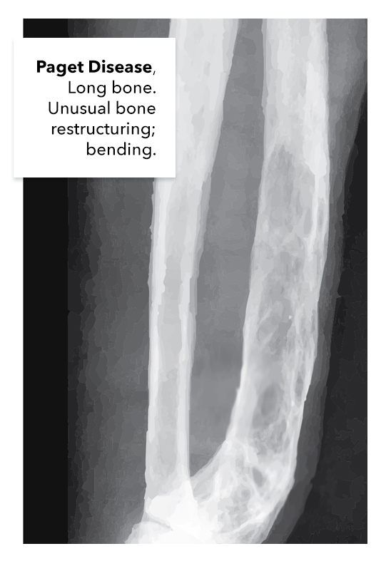
The disease can affect almost every bone in the skeleton but has an affinity for the long bones, axial skeleton, and skull. The involvement of the feet and hands is very rare.
Histopathology
The key histopathological feature of Paget disease involveS the bone architecture and includes the three phases of the disease: mixed, osteolytic, and osteosclerotic. These phases may occur at the same time or separately. The osteolytic phase has areas of resorption due to a large increase in the number of abnormal osteoclasts that contain dozens of nuclei. The osteoblastic phase that follows is disorganized. The bone development is fragmented and irregular. The presence of irregularly shaped bone particles appears like a jigsaw and is the hallmark feature of Paget disease. As the disorder advances, the osteoblastic phase becomes dominant, resulting in excessive bone formation which is fibrous and coarse. The marrow space is filled with vascularized fibrous tissue, which accounts for the persistent warmth and fever.
The bone in Paget disease does not have centralized blood vessels or Haversian systems. Once the osteoblastic phase subsides, the new bone is poorly mineralized and is devoid of any structural integrity.[5][6][7]
History and Physical
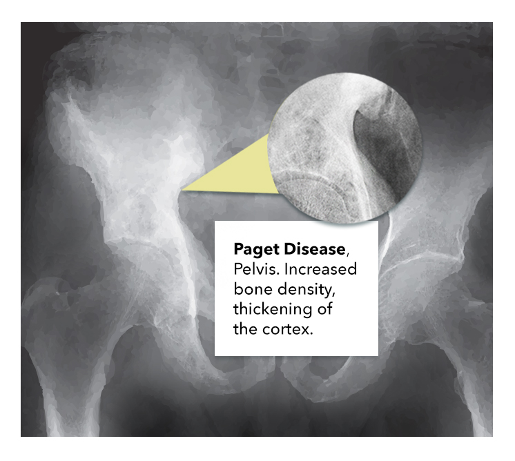
Many patients that present to the clinic with pathognomonic features associated with Paget disease are usually asymptomatic. The majority of patients with the condition are often diagnosed by an incidental finding on an x-ray study. The disease will present with one bone affected in 1/3 of cases. The spine and pelvis are commonly affected and among the long bones, the femur is often involved. Symptomatic patients can present with the following:
- Pain involving the bones and joints
- Diffuse joint stiffness
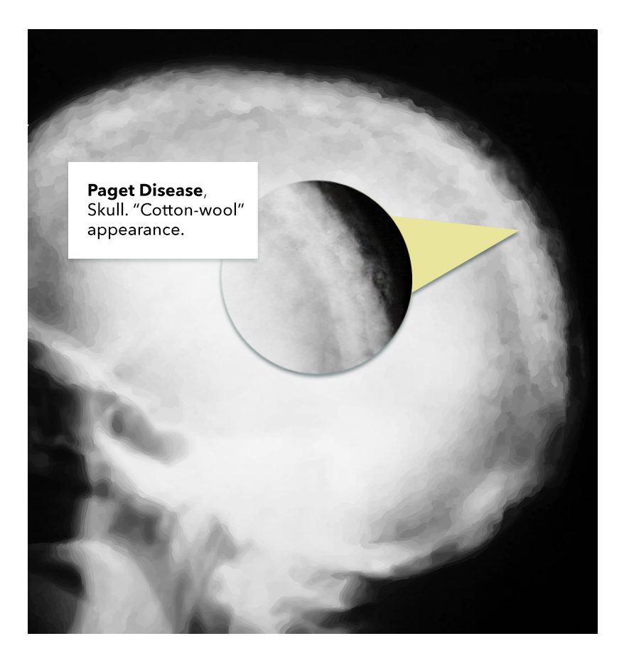
- Abnormally enlarged skull
- Musculoskeletal deformities
- Loss of hearing (due to the involvement of the petrous temporal bone)
- Migraines
- Fractures
- Heart failure
- Cranial nerve neuropathies
- Headaches
- Enlarged skull
- Skull and jaw deformity
The lumbar spine, sacrum, and skull are involved in most cases. Pain is a common feature and is worse with weight-bearing.
The physical exam may show bone deformity or angulation, localized pain to palpation and increased warmth. The gait may be altered and there may be balance problems.
Incomplete fractures are common in Paget disease and seen in the tibia and femur. Even mild injuries can result in fractures. Femur fractures often involve the subtrochanteric region.
Osteosarcoma is a rare complication but should be suspected in a patient with sudden increase in swelling or bone pain. The disorder is fatal. Giant cell tumors may also arise in pagetic bone and involve the facial bones. With vertebral fractures, acute spinal cord compression can occur.
An increase in cardiac output is noted in 20% of patients when the axial skeleton is involved. In addition, calcified aortic stenosis is also common in this population.
Evaluation
Tests to assist in the diagnosis of Paget disease include:
- Bone scan
- Bone x-ray
- Elevated markers of bone breakdown like N-telopeptide
This disease also may also present with the following findings:
- Elevated ALP (alkaline phosphatase)
- Normal Serum calcium and Phosphate
Measurement of serum alkaline phosphatase is useful as well as urine levels of hydroxyproline, C-telopeptide, and N telopeptide.
Procollagen N terminal peptide is also a sensitive serum marker for bone formation.
Hyperuricemia is common and is due to a high turnover of bone.
Secondary hyperparathyroidism occurs in about 10% of patients due to inadequate calcium in the face of increased demand.
Plain x-rays may reveal arthritis or fractures of gross bony lesions.
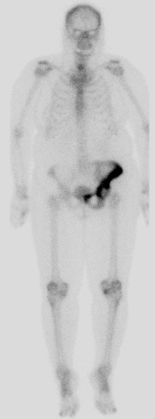
Bone scans can help document the extent of disease and should be used to follow treatment. In addition, a bone scan can pick up early changes in bone even before the patient develops symptoms.
Treatment / Management
Some patients diagnosed with Paget disease may not require treatment. [8][9][10] This patient cohort includes:
- Patients with no abnormal blood tests
- Patients who have no active signs of the disease and those who are asymptomatic
The most commonly treated patients diagnosed with Paget disease include:
- Those with abnormal bone defects
- When weight-bearing bones are involved
- Those with skull deformities
- When there’s evidence of a rapidly progressing bone change
- Those with complaints of diffuse pain
There are several treatment regimens that aid in prophylactically preventing bone breakdown and the subsequent formation.[11] Some of the more common drug therapies include:
- Bisphosphonates that have been approved as the first-line treatment option, secondary to its influence in bone remodeling.
- Calcitonin that is usually a second-line treatment. This drug is assisting in bone absorption
- Supplements such as calcium and vitamin D that have been known to provide some symptomatic benefit
- Pain management that is usually achieved by either NSAIDs or acetaminophen
Surgery is only offered as an option to patients diagnosed with Paget disease when there is a progression into osteosarcoma. The majority of patients diagnosed with osteosarcoma are often offered palliative options such as amputation of the affected limb. In many cases, clinicians are tasked with the job of making judgment calls about which treatment options to offer to the wide spectrum of patients that are diagnosed with Pagte disease. For example, younger patients are usually offered a surgical procedure where they could potentially salvage the limb by resecting the tumor with wide margins. This may not be a viable alternative for an elderly patient with multiple comorbidities and risk factors. Patients may also develop pathological fractures that may need radiation and internal fixation to relieve pain burden. Chemotherapy has been shown to be an ineffective option for patients diagnosed with a sarcoma. It is important to note that surgical failure rates are high in this group of patients. Often, revision surgery is indicated.
Patients with cauda equina and other nerve compression complications will frequently require laminectomy.
Differential Diagnosis
The differential diagnosis includes:
- Osteomalacia
- Osteoporosis
- Malignancy of the bone, primary or metastatic
- Renal osteodystrophy
- Osteoarthritis
- Osteopenia
- Fibrous dysplasia
Prognosis
The prognosis for patients who are treated is good, especially if the disease is in its early stages. There is no cure for Paget disease but the disorder can be controlled from progressing. Patients with polyostotic disease tend to have poor outcomes compared to monostotic disease. The morbidity is usually due to fractures, chronic pain, bone deformity, and neurological complications. Once the patient develops sarcomatous degeneration, the survival rate is very poor.
Complications
Complications include:
- Secondary osteoarthritis
- Vertigo
- Deafness
- Dental malocclusion
- Tinnitus
- Cranial nerve compression
- Basilar invagination
- Cauda equina syndrome
- Fractures
- Hashimoto thyroidits
- Osteopetrosis
- Dupuytren contracture
- Development of osteosarcoma
- High output failure
Pearls and Other Issues
Diet and Activity
- While there is no specific diet for patients with Paget disease, those who are prescribed bisphosphonates should ensure adequate intake of calcium and vitamin D.
- Aggressive physical activity is not recommended, as the risk of fracture is high. However, muscle-strengthening exercise at a low level is recommended.
Deterrence
To date, there is no way to prevent Paget disease since the cause remains unknown. For family members of a patient with Paget disease, some physicians do recommend monitoring levels of alkaline phosphatase levels every 2 years. If the levels are within the normal range, then imaging of the bone may also be performed.
Guidelines Summary[12]
Current endocrine guidelines for Paget disease:
- Obtain plain x-rays of the affected body part.
- Determine the extent of bone involvement with a radionuclide scan.
- Measure levels of serum alkaline phosphatase to evaluate bone formation/resorption amd also assess response to treatment or follow untreated patients.
- Patients at risk for complications like fracture should be started on bisphosphonates with Alendronate 40mg daily being the first choice in the oral category.
- Another easy option is a single 5 mg dose of intravenous zoledronate if there are no contraindications.
- If a patient has normal alkaline phosphatase levels, monitor disease with a specific marker for bone formation.
- One can follow patients with serial bone scans to assess the disease if bone markers are all normal.
- Use of bisphosphonates is effective in slowing down the progression of the disease or the hearing loss.
- If patients with Paget disease need surgery, should consider pre-treatment with bisphosphonates.
Enhancing Healthcare Team Outcomes
Paget disease is the second most common bone disorder in the elderly and is associated with very high morbidity and mortality. There is no cure for the disorder and early diagnosis is key.
The diagnosis and management of Paget disease are better made with an interprofessional team that consists of a rheumatologist or endocrinologist, neurologist, audiologist, internist, nurse practitioner, and a pathologist. Asymptomatic patients do not require treatment.
Patients need to be referred to a physical therapist as they will benefit from learning about body mechanics, proper posture and avoidance of trauma. Because the patients have weak bones, they need to be educated about protected weight-bearing and the use of ambulatory devices. The nurse should reinforce education about safe ambulation to prevent fractures. Immobility should be avoided as it also increases morbidity. The pharmacist should educate the patient on medication compliance and potential adverse effects, and nursing should watch for signs of adverse drug effects and monitor treatment progress on subsequent visits, reporting any findings to the clinician staff.
Symptomatic patients usually can be managed by bisphosphonates, calcitonin and vitamin D supplements. The pharmacist should be involved in the decision of best medication and dosing choice to optimize the therapeutic effect. A pain specialist should also be involved as these patients have moderate to severe bony pain that is often disabling.
Indications for surgery are usually offered as an option to patients diagnosed with Paget disease when there is a progression into osteosarcoma. The majority of patients diagnosed with osteosarcoma are usually offered palliative options such as amputation of the affected limb. Patients may also develop pathologic fractures that may need radiation and internal fixation to relieve pain burden. Chemotherapy has been shown to be an ineffective option for patients diagnosed with a sarcoma. It is important to note that surgical failure rates are high in this group of patients.[2][13]
These examples of interprofessional teamwork demonstrate how such collaboration between disciplines can result in optimal patient outcomes. [Level 5]