Peroneal Nerve Injury

Earn CME/CE in your profession:


Continuing Education Activity

The common peroneal nerve is a major nerve that innervates the lower extremity. There are numerous traumatic and non-traumatic causes of peroneal nerve injuries, including knee dislocation, direct impact or cut on the fibular neck, fracture of the proximal fibula, compression by use of a tight plaster cast, or compression wrapping, prolonged bed rest, and regular crossing of the legs. Peroneal nerve injury is typically managed conservatively with orthotics and/or physical therapy. However, there are also surgical options for more severe cases. This activity outlines the evaluation and treatment of peroneal nerve injuries and highlights the role of an interprofessional team in evaluating, treating, and managing patients with this injury.

Objectives:

  • Identify the etiology of peroneal nerve Injuries.
  • Review the appropriate steps for evaluation of peroneal nerve Injuries.
  • Describe the management options available for peroneal nerve injuries.
  • Summarize interprofessional team strategies for improving care coordination and communication to advance peroneal nerve Injuries and improve outcomes.

Introduction

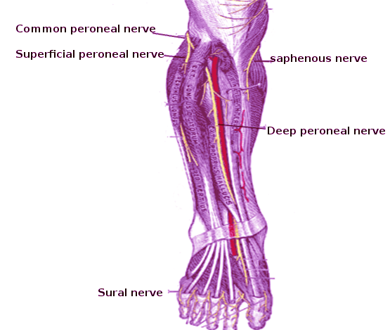
The common peroneal nerve often referred to as the common fibular nerve, is a major nerve that innervates the lower extremity. It is one of the two major branches off the sciatic nerve and receives fibers from the posterior divisions of L4 through S2 nerve roots.[1] The common peroneal nerve separates from the sciatic nerve in the distal posterior thigh proximal to the popliteal fossa.[1] After branching off of the sciatic nerve, it continues down the thigh, running posteroinferior to the biceps femoris muscle, and crosses laterally to the head of the lateral gastrocnemius muscle through the posterior intermuscular septum. The nerve then curves around the fibular neck before dividing into two branches, the superficial peroneal nerve (SPN) and the deep peroneal nerve (DPN).[1] The common peroneal nerve does not have any motor innervation before dividing; however, it provides sensory innervation to the lateral leg via the lateral sural nerve.[1]

The superficial peroneal nerve innervates the lateral compartment of the leg, and the deep peroneal nerve innervates the anterior compartment of the leg and the dorsum of the foot.[2] These two nerves are essential in the eversion of the foot and dorsiflexion of the foot, respectively. The superficial peroneal nerve provides both motor and sensory innervation. The innervation of the superficial peroneal nerve appears below:

  • Motor
    • Lateral compartment
      • Peroneus longus
      • Peroneus brevis
  • Sensory
    • Anterolateral leg

The deep peroneal nerve also provides both motor and sensory innervation in the leg. Deep peroneal nerve innervation is as follows:

  • Motor
    • Anterior compartment
      • Tibialis anterior
      • Extensor hallucis longus
      • Extensor digitorum longus
      • Peroneus tertius
  • Sensory
    • First dorsal webspace

The following tests can quickly assess the common peroneal nerve and its respective branches for both motor and sensory functions:

  • Sensory
    • Common peroneal nerve: lateral aspect of the leg just distal to the knee
    • Superficial peroneal nerve: the anterolateral distal third of the leg and the majority of the dorsum of the foot (except the first webspace)
    • Deep peroneal nerve: first dorsal webspace
  • Motor
    • Common peroneal nerve: none
    • Superficial peroneal nerve: eversion of the foot
    • Deep peroneal nerve: dorsiflexion of the foot and great toe extension

This paper will focus on peroneal nerve injuries, including etiologies, epidemiology, history, and physical exam findings, as well as diagnosis and treatment.

Etiology

There have been numerous traumatic and atraumatic causes of peroneal nerve injury. Common causes of damage to the peroneal nerve include the following:[3][4][5]

  • Trauma or injury to the knee
    • Knee dislocation
      • Common peroneal nerve injury can occur in up to 40% of patients[6][7]
    • A direct impact, penetrating trauma, or laceration
      • Injury predisposition as the nerve courses around the fibular head/neck
    • Fracture of the fibula, especially at the proximal fibula
  • Anatomic risk factors
    • Common peroneal nerve entrapment can occur secondary to a fibrous band at the origin of the peroneus longus
  • External compression sources
    • Tight splint/cast
    • Compression wrapping/bandage
    • Habitual leg crossing
    • Prolonged bed rest
      • Can be seen in comatose patients or those intubated for prolonged periods
    • Positioning during anesthesia and surgery
      • Important to always pad bony prominences, including the area of the fibular head and neck at the lateral aspect of the knee
  • Intraneural ganglion
  • Peripheral nerve tumor
  • Iatrogenic injury following surgery about the hip, knee, and ankle

The common peroneal nerve is most vulnerable to injury as it passes around the fibular neck. Therefore it is most susceptible to injury at the lateral aspect of the knee.[1][8]

The overall incidence of peroneal nerve injury secondary to closed fractures is challenging to elucidate, given the lack of prospectively acquired data. Fractures of the tibia (including tibial plateau) and/or fibula are reportedly associated with approximately a 1 to 2% rate of peroneal nerve injury.[9] There are reports of even rarer incidences following total knee arthroplasty (TKA) or arthroscopic lateral meniscal repair.[8]

Significant trauma around the knee can also result in peroneal nerve injury due to the nerve’s proximity to the knee joint itself as well as its superficial location; this can include a direct impact to the fibular neck, lacerations, and knee dislocations. Common peroneal nerve injuries are commonly encountered in athletes, especially football or soccer players, either in association with knee dislocation and ligamentous injury or in isolation.[8]

Several systemic illnesses can cause compressive peroneal neuropathy and injury to the common peroneal nerve, including:[8]

  • Diabetes mellitus
  • Inflammatory conditions
  • Motor neuron diseases
  • Anorexia nervosa (exceedingly thin states) due to loss of subcutaneous fat at this level causing compression of the nerve

Epidemiology

Common peroneal neuropathy is the commonest mononeuropathy encountered in the lower limbs and the third most common focal neuropathy encountered overall, after median (carpal tunnel syndrome) and ulnar neuropathies.[8][10][11] Traumatic injuries to the common peroneal nerve (CPN) most commonly afflict young athletic patients (e.g., football, soccer) and adult patients following high energy trauma (e.g., motor vehicle collisions (MVCs)), with CPN injury reportedly occurring in 16 to 40% of patients following knee dislocations.[7] Low energy knee dislocations can occur in obese patients during basic activities of daily living.

History and Physical

A thorough history and physical exam to assess the status and function of the common peroneal nerve is always required. In patients with a peroneal nerve injury, clinical presentation varies based on the location and severity of the injury and the presence of anatomic variations. The most common presentation of a common peroneal nerve injury is a weakness of ankle dorsiflexion and the classic resultant foot drop or catching the toes while ambulating.[8] Foot drop can develop acutely or over days to weeks, depending on the etiology. It can also be complete or partial in severity. There may also be accompanying numbness or paresthesia present along the lateral leg, dorsal foot, and/or the first toe webspace. Pain may also be present in traumatic cases but is not always present.[8]

The clinician directs the clinical physical examination by the patient’s reported symptoms and requires an understanding of the relevant anatomy and possible underlying etiology. For example, in cases of suspected chronic peroneal nerve palsy with resultant foot drop, the clinician should consider a primary documented part of the examination focusing on gait assessment as it may provide significant clues to the etiology of the symptoms.[8] A patient with weakened or paralyzed dorsiflexors may ambulate with a high steppage gait to prevent dragging their toes on the ground due to nerve injury. Furthermore, numbness or dysesthesia in the upper lateral leg indicates a lesion proximal to the fibular head, which may involve the sciatic nerve or lumbosacral nerve roots. In contrast, if there is a decreased or abnormal sensation in the lower lateral leg and dorsum of the foot, the superficial peroneal nerve may be involved. If there is also altered sensation in the dorsal aspect of the first web space of the foot, the deep peroneal nerve may also be involved.[8]

In the setting of traumatic injuries to the knee, including knee dislocations, the initial examination testing is done to rule out an acute neurovascular injury that could potentially compromise the lower extremity. Knee dislocations can notoriously compromise the neurovascular structures passing from the thigh and knee to the lower limb and, if not addressed urgently, can lead to acute compartment syndrome (ACS), which can result in long-term compromise to the lower limb requiring operative amputation.[12][13]

To test for the motor involvement of the superficial peroneal nerve and deep peroneal nerve, one must assess foot eversion (SPN) and foot/toe dorsiflexion (DPN). A finding of weakness of both foot eversion as well as foot/toe dorsiflexion suggests a lesion involving the common peroneal nerve.[8] Proximal lesions, for example, secondary to traumatic knee dislocations, will often present with varying degrees of numbness in both superficial peroneal nerve and deep peroneal nerve distributions. A careful and detailed examination detecting the presence or absence of dorsiflexion of the ankle and/or great toe past neutral is imperative for motor examination testing. The subtle distinction is important in differentiating the ability to actively dorsiflex the great toe from a maximally plantarflexed position to a near-neutral position. Such findings do not rule out deep peroneal nerve injury.

Tinel sign is also a reliable clinical sign to localize the area of nerve irritation or entrapment. The examiner performs the Tinel test by tapping along the course of the nerve – particularly around the fibular neck. If tingling or paresthesia is elicited distally to the point of compression by tapping, it denotes a positive Tinel sign.[8]

Evaluation

When the history and physical examination is indicative of a potential injury to the common peroneal nerve, plain radiography should be part of the initial workup. Because of the proximity of the common peroneal nerve to the fibular neck as well as its superficial location, it is particularly susceptible to direct trauma injury as well as entrapment by soft and bony tissue. In addition to plain radiography, CT scans can be used to evaluate further osseous abnormalities, and MRI/ultrasound can be used to assess soft-tissue sources or masses.[4] The former should be considered in the setting of tibial plateau fractures, while the latter (i.e., MRI) is prudent in cases of traumatic knee dislocations.

Electrodiagnostic studies, including nerve conduction velocity (NCV) tests and electromyography tests (EMG), can be used to diagnose peroneal nerve palsy. These tests help in the evaluation of the motor and sensory axons of the peroneal nerve and its branches. They are also helpful in the localization of nerve injury. These tests are useful in patients presenting with new-onset of peroneal nerve symptoms such as foot drop without a traumatic mechanism in addition to evaluating patients in the postoperative setting of a known traumatic peroneal nerve injury to plan long-term management as well as to provide patient.

Treatment / Management

There are operative and non-operative options for the treatment of common peroneal nerve injuries. Non-operative treatments include ankle-foot orthoses and physical therapy. An ankle-foot orthotic (AFO) may be used for foot drop when surgery is not warranted or during surgical recovery. The specific purpose of an ankle-foot orthotic is to provide toe dorsiflexion during the swing phase, medial or lateral stability at the ankle during stance, and, if necessary, some push off stimulation during the late stance phase. There have been advancements in ankle-foot orthotics in recent years, including carbon fiber that provides even more energy for push-off during the stance phase as well as being lighter and more comfortable to use for patients. Physical therapy for common peroneal nerve injury includes a program of stretching, strengthening, mobilization, manipulation, and proprioceptive and balancing exercises. It may also include icing, ankle bracing, and/or taping.

Surgical indications for common peroneal nerve injuries include a rapidly deteriorating lesion and no signs of improvement within three months and open injuries with suspected nerve laceration.[4] Open lacerations should undergo exploration and surgical repair within 72 hours.[4]

Differential Diagnosis

Critical elements pertinent to the differential diagnosis of CPN injury entail discerning the following:

  • Chronicity of the injury
    • Acute, subacute, or chronic
  • Injury location
    • CPN injury can occur proximally about the hip (e.g., acetabular fracture, posterior hip dislocation), at the knee/fibular head/neck, or more distally in the SPN or DPN distributions
  • Underlying etiology
    • Traumatic injuries (fracture/dislocations)
    • Atraumatic mechanisms
      • Systemic or inflammatory conditions (e.g., diabetes mellitus)
    • Iatrogenic causes
    • External (splint/cast) or internal (anatomic) sources of compression

Other clinical entities/pathologies that can mimic these types of presentations can include, but are not limited to:

  • Peroneal tendon pathology
  • Other compressive neuropathies
    • Tarsal tunnel syndrome
    • Anterior tarsal tunnel syndrome
    • Nonspecific tendinitis affecting lower limb muscles/tendons
  • Chronic ankle pain

Prognosis

Prognosis depends on etiology. While some transient common peroneal nerve neuropathies often improve or resolve over time with nonsurgical measures, common peroneal nerve palsy following traumatic knee dislocations results in inferior outcomes with a poor prognosis long-term recovery of nerve function.[14] Surgical intervention and tendon transfers are often necessary for refractory common peroneal nerve palsy, dictated by follow-up physical examination and serial EMG/NCS testing.

Prognosis has been described extensively in patients who experience common peroneal nerve injury following total knee arthroplasty. In one particular study, 62% of patients had a maximal neurologic recovery, and 38% had complete recovery at 12 months.[15] In another large case series study including 318 operatively-managed peroneal nerve lesions associated with various mechanisms, 84% of patients who underwent end-to-end suture repair received good recovery by 24 months.[4] In patients who required grafts, those with grafts less than 6 cm had a 75% recovery of function. Longer grafts were associated with worse outcomes.[4]

Complications

Common peroneal nerve injuries typically receive treatment with conservative modalities, including splinting and physical therapy. In patients with foot drop, the clinician must ensure the physical therapist and rehabilitation care team are ensuring that the risks of a fixed equinus contracture are mitigated by performing passive stretching exercises to ensure the ankle can achieve at least passive dorsiflexion to a neutral position.[8]

Deterrence and Patient Education

It is the physician's responsibility to provide patient education on the treatment options available for common peroneal nerve injuries, including the prognosis and complications that go along with each. In turn, patients should follow their individualized treatment plans for the best results. Patients should also maintain regular contact and follow up with their physician to ensure appropriate care. 

Pearls and Other Issues

  • The most common presentation with common peroneal nerve injury or palsy is acute foot drop, although symptoms may be progressive and can include sensory loss or pain.
  • Weakness in foot eversion may occur if the superficial peroneal nerve component is involved.
  • Weakness in dorsiflexion of the foot and great toe extension suggests deep peroneal nerve involvement.
  • The pattern of sensory loss suggests either superficial or deep peroneal nerve involvement or both.
  • The prognosis for common peroneal nerve injury is good following end-to-end suture repair as well as grafting.

Enhancing Healthcare Team Outcomes

Patients with common peroneal nerve injury/palsy experience a wide range of prognoses along a spectrum depending on the underlying etiology. For example, in the setting of knee dislocations with presenting foot drop and peroneal nerve palsy, outcomes across the board are poor in general in regards to the return of native function.

To adequately treat patients with common peroneal nerve injuries, specialists including neurologists and orthopedic surgeons must coordinate and work closely with primary care physicians, nurse practitioners, physician assistants, and physical therapists to ensure appropriate management and outcomes. 

The key to the management of the common peroneal nerve is early recognition and appropriate referral to the specialist. The emergency department physician should be able to recognize common peroneal nerve injuries and consult with the orthopedic surgeon. For those managed with non-surgical methods, the nurse has to educate the patient about foot drop and the potential to cause more injury to the tissues. The physical therapist should educate the patient on ambulation, ankle bracing, and the use of an assistive device. The patient requires monitoring for recovery, which in many cases, may take weeks or months. A wound care nurse should follow patients with open wounds until complete healing has occurred. Post-surgical pain control can benefit from pharmacist involvement, assisting with agent selection as well as minimizing opioid use. Communication between all these disciplines is paramount so that everyone on the interprofessional team has the full clinical picture and can make decisions based on the current status.

Peroneal nerve injuries require an interprofessional team approach, including physicians, specialists, specialty-trained nurses, physical therapists, and pharmacists, all collaborating across disciplines to achieve optimal patient results. [Level 5]


(Click Image to Enlarge)
Peroneal nerve
Peroneal nerve
Image courtesy O.Chaigasame
Details

Author

Bradley Lezak

Updated:

11/14/2022 11:50:33 AM

References


[1]

Hardin JM, Devendra S. Anatomy, Bony Pelvis and Lower Limb: Calf Common Peroneal Nerve (Common Fibular Nerve). StatPearls. 2023 Jan:():     [PubMed PMID: 30422563]


[2]

Garrett A, Geiger Z. Anatomy, Bony Pelvis and Lower Limb: Superficial Peroneal Nerve (Superficial Fibular Nerve). StatPearls. 2023 Jan:():     [PubMed PMID: 30521214]


[3]

Baima J, Krivickas L. Evaluation and treatment of peroneal neuropathy. Current reviews in musculoskeletal medicine. 2008 Jun:1(2):147-53. doi: 10.1007/s12178-008-9023-6. Epub     [PubMed PMID: 19468889]


[4]

Poage C, Roth C, Scott B. Peroneal Nerve Palsy: Evaluation and Management. The Journal of the American Academy of Orthopaedic Surgeons. 2016 Jan:24(1):1-10. doi: 10.5435/JAAOS-D-14-00420. Epub     [PubMed PMID: 26700629]


[5]

Gloobe H, Chain D. Fibular fibrous arch. Anatomical considerations in fibular tunnel syndrome. Acta anatomica. 1973:85(1):84-7     [PubMed PMID: 4713100]


[6]

LaPrade RF, Terry GC. Injuries to the posterolateral aspect of the knee. Association of anatomic injury patterns with clinical instability. The American journal of sports medicine. 1997 Jul-Aug:25(4):433-8     [PubMed PMID: 9240974]


[7]

Moatshe G, Dornan GJ, Løken S, Ludvigsen TC, LaPrade RF, Engebretsen L. Demographics and Injuries Associated With Knee Dislocation: A Prospective Review of 303 Patients. Orthopaedic journal of sports medicine. 2017 May:5(5):2325967117706521. doi: 10.1177/2325967117706521. Epub 2017 May 22     [PubMed PMID: 28589159]


[8]

Marciniak C. Fibular (peroneal) neuropathy: electrodiagnostic features and clinical correlates. Physical medicine and rehabilitation clinics of North America. 2013 Feb:24(1):121-37. doi: 10.1016/j.pmr.2012.08.016. Epub 2012 Oct 26     [PubMed PMID: 23177035]


[9]

Noble J, Munro CA, Prasad VS, Midha R. Analysis of upper and lower extremity peripheral nerve injuries in a population of patients with multiple injuries. The Journal of trauma. 1998 Jul:45(1):116-22     [PubMed PMID: 9680023]


[10]

Sevy JO, Varacallo M. Carpal Tunnel Syndrome. StatPearls. 2023 Jan:():     [PubMed PMID: 28846321]


[11]

Aleksenko D, Varacallo M. Guyon Canal Syndrome. StatPearls. 2023 Jan:():     [PubMed PMID: 28613717]


[12]

Varacallo M, Shirey L, Kavuri V, Harding S. Acute compartment syndrome of the hand secondary to propofol extravasation. Journal of clinical anesthesia. 2018 Jun:47():1-2. doi: 10.1016/j.jclinane.2018.01.020. Epub 2018 Feb 21     [PubMed PMID: 29476968]


[13]

Chandwani D, Varacallo M. Exertional Compartment Syndrome. StatPearls. 2023 Jan:():     [PubMed PMID: 31335004]


[14]

O'Malley MP, Pareek A, Reardon P, Krych A, Stuart MJ, Levy BA. Treatment of Peroneal Nerve Injuries in the Multiligament Injured/Dislocated Knee. The journal of knee surgery. 2016 May:29(4):287-92. doi: 10.1055/s-0035-1570019. Epub 2015 Dec 18     [PubMed PMID: 26683981]


[15]

Park JH, Restrepo C, Norton R, Mandel S, Sharkey PF, Parvizi J. Common peroneal nerve palsy following total knee arthroplasty: prognostic factors and course of recovery. The Journal of arthroplasty. 2013 Oct:28(9):1538-42. doi: 10.1016/j.arth.2013.02.025. Epub 2013 Apr 4     [PubMed PMID: 23562462]