Posterior Tibial Tendon Dysfunction

Earn CME/CE in your profession:


Continuing Education Activity

Posterior tibial tendon dysfunction (PTTD) represents an acquired, progressive disease of the foot and ankle that is seen commonly in middle-aged patients. It is the most common cause of adult acquired flatfoot deformity. Treatments involve conservative and surgical options depending on the severity of the disease. This activity outlines the evaluation and treatment of PTTD and highlights the role of the interprofessional team in managing patients with this condition.

Objectives:

  • Summarize the etiology of posterior tibial tendon dysfunction.
  • Describe the physical exam findings associated with posterior tibial tendon dysfunction.
  • Identify treatment and management options available for all stages of posterior tibial tendon dysfunction.
  • Outline interprofessional team strategies for improving care coordination and communication to advance the treatment of posterior tibial tendon dysfunction and improve patient outcomes.

Introduction

Posterior tibial tendon dysfunction (PTTD) insufficiency is the most common cause of adult acquired flatfoot deformity. Failure of the tendon affects surrounding ligamentous structures and will eventually lead to bony involvement and deformity. The extent this disease progression will be explained in this review. PTTD is a progressive and debilitating disorder, which can be detrimental to patients due to limitations in mobility, significant pain, and weakness.[1] Risk factors for the disease have been studied and may include hypertension, obesity, diabetes, previous trauma, or steroid exposure.[2]

Etiology

Researchers have proposed numerous mechanisms for the degeneration of the posterior tibial tendon (PTT). The most commonly the cause for PTT degeneration is credited to a repetitive loading causing microtrauma and progressive failure. A retromalleolar, hypovascular region does exist and may also contribute to the disease. In a study by Manske et al., cadaver specimens showed a region of decreased blood supply from 2.2 +/- 0.8cm proximal to the medial malleolus to a region 0.6 +/- 0.6cm proximal to the medial malleolus.[3]

The anatomic course of the posterior tibial tendon also likely contributes as the tendon does make an acute turn around the medial malleolus. This location puts a significant amount of tension on the tendon in the region distal and posterior to the medial malleolus. The adjacent tendons, namely the flexor hallucis longus and the flexor digitorum longus, do not take this sharp turn.[4]

Other potential culprits include constriction beneath the flexor retinaculum,[5] abnormal anatomy of the talus,[6] degenerative changes associated with osteoarthritis,[7] and preexisting pes planus.[8]

Epidemiology

Although no significant large-scale studies have been performed about the overall incidence of this disease, it is believed that the prevalence is anywhere from 3.3 to 10%, depending on the sex and age of the patient. This disorder is associated with adult-acquired flatfoot deficiency which can cause it to be misdiagnosed, meaning the actual prevalence may be much higher than shown in the literature. The underreporting may also be due to early, asymptomatic stages of PTTD. The classic PTTD patient is an obese woman in her sixth decade of life.[9]

Histopathology

The normal posterior tibial tendon should be comprised of linear collagen bundles, normal fibroblast cellularity, and low proportion of vascular density. In posterior tibial tendon dysfunction, the tendon undergoes microtrauma and subsequent tendinitis characterized by increased mucin contentment, neovascularization, and fibroblast hypercellularity.[7]

History and Physical

The majority of posterior tibial tendon dysfunction patients will demonstrate medial ankle and foot pain, especially in the early stages of the disease. At later stages, lateral pain can be seen as well, due to sub-fibular impingement or peroneal tendon injury.[10] A thorough physical examination can help to determine the grading and severity of the disease. Attention should first be turned to the gross inspection of the feet while standing. A weight-bearing examination is of importance as a flexible deformity may present normally, while the patient is non-weight bearing. The medial longitudinal arch collapse leads to pes planus, an easily visualized condition. While inspecting the patient, one will also commonly see a “too many toes” sign, which is visible when looking from behind the patient. Due to the valgus alignment of the foot, you will see flaring out of more than two toes from the lateral hindfoot. Equinus contracture is also a common finding, which can limit ankle dorsiflexion.

Single-limb heel raise is an important clinical test that can differentiate stage 1 disease from stage 2 and higher. A patient in stage 1 disease should be able to perform this test without pain. In stage 2 disease, patients may be able to perform the test but will likely have pain. In later stages, the rigid deformity may prevent the patient from completing the test. The flexibility of the foot also requires evaluation on an exam.[10]

Evaluation

Imaging is critical in determining the severity of disease and subsequent treatment. Anteroposterior (AP) and lateral radiographs are necessary. Increased talonavicular uncoverage and increased talo-first metatarsal angle (or Simmons angle) will present on the AP foot radiograph. The normal talo-first metatarsal angle is around 7 degrees, and angles over 16 degrees indicate flatfoot deformity.[11] The talonavicular coverage expresses itself as the amount of the talus that is not in contact with the navicular medially, with values over 30 to 40% typically indicated forefoot abduction seen in stage II-IV PTTD.[9] On the weight-bearing lateral radiograph, one should evaluate for increased talo-first metatarsal angle (or Meary angle) which normally measures 0 degrees +/- 4 degrees but typically measures over 20 degrees in flatfoot deformity.[10]

Treatment / Management

Treatment for posterior tibial tendon dysfunction is a complicated subject, so this review will attempt to simplify by each stage for the reader:

  • All Stages initially:
    • Conservative management with NSAIDs and activity modification. Also meant for non-surgical candidates or low demand, elderly patients.[12]
  • Stage 1:
    • Conservative management through immobilization in a walking boot or cast for up to 3 to 4 weeks to allow for healing of the posterior tibial tendon may be warranted followed eccentric strengthening with physical therapy.[1]
    • If immobilization and physical therapy are successful, transitioning into custom-molded orthotics or AFO is appropriate to maintain relief. Emphasis on medial forefoot posting is critical. University of California Biomechanics Laboratory (UCBL) orthoses are a form of a custom insert with a lace-up component that can help maintain midfoot height.[13]
    • Conservative therapy should be for 3 to 4 months, and if it fails, then surgical intervention may be warranted. A tenosynovectomy, with tubularization, may be indicated
  • Stage 2A:
    • Conservative immobilization and physical therapy with orthotics or ankle-foot orthosis (AFO) as recommended in stage 1
    • Surgical treatment involves medial calcaneal osteotomy with posterior tendon debridement and repair. Ancillary procedures may include any or all of the following: flexor digitorum tendon (FDL) transfer, spring ligament reconstruction, or Achilles tendon lengthening.[14]
  • Stage 2B:
    • All of the previously listed procedures in Stage 2A +/- lateral column lengthening, or isolated subtalar joint arthrodesis
  • Stage 3:
    • Conservative therapies, as mentioned above.
    • Surgical treatment is often warranted as it involves rearfoot arthritic changes and a medial double arthrodesis or triple arthrodesis common (subtalar, calcaneocuboid, and talonavicular arthrodesis) is indicated with or without deltoid ligament repair.[15]
  • Stage 4
    • Conservative therapies, as mentioned above.
    • Surgical treatment is often necessary as it involves arthritic changes in the ankle, as well as rearfoot. Surgical options include any combination of Triple arthrodesis with Achilles tendon lengthening, deltoid ligament reconstruction, and total ankle arthroplasty with replacement.[15] Medial double arthrodesis, deltoid ligament reconstruction, and total ankle arthroplasty with a replacement if ankle deformity is reducible. If the patient is a candidate for total ankle arthroplasty, then it is recommended to stage procedure by performing rearfoot arthrodesis and soft tissue balancing followed by a second stage 4 to 8 weeks later for total ankle arthroplasty 
    • Tibiotalocalcaneal (TTC) arthrodesis for rigid hindfoot with significant valgus alignment of the talus within the ankle mortise.[16][17]. Pantalar arthrodesis (triple arthrodesis with the addition of ankle arthrodesis).

Differential Diagnosis

Although posterior tibial tendon dysfunction is the most common cause of adult acquired flatfoot deformity, there are many other related conditions. Diagnoses listed below can present very similarly to PTTD and should merit consideration during evaluation [18]:

  • Tarsal coalition
  • Inflammatory arthritis
  • Charcot arthropathy
  • Neuromuscular disease
  • Traumatic disruption of midfoot ligaments

Staging

Stage 1

  • Normal radiographs, able to perform single-heel raise, and mild tenosynovitis

Stage 2A

  • Arch collapse on a radiograph, unable to perform single-heel raise, and a flexible flatfoot deformity

Stage 2B[19]

  • Arch collapse and talonavicular uncoverage (over 40%) on a radiograph, unable to perform single heel raise, flexible flatfoot deformity, and characteristic forefoot abduction or “too many toes” sign

Stage 3

  • Subtalar arthritis on a radiograph, unable to perform single heel raise, flatfoot deformity with rigid forefoot abduction, and hindfoot valgus

Stage 4 [19]

  • Valgus deformity of talus in the ankle mortise visualized on AP radiograph of the ankle - talar tilt due to deltoid ligament compromise, subtalar arthritis on radiographs, unable to perform single heel raise, flatfoot deformity with rigid forefoot abduction, and hindfoot valgus

Prognosis

Posterior tibial tendon dysfunction is a progressive disorder that will continue to deteriorate without treatment. Early detection and intervention will help to slow progression. Patients provided with custom orthotics and rehabilitation have been shown to have significant improvement. In a recent study by Alvarez et al., about 89% of their patients with stage I and II PTTD responded to orthotics and PT. Nearly all of these patients were back to full strength by 4 months.[20] According to analyses of outcomes of surgical treatment, results are much less predictable, and a return to the pre-disease state should not be guaranteed. Patients may continue to have some residual effects after reconstructive surgeries.[21]

Complications

General complications include thromboembolic events, infection, wound dehiscence, neurologic injury, and/or painful hardware. Reports exist of wound healing complications in up to one-third of patients undergoing flatfoot reconstruction, so proper wound care is paramount.[22]

Postoperative and Rehabilitation Care

Postoperative care is crucial to the success of the procedures. Typically, patients will receive a type of non-weight bearing cast or splint for around 6 to 8 weeks. Followup should be between 10 to 14 days for suture removal evaluation. Physical therapy may be necessary for certain patients.

Deterrence and Patient Education

Patient education and emphasis of conservative approach can help patient outcomes: 

  • Activity restriction and modification is a significant initial step in non-operative management.
  • Evidence supports conservative therapy in the early stages of the disease, such as the use of non-steroidal anti-inflammatory drugs. 
  • Providing prescriptions for medial arch support insoles or custom orthotics is necessary in many cases.
  • Moreover, surgery is usually the last resort if recovery is prolonged.
  • When performing surgery, temper patient expectations, as reconstruction options commonly have residual symptoms.

Pearls and Other Issues

  • Staging of posterior tibial tendon dysfunction is critical to surgical plan/outcome
  • Difference between Stage 2 and 3 is the presence of rigid deformity
  • Identification of the specific location of arthritic changes on radiograph will help to differentiate between stage 3 and 4 PTTD: Stage 3 demonstrates subtalar joint arthritis, Stage 4 demonstrates talar tilt and ankle arthritis

Enhancing Healthcare Team Outcomes

The importance of finding posterior tibial tendon dysfunction at early stages may prevent rapid deterioration with conservative measures. Primary care physicians and nurse practitioners may be able to refer patients to sub-specialists, such as foot and ankle orthopedic surgeons for a more thorough workup/evaluation, which can lead to significantly better patient outcomes. 

PTTD requires an interprofessional team approach, including physicians, specialists, specialty-trained nurses, and pharmacists, all collaborating across disciplines to achieve optimal patient results. [Level V]


(Click Image to Enlarge)
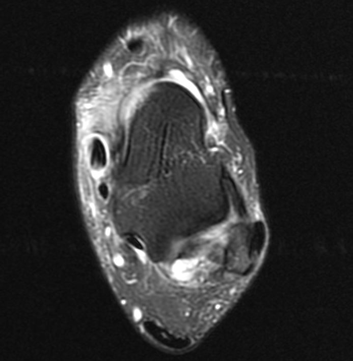
Posterior Tibial Tendon Dysfunction- MRI demonstrating extensive tenosynovitis of the posterior tibial tendon (PTT)
Posterior Tibial Tendon Dysfunction- MRI demonstrating extensive tenosynovitis of the posterior tibial tendon (PTT). No tears noted. Note the PTT is over double the size of the FDL tendon.
Contributed by Mark A. Dreyer, DPM, FACFAS
Details

Author

Paul W. Knapp

Editor:

Dustin Constant

Updated:

5/23/2023 12:30:32 PM

References


[1]

Ross MH, Smith MD, Mellor R, Vicenzino B. Exercise for posterior tibial tendon dysfunction: a systematic review of randomised clinical trials and clinical guidelines. BMJ open sport & exercise medicine. 2018:4(1):e000430. doi: 10.1136/bmjsem-2018-000430. Epub 2018 Sep 19     [PubMed PMID: 30271611]

Level 1 (high-level) evidence

[2]

Holmes GB Jr, Mann RA. Possible epidemiological factors associated with rupture of the posterior tibial tendon. Foot & ankle. 1992 Feb:13(2):70-9     [PubMed PMID: 1349292]

Level 2 (mid-level) evidence

[3]

Manske MC, McKeon KE, Johnson JE, McCormick JJ, Klein SE. Arterial anatomy of the tibialis posterior tendon. Foot & ankle international. 2015 Apr:36(4):436-43. doi: 10.1177/1071100714559271. Epub 2014 Nov 19     [PubMed PMID: 25411117]


[4]

Uchiyama E, Kitaoka HB, Fujii T, Luo ZP, Momose T, Berglund LJ, An KN. Gliding resistance of the posterior tibial tendon. Foot & ankle international. 2006 Sep:27(9):723-7     [PubMed PMID: 17038285]


[5]

Jahss MH. Spontaneous rupture of the tibialis posterior tendon: clinical findings, tenographic studies, and a new technique of repair. Foot & ankle. 1982 Nov-Dec:3(3):158-66     [PubMed PMID: 7152401]


[6]

Anderson JG, Harrington R, Ching RP, Tencer A, Sangeorzan BJ. Alterations in talar morphology associated with adult flatfoot. Foot & ankle international. 1997 Nov:18(11):705-9     [PubMed PMID: 9391815]


[7]

Mosier SM, Lucas DR, Pomeroy G, Manoli A 2nd. Pathology of the posterior tibial tendon in posterior tibial tendon insufficiency. Foot & ankle international. 1998 Aug:19(8):520-4     [PubMed PMID: 9728698]


[8]

Mann RA, Thompson FM. Rupture of the posterior tibial tendon causing flat foot. Surgical treatment. The Journal of bone and joint surgery. American volume. 1985 Apr:67(4):556-61     [PubMed PMID: 3980501]


[9]

Deland JT, Page A, Sung IH, O'Malley MJ, Inda D, Choung S. Posterior tibial tendon insufficiency results at different stages. HSS journal : the musculoskeletal journal of Hospital for Special Surgery. 2006 Sep:2(2):157-60. doi: 10.1007/s11420-006-9017-0. Epub     [PubMed PMID: 18751830]


[10]

Ellis SJ, Deyer T, Williams BR, Yu JC, Lehto S, Maderazo A, Pavlov H, Deland JT. Assessment of lateral hindfoot pain in acquired flatfoot deformity using weightbearing multiplanar imaging. Foot & ankle international. 2010 May:31(5):361-71. doi: 10.3113/FAI.2010.0361. Epub     [PubMed PMID: 20460061]


[11]

Younger AS, Sawatzky B, Dryden P. Radiographic assessment of adult flatfoot. Foot & ankle international. 2005 Oct:26(10):820-5     [PubMed PMID: 16221454]


[12]

Nielsen MD, Dodson EE, Shadrick DL, Catanzariti AR, Mendicino RW, Malay DS. Nonoperative care for the treatment of adult-acquired flatfoot deformity. The Journal of foot and ankle surgery : official publication of the American College of Foot and Ankle Surgeons. 2011 May-Jun:50(3):311-4. doi: 10.1053/j.jfas.2011.02.002. Epub 2011 Mar 31     [PubMed PMID: 21458301]


[13]

Imhauser CW, Abidi NA, Frankel DZ, Gavin K, Siegler S. Biomechanical evaluation of the efficacy of external stabilizers in the conservative treatment of acquired flatfoot deformity. Foot & ankle international. 2002 Aug:23(8):727-37     [PubMed PMID: 12199387]


[14]

Myerson MS, Badekas A, Schon LC. Treatment of stage II posterior tibial tendon deficiency with flexor digitorum longus tendon transfer and calcaneal osteotomy. Foot & ankle international. 2004 Jul:25(7):445-50     [PubMed PMID: 15319100]


[15]

Kadakia AR, Haddad SL. Hindfoot arthrodesis for the adult acquired flat foot. Foot and ankle clinics. 2003 Sep:8(3):569-94, x     [PubMed PMID: 14560906]


[16]

Pellegrini MJ, Schiff AP, Adams SB Jr, DeOrio JK, Easley ME, Nunley JA 2nd. Outcomes of Tibiotalocalcaneal Arthrodesis Through a Posterior Achilles Tendon-Splitting Approach. Foot & ankle international. 2016 Mar:37(3):312-9. doi: 10.1177/1071100715615398. Epub 2015 Nov 17     [PubMed PMID: 26578482]


[17]

Smith JT, Bluman EM. Update on stage IV acquired adult flatfoot disorder: when the deltoid ligament becomes dysfunctional. Foot and ankle clinics. 2012 Jun:17(2):351-60. doi: 10.1016/j.fcl.2012.03.011. Epub 2012 Apr 10     [PubMed PMID: 22541531]


[18]

Pomeroy GC, Pike RH, Beals TC, Manoli A 2nd. Acquired flatfoot in adults due to dysfunction of the posterior tibial tendon. The Journal of bone and joint surgery. American volume. 1999 Aug:81(8):1173-82     [PubMed PMID: 10466651]


[19]

Iossi M, Johnson JE, McCormick JJ, Klein SE. Short-term radiographic analysis of operative correction of adult acquired flatfoot deformity. Foot & ankle international. 2013 Jun:34(6):781-91. doi: 10.1177/1071100713475432. Epub 2013 Feb 5     [PubMed PMID: 23386748]


[20]

Alvarez RG, Marini A, Schmitt C, Saltzman CL. Stage I and II posterior tibial tendon dysfunction treated by a structured nonoperative management protocol: an orthosis and exercise program. Foot & ankle international. 2006 Jan:27(1):2-8     [PubMed PMID: 16442022]


[21]

Pinney SJ, Lin SS. Current concept review: acquired adult flatfoot deformity. Foot & ankle international. 2006 Jan:27(1):66-75     [PubMed PMID: 16442033]


[22]

Coetzee JC, Hansen ST. Surgical management of severe deformity resulting from posterior tibial tendon dysfunction. Foot & ankle international. 2001 Dec:22(12):944-9     [PubMed PMID: 11783917]