Anatomy, Shoulder and Upper Limb, Radial Nerve


Introduction

The radial nerve is a peripheral nerve that provides motor and sensory function to the arm. The motor function innervates the posterior compartment of the arm, including the medial and lateral heads of the triceps brachii muscles in addition to all 12 muscles in the posterior forearm compartment, as well as the extrinsic extensor muscles found in the wrist and fingers. The sensory function provides cutaneous innervation to a portion of the anterolateral arm, distal posterior arm, posterior forearm, posterior aspects of the thumb, index finger, middle finger, and the lateral half of the ring finger. The radial nerve originates from the ventral roots of the spinal nerves C5-T1 of the brachial plexus, which eventually forms the posterior cord. Disruption of the radial nerve can have motor consequences such as an inability to extend the arm, wrist, and fingers and paresthesias about its sensory distribution.[1]

Structure and Function

Structure

The radial nerve is formed as a continuation of the posterior cord of the brachial plexus and arises from the C5-T1 nerve fibers. It courses from the axilla to the posterior compartment of the arm, then into the anterior compartment of the arm, and continues into the posterior compartment of the forearm.[2]

Arm

The radial nerve derives from the posterior cord of the brachial plexus and exits the axilla posteriorly to the brachial artery. It passes with the deep brachial artery and gives two motor branches and one sensory branch before traversing the triangular interval. These motor branches innervate the medial and long heads of the triceps. This sensory branch is called the posterior cutaneous nerve of the arm, which supplies cutaneous sensory innervation to a portion of the distal posterior arm. After passing through the triangular interval, the radial nerve descends the radial groove before laterally wrapping around the humerus. At this point, the radial nerve gives a motor branch to the lateral head of the triceps brachii, followed by two sensory branches: the inferior lateral cutaneous nerve of the arm, which perforates through the lateral head of the triceps, and the posterior cutaneous nerve of the forearm.[3]

Forearm

The posterior cutaneous nerve of the antebrachium also perforates through the lateral head of the triceps but continues to innervate a posterior strip of the forearm. After giving these two sensory branches, the radial nerve passes through the lateral intermuscular septum to infiltrate the anterior compartment of the forearm between the brachialis and brachioradialis muscles. The radial nerve then passes over the lateral epicondyle into the cubital fossa and forearm. Here, the radial nerve separates into deep and superficial branches. The deep branch is a motor branch that passes between the heads of the supinator muscle and becomes the posterior interosseous nerve to innervate the muscles of the posterior compartment of the forearm. The superficial branch follows the radial artery inferiorly to the anterolateral portion of the radius, deep to the brachioradialis muscle. The superficial branch then courses dorsally over the distal radius over the anatomical snuffbox to innervate the posterior lateral three and a half digits (the thumb, index, middle, and lateral half of the ring fingers) and the associated hand area.[3] 

Function

The following is a list of the motor and cutaneous sensory functions of the radial nerve.

Sensory

Anterior aspect:

  • The inferior lateral cutaneous nerve of the arm - provides sensation to the anterior lateral aspect of the mid-arm.

Posterior aspect:

  • The posterior cutaneous nerve of the arm - sensation to the posterior distal arm.
  • The posterior cutaneous nerve of the forearm - sensation to a strip posterior aspect of the forearm
  • Superficial branch - sensation to the posterior aspect of the thumb, index, middle, and lateral half of the ring fingers, as well as the associated dorsal hand area.

Motor

The Radial Nerve branches off to the Deep Branch after it passes through the cubital fossa and then continues as the Posterior Interosseous Nerve after it passes between the supinator muscle heads.

Radial Nerve:

  • Triceps brachii (medial and lateral heads) — provides the extension of the forearm.
  • Extensor carpi radialis longus — provides for the extension of the wrist.
  • Brachioradialis — provides flexion of the elbow as well as pronation and supination, depending on the position of the forearm.
  • Anconeus — provides for elbow extension.

Deep Branch of the Radial Nerve:

  • Extensor carpi radialis brevis — extends and abducts the wrist.
  • Supinator — supinates the forearm.

Posterior interosseous nerve:

  • Abductor pollicis longus — abduct the thumb at the wrist.
  • Extensor carpi ulnaris — extends and adducts the wrist.
  • Extensor digiti minimi — extends the wrist and small finger.
  • Extensor digitorum — extends the medial four digits of the hand.
  • Extensor indicis — extends the index finger and, to some extent, wrist extension.
  • Extensor pollicis brevis — extends and abducts the thumb at the carpometacarpal and metacarpophalangeal joints.
  • Extensor pollicis longus — extends the terminal phalanx of the thumb.

Embryology

The radial nerve is a peripheral nerve that arises from the bilaterally paired neural crest, which are strips of cells arising from the ectoderm at the margins of the neural tube during embryonic development. The migration of the neural crest cells eventually forms what becomes the radial nerve in a fully developed body.[4]

Blood Supply and Lymphatics

The radial nerve exits the axilla with the axillary artery and follows it posteriorly with the brachial artery. It then continues with the deep brachial artery into the posterior compartment via the triangular interval. The radial nerve follows the radial collateral artery until it wraps anteriorly over the cubital fossa. At this point, the radial nerve branches into the superficial branch of the radial nerve, which passes with the radial artery, the deep branch of the radial artery, and the posterior interosseous artery.[5]

Nerves

The radial nerve forms as a continuation of the posterior cord of the brachial plexus with nerve fibers from the C5-T1 nerve roots.[6]

The Radial Nerve branches:

Sensory:

  • The inferior lateral cutaneous nerve of the arm
  • The posterior cutaneous nerve of the arm
  • The posterior cutaneous nerve of the forearm
  • The superficial branch of the radial nerve

Motor:

  • Deep branch of the radial nerve
  • Posterior interosseous nerve

Muscles

The radial nerve and its branches provide innervation of the following muscles (See Structure and Function for specific nerve branch innervations and muscle actions)[7]:

  • Abductor pollicis longus
  • Anconeus
  • Brachioradialis
  • Extensor carpi radialis brevis
  • Extensor carpi radialis longus
  • Extensor carpi ulnaris
  • Extensor digiti minimi
  • Extensor digitorum
  • Extensor pollicis brevis
  • Extensor indicis
  • Extensor pollicis longus
  • Triceps brachii (medial and lateral heads)
  • Supinator

Physiologic Variants

The deep branch of the radial nerve normally passes between the heads of the supinator muscle as it becomes the posterior interosseous nerve to innervate the muscles of the posterior compartment of the forearm. A variant can occur where the deep branch of the radial nerve passes through the Arcade of Frohse (supinator arch), which can increase the likelihood of impingement.[8][9]

Surgical Considerations

Any surgery near the radial nerve holds a potential risk for an injury that will cause adverse downstream effects. 

The brachial plexus injury with radial nerve involvement can occur with the use of sternal retraction after sternotomy, particularly with internal mammary dissection during cardiac surgery.[10]

Damage to the superficial radial nerve is a potential complication of surgery for de Quervain tendinopathy.

There is a theoretical risk of radial nerve injury with hyperextension of the elbow, and the forearm should be placed in a slightly flexed position during anesthesia.[3]

Clinical Significance

Radial Tunnel Syndrome 

Radial Tunnel Syndrome presents with symptoms including fatigue or dull, aching pain at the proximal portion of the forearm during use. Less commonly, these symptoms can occur at the dorsal aspect of the wrist or hand. Radial Tunnel Syndrome typically occurs secondary to overuse or repetitive movements from pushing, pulling, gripping, pinching, or bending at the wrist, typically from a job or playing sports. Muscle overuse may cause compression of the radial nerve anywhere along its path but most commonly occurs over the elbow as it passes through the radial tunnel. Treatment for radial tunnel syndrome can be conservative or surgical if non-operative therapy fails.[11]

Radial Nerve Palsy

Radial neuropathies occur from injury to the radial nerve due to compression, ischemia, fractures to the arm, or penetrating wounds. Wrist drop is the most common presentation. The severity of the neuropathy depends on the level of the injury. Surgical procedures such as the stabilization of an acute humeral fracture with humeral nailing can also cause radial neuropathies. Palsy of the radial nerve is also known as crutch palsy, Saturday night palsy, and honeymooner’s palsy, conditions which may occur after placing one’s arm over a chair (or crutches) for an extended period causing a pressure injury to the radial nerve.[12][11][13]

Radial Nerve Entrapment

Radial nerve injury or compression can occur anywhere along the nerve's path, which can cause extensor or supinator muscle denervation. This can result in pain, weakness, dysfunction, or paresthesias and numbness along the sensory distribution of the radial nerve. The proximal forearm is the most common area of compression where the posterior interosseous branch of the radial nerve passes between the supinator heads. Other, less common, sites of compression can occur due to fractures of the humerus about the middle and proximal thirds of the shaft. The radial aspect of the wrist is another site of possible compression of the radial nerve. Treatment for radial nerve entrapment depends on the pathology and may be treated conservatively or surgically if conservative measures fail.[12][11] 

Cheiralgia Paresthetica (Wartenberg syndrome) 

Cheiralgia paresthetica is a hand neuropathy commonly caused by trauma or compression of the superficial branch of the radial nerve. The dorsum of the hand near the base of the thumb (in the vicinity of the anatomical snuffbox) is typically affected; however, it may affect the dorsum of the thumb, index finger, and hand. Symptoms include pain, numbness, tingling, or a burning sensation. There is no motor involvement since the superficial branch is purely sensory. Its etiology is thought to be caused by a constriction of the wrist as with a watch band or bracelet. It is associated with handcuff use and is also commonly referred to as handcuff neuropathy.[14]

Other Issues

The radial nerve passes through the groove of the humerus and therefore is vulnerable to injury in humeral fractures.[15]

Injury to the radial nerve can cause a wrist drop.[16]


(Click Image to Enlarge)
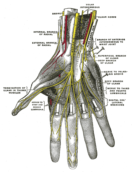
The Anterior Division, Deep palmar nerves, Ulnar and Radial Nerve
Contributed by Gray's Anatomy Plates

(Click Image to Enlarge)
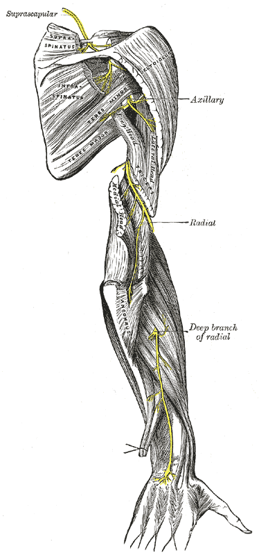
The suprascapular, axillary, and radial nerves.
Contributed by Henry Vandyke Carter - Henry Gray (1918) Anatomy of the Human Body (See "Book" section below) Bartleby.com: Gray's Anatomy, Plate 818
Details

Updated:

8/29/2022 2:03:45 PM

References


[1]

Cho SH, Chung IH, Lee UY. Relationship between the ulnar nerve and the branches of the radial nerve to the medial head of the triceps brachii muscle. Clinical anatomy (New York, N.Y.). 2019 Jan:32(1):137-142. doi: 10.1002/ca.23207. Epub 2018 Dec 3     [PubMed PMID: 29770497]


[2]

Musso D, Klaastad Ø, Wilsgaard T, Ytrebø LM. Brachial plexus block of the posterior and the lateral cord using ropivacaine 7.5 mg/mL. Acta anaesthesiologica Scandinavica. 2019 Mar:63(3):389-395. doi: 10.1111/aas.13277. Epub 2018 Oct 18     [PubMed PMID: 30338518]


[3]

Zhu W, Zhou R, Chen L, Chen Y, Huang L, Xia Y, Papadimos TJ, Xu X. The ultrasound-guided selective nerve block in the upper arm: an approach of retaining the motor function in elbow. BMC anesthesiology. 2018 Oct 19:18(1):143. doi: 10.1186/s12871-018-0584-7. Epub 2018 Oct 19     [PubMed PMID: 30340524]


[4]

Fraher JP,Dockery P,O'Donoghue O,Riedewald B,O'Leary D, Initial motor axon outgrowth from the developing central nervous system. Journal of anatomy. 2007 Nov     [PubMed PMID: 17850285]


[5]

Okwumabua E, Thompson JH. Anatomy, Shoulder and Upper Limb, Axillary Nerve. StatPearls. 2023 Jan:():     [PubMed PMID: 29630264]


[6]

Noland SS, Bishop AT, Spinner RJ, Shin AY. Adult Traumatic Brachial Plexus Injuries. The Journal of the American Academy of Orthopaedic Surgeons. 2019 Oct 1:27(19):705-716. doi: 10.5435/JAAOS-D-18-00433. Epub     [PubMed PMID: 30707114]


[7]

Puffer RC, Bishop AT, Spinner RJ, Shin AY. Bilateral Brachial Plexus Injury After MiraDry Procedure for Axillary Hyperhidrosis. World neurosurgery. 2019 Apr:124():370-372. doi: 10.1016/j.wneu.2019.01.093. Epub 2019 Jan 29     [PubMed PMID: 30703585]

Level 3 (low-level) evidence

[8]

Parra S,Orenga JV,Ghinea AD,Estarelles MJ,Masoliver A,Barreda I,Puertas FJ, Neurophysiological study of the radial nerve variant in the innervation of the dorsomedial surface of the hand. Muscle     [PubMed PMID: 29896804]


[9]

Babaei-Ghazani A, Roomizadeh P, Sanaei G, Najarzadeh-Mehdikhani S, Habibi K, Nikmanzar S, Kheyrollah Y. Ultrasonographic reference values for the deep branch of the radial nerve at the arcade of Frohse. Journal of ultrasound. 2018 Sep:21(3):225-231. doi: 10.1007/s40477-018-0303-8. Epub 2018 Jun 16     [PubMed PMID: 29909505]


[10]

Bunnell AE, Kao DS. Planning Interventions to Treat Brachial Plexopathies. Physical medicine and rehabilitation clinics of North America. 2018 Nov:29(4):689-700. doi: 10.1016/j.pmr.2018.06.005. Epub 2018 Sep 5     [PubMed PMID: 30293624]


[11]

Latef TJ, Bilal M, Vetter M, Iwanaga J, Oskouian RJ, Tubbs RS. Injury of the Radial Nerve in the Arm: A Review. Cureus. 2018 Feb 16:10(2):e2199. doi: 10.7759/cureus.2199. Epub 2018 Feb 16     [PubMed PMID: 29666777]


[12]

Akhavan-Sigari R,Mielke D,Farhadi A,Rohde V, Study of Radial Nerve Injury Caused By Gunshot Wounds and Explosive Injuries among Iraqi Soldiers. Open access Macedonian journal of medical sciences. 2018 Sep 25     [PubMed PMID: 30337976]

Level 2 (mid-level) evidence

[13]

Heiling B, Waschke A, Ceanga M, Grimm A, Witte OW, Axer H. Not your average Saturday night palsy-High resolution nerve ultrasound resolves rare cause of wrist drop. Clinical neurology and neurosurgery. 2018 Sep:172():160-161. doi: 10.1016/j.clineuro.2018.07.006. Epub 2018 Jul 9     [PubMed PMID: 30015054]


[14]

Ehrlich W, Dellon AL, Mackinnon SE. Classical article: Cheiralgia paresthetica (entrapment of the radial nerve). A translation in condensed form of Robert Wartenberg's original article published in 1932. The Journal of hand surgery. 1986 Mar:11(2):196-9     [PubMed PMID: 3514740]


[15]

Sukegawa K, Kuniyoshi K, Suzuki T, Matsuura Y, Onuma K, Kenmoku T, Takaso M. Effects of the Elbow Flexion Angle on the Radial Nerve Location around the Humerus: A Cadaver Study for Safe Installation of a Hinged External Fixator. The journal of hand surgery Asian-Pacific volume. 2018 Sep:23(3):388-394. doi: 10.1142/S242483551850042X. Epub     [PubMed PMID: 30282553]


[16]

Alam M, Haq AU. Wrist drop and focal seizures in a 60-year-old man. Postgraduate medical journal. 2018 Dec:94(1118):729. doi: 10.1136/postgradmedj-2018-136115. Epub 2018 Oct 19     [PubMed PMID: 30341228]