Anatomy, Abdomen and Pelvis, Rectus Femoris Muscle


Introduction

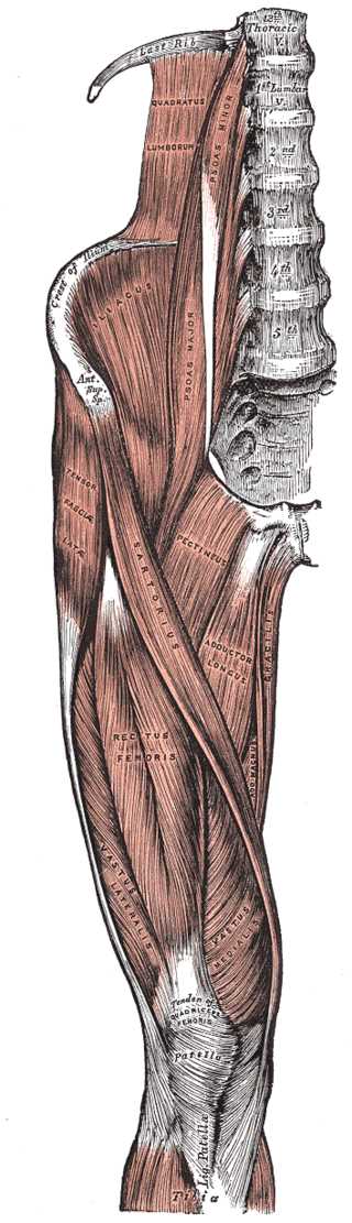
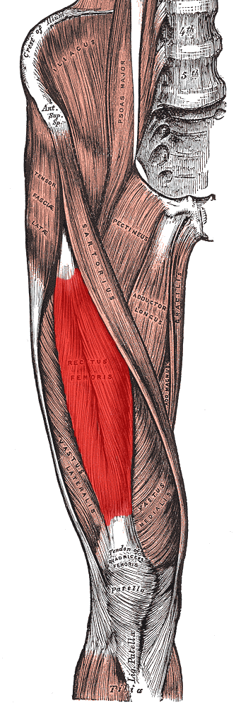
The rectus femoris is a long, fleshy muscle located in the anterior compartment of the thigh. The rectus femoris is fusiform in shape with superficial fibers that are bipenniform and deep fibers that run straight (rectus) to the deep aponeurosis.[1] The rectus femoris is the most superficial of the quadriceps muscles, alongside the vastus lateralis, vastus intermedius, and vastus medialis. These four muscles conjoin to attach to the patella as the quadriceps tendon. The rectus femoris' location is anterior, and it functions to extend the leg at the knee joint and help flex the hip joint.[1][2] 

The rectus femoris has two heads of origin: the direct (straight) head and the indirect (reflected) head. The direct head arises from the anterior aspect of the inferior iliac spine (AIIS; a common site of avulsion), while the indirect head originates from the acetabular ridge. The two heads merge to become the conjoined tendon below their origin.[3]

Structure and Function

Structure: The rectus femoris is fusiform in shape with superficial fibers that are bipenniform and deep fibers that run straight (rectus) to the deep aponeurosis. The rectus femoris arises from two tendons that originate from the anterior inferior iliac spine and the other from the ridge of the acetabulum. The rectus femoris tendons conjoin at an acute angle spreading into the aponeurosis. The end of the muscle inserts into the base of the patella, forming the patellar tendon with the rest of the quadriceps muscles.

Function: The rectus femoris flexes the hip along with the sartorius and iliopsoas and extends the lower leg at the knee, working in conjunction with the other three quadriceps muscles. The rectus femoris muscle antagonizes the hamstring muscles at the knee and the hip. When seated, the rectus femoris is one of the weaker extensors of the knee. With the knee fully extended, the rectus femoris is relatively short. Consequently, flexion of the hip is limited, which is referred to as active insufficiency. In contrast, when the knee is fully flexed, the rectus femoris is at its longest length, and extension of the hip is subsequently limited; this is passive insufficiency.[1]

Embryology

During the fifth week of development, the lower limb bud appears. It originates laterally from the L2 through S2 spinal segments and contains all primary germ layers: ectoderm, endoderm, and mesoderm. The musculature of the rectus femoris derives from the myotomic portions of somites (part of the mesoderm), while the skeletal elements supporting the lower limb come from the lateral plate somatic mesoderm. During embryogenesis, the lower limb rotates 90 degrees medially about the longitudinal axis, bringing the knee to the anterior aspect of the fetus.[2]

Blood Supply and Lymphatics

The femoral artery supplies the quadriceps muscle; the descending branch of the lateral femoral circumflex artery specifically nourishes the rectus femoris. The venous drainage is by the femoral vein and its branches.

The lumbar aortic lymph nodes facilitate the lymphatic vessels of the lower limb, including the rectus femoris muscle, and the drainage will flow into the cisterna chyli.[1]

Nerves

The neuronal mechanism of the quadriceps muscle (including the rectus femoris muscle) movement begins at the apex of the precentral gyrus. The neuronal signal travels down the corticospinal tract, which decussates in the medullary pyramid. After decussation, the corticospinal tract continues as the lateral corticospinal tract. Once the neuronal signal reaches spinal nerve L4, upper motor neurons synapse to lower motor neurons. The neuronal signal propagates through the anterior root of L4 until it reaches the anterior rami of L4, where it exits the spinal cord by way of the lumbar plexus. The posterior division of the L4 root, also known as the femoral nerve, supplies the quadriceps muscles. The lumbar nerves L2 and L3 also contribute to the femoral nerve. Once the signal reaches the quadriceps muscle via the femoral nerve, the quadriceps muscles work together to extend the knee and flex the thigh at the hip.[1]

Physiologic Variants

Accessory muscles can arise from the rectus femoris. These variations include a muscle slip from the acetabulum directly into the vastus lateralis. Variations in the origin of the rectus femoris can change from the upper anterior iliac spine to the lower anterior iliac spine with no acetabular origin in different individuals.[1][4]

Surgical Considerations

In acute rectus femoris muscle injuries, the patient may feel a tearing sensation with abrupt onset of pain. Subacute injuries may present with gradual onset of pain while running. Evaluation of a suspected rectus femoris injury generally includes imaging (ultrasonography and MRI) to diagnose and monitor the resolution of the injury.[5]

Distal rectus tear is more commonly associated with and involved in quadriceps tendon ruptures; isolated tears are uncommon. Proximal rectus femoris tears are typically managed conservatively initially. Should conservative management fail (exceedingly rare), surgery is a consideration. In keeping with the principles of surgical tendon management, repair of an avulsed tendon involves sewing the tendon into sutures anchored into the bony origin. If the torn tendon has sufficient tissue for surgical sewing, it may be sewn to the tendon stump. If the tendon edges are not suitable for repair or, more commonly, cannot be adequately mobilized (typical in chronic cases). Reconstruction with the use of tendon allografts (i.e., from a cadaver) is usually performed.[6]

Clinical Significance

The rectus femoris muscle is the most frequently injured quadriceps muscle and is particularly common in soccer players.[5][7][8][9] During intense exercise or trauma, the rectus femoris can tear at either the proximal or distal end, resulting in a debilitating injury that causes severe pain.[10] This injury can either be a partial tear of the tendon at the patella or the muscle itself. This type of strain injury may occur during sprinting, kicking, or jumping. When strain injury occurs, patients typically present with moderate to severe pain in the groin or anterior aspect of the hip and swelling surrounding the affected area. The patient may also be unable to extend the knee. MRI detects rectus femoral strains. As mentioned previously, a partial tear is usually treated with conservative care through activity modification (rest, crutch use) and positional restriction (a short period of avoiding hip extension).[11] Physical therapy can also play a role in rehabilitating torn muscles, ensuring a proper range of motion while healing occurs, which can help prevent re-injury later on.


(Click Image to Enlarge)
Tensor Fasciae Latae Muscles, Abductor of the Thigh, Thoracic Vertebrae, Quadratus Lumborum, Psoas Minor and Major, Crest of
Tensor Fasciae Latae Muscles, Abductor of the Thigh, Thoracic Vertebrae, Quadratus Lumborum, Psoas Minor and Major, Crest of Ilium, Anterior Superior Spine, Iliacus, Tensor Fascia Latae, Sartorius, Pectineus, Adductor Longus, Gracilis, Adductor Magnus, Rectus Femoris, Vastus Lateralis and Medialis, Tibia, Patella, Tendon of Quadriceps
Contributed by Gray's Anatomy Plates

(Click Image to Enlarge)
Rectus Femoris Muscle
Contributed by Grey's Anatomy Plates (Public Domain)

(Click Image to Enlarge)
Femoral muscles, Gluteus medius, Gluteus minimus, Piriformis, Superior gemellus, Tensor fasciae latae, Obturator internus, In
Femoral muscles, Gluteus medius, Gluteus minimus, Piriformis, Superior gemellus, Tensor fasciae latae, Obturator internus, Inferior gemellus, Quadratus femoris, Semitendinosus, Semimembranosus, Biceps femoris, Illiacus, Psoas major, Psoas minor, Iliopsoas, Pectineus, Adductor brevis, Adductor longus, Adductor magnus, Gracilis, Vastus lateralis, Rectus femoris, Vastus medialis, Sartorius
Illustration by Emma Gregory
Details

Editor:

Kofi Agyeman

Updated:

11/4/2022 8:11:11 PM

References


[1]

Bordoni B,Varacallo M, Anatomy, Bony Pelvis and Lower Limb, Thigh Quadriceps Muscle . 2018 Jan     [PubMed PMID: 30020706]


[2]

Lew V,Kang M, Anatomy, Abdomen and Pelvis, Femoral Sheath . 2018 Jan     [PubMed PMID: 29494010]


[3]

Kumaravel M,Bawa P,Murai N, Magnetic resonance imaging of muscle injury in elite American football players: Predictors for return to play and performance. European journal of radiology. 2018 Nov     [PubMed PMID: 30396649]


[4]

Biondi NL,Varacallo M, Anatomy, Bony Pelvis and Lower Limb, Vastus Lateralis Muscle . 2018 Jan     [PubMed PMID: 30335342]


[5]

Mendiguchia J,Alentorn-Geli E,Idoate F,Myer GD, Rectus femoris muscle injuries in football: a clinically relevant review of mechanisms of injury, risk factors and preventive strategies. British journal of sports medicine. 2013 Apr     [PubMed PMID: 22864009]


[6]

Volk WR,Yagnik GP,Uribe JW, Complications in brief: Quadriceps and patellar tendon tears. Clinical orthopaedics and related research. 2014 Mar     [PubMed PMID: 24338040]


[7]

Cross TM,Gibbs N,Houang MT,Cameron M, Acute quadriceps muscle strains: magnetic resonance imaging features and prognosis. The American journal of sports medicine. 2004 Apr-May     [PubMed PMID: 15090389]


[8]

Gyftopoulos S,Rosenberg ZS,Schweitzer ME,Bordalo-Rodrigues M, Normal anatomy and strains of the deep musculotendinous junction of the proximal rectus femoris: MRI features. AJR. American journal of roentgenology. 2008 Mar     [PubMed PMID: 18287410]


[9]

Pesquer L,Poussange N,Sonnery-Cottet B,Graveleau N,Meyer P,Dallaudiere B,Feldis M, Imaging of rectus femoris proximal tendinopathies. Skeletal radiology. 2016 Jul     [PubMed PMID: 26956398]


[10]

Esser S,Jantz D,Hurdle MF,Taylor W, Proximal Rectus Femoris Avulsion: Ultrasonic Diagnosis and Nonoperative Management. Journal of athletic training. 2015 Jul     [PubMed PMID: 25978099]


[11]

Colombelli A,Polidoro F,Guerra G,Belluati A, Patellar and quadriceps tendons acute repair with suture anchors. Acta bio-medica : Atenei Parmensis. 2019 Jan 14     [PubMed PMID: 30715027]