Respiratory Distress Syndrome

Earn CME/CE in your profession:


Continuing Education Activity

Acute respiratory distress syndrome (ARDS) is a common condition which is caused by pulmonary and extra-pulmonary pathologies, and it carries high rates of mortality and morbidity. It was first described 50 years ago. The definition is non-cardiogenic pulmonary edema that occurs because of an inflammatory process that increases capillary membranes permeability leading to impairment in oxygenation and gas exchange presenting with hypoxemia and bilateral pulmonary infiltrates. This activity examines when this condition should be considered on a differential diagnosis and how to properly evaluate for it. This activity highlights the role of the interprofessional team in caring for patients with this condition.

Objectives:

  • Identify the etiology of ARDs
  • Review the presentation of a patient with ARDs.
  • Outline the treatment and management options available for ARDs
  • Describe interprofessional team strategies for improving care and outcomes in patients with ARDs.

Introduction

Acute respiratory distress syndrome (ARDS) is a common condition which is caused by pulmonary and extra-pulmonary pathologies, and it carries high rates of mortality and morbidity. It was first described 50 years ago. The definition is non-cardiogenic pulmonary edema that occurs because of an inflammatory process that increases capillary membranes permeability leading to impairment in oxygenation and gas exchange presenting with hypoxemia and bilateral pulmonary infiltrates.[1][2]

Etiology

The underlying etiology of ARDS divides into pulmonary and extra-pulmonary pathologies. Common causes include bacteremia, sepsis, trauma, burns, reaction to massive transfusion, pneumonia, aspiration, severe pancreatitis, near drowning and fat embolism. Patients with increased severity scores for critical illness (e.g., Acute Physiology And Chronic Health Evaluation (APACHE) II score) are more prone to develop ARDS manifestations.

Several risk factors correlate with an increased risk of development of ARDS including female sex, advanced age, cigarette smoking, and alcohol abuse. Around 40 genes have links with ARDS, including genes encoding for angiotensin-converting enzyme inhibitor, tumor necrosis factor, interleukin 10, and others. However, the correlation between these genes, susceptibility for ARDS and its effect on clinical outcomes remained limited.[3]

Epidemiology

ARDS is a common condition in critical illness settings affecting 200000 patients in the United States and 3 million patients worldwide every year. It accounts for 10% admissions to intensive care units and is responsible for 24% of patients requiring invasive mechanical ventilation. Mortality rates range between 35 to 45% with 75000 deaths annually in the United States, a number which is more than mortality caused by breast cancer and HIV.[4][5]

Pathophysiology

There are three distinct phases in the development of ARDS: exudative, proliferative and fibrotic phase. The first exudative phase that occurs over the first 7 to 10 days occurred after lung exposure to injury with subsequent activation of an inflammatory cascade leading to the accumulation of protein-rich fluid and hemorrhage secondary to alveolar endothelial and epithelial barriers damage. In the second proliferative phase, the repair process takes place by restoration of epithelial and endothelial barriers and reestablishing the epithelial integrity with the absorption of the intra-alveolar fluid that enhances functional recovery. The fibrotic phase which does not occur in all patients is associated with fibrous tissue formation and linked to increased mortality and prolonged duration of mechanical ventilation.[1]

History and Physical

Dyspnea is the common presenting symptom usually occurring immediately after exposure to the inciting event. Physical examination shows signs of respiratory distress with tachycardia, tachypnea, and diffuse crackles. In severe cases, patients are somnolent, cyanosed and diaphoretic.

Evaluation

ARDS is diagnosed clinically based on the diagnostic Berlin criteria[6]:

  • Development of new-onset respiratory symptoms within one week of known clinical insult
  • Bilateral opacities are apparent on chest radiograph or computed tomography which are not fully explained by other condition such as lobar or lung collapse, pleural effusions, and pulmonary nodules
  • Respiratory failure not fully explainable by cardiogenic pulmonary edema or fluid overload - objective assessment such as echocardiography is needed to rule out left ventricular dysfunction and hydrostatic edema in the absence of risk factors for ARDS
  • Hypoxemia defined as partial arterial oxygen pressure to a fraction of inspired oxygen (PaO2/FiO2) ratio is less than or equal to 300 mmHg

The severity of ARDS is classified according to the PaO2/FiO2 ratio on a mechanical ventilator with minimum positive end-expiratory pressure (PEEP) or continuous positive airway pressure of 5 cm water.  Classifications are mild (PaO2/FiO2 ratio less than or equal to 300 but greater than 200), moderate (PAO2/FiO2 ration less than or equal to 200 but greater than100) and severe (PaO2/FiO2 less than or equal to 100mmHg). 

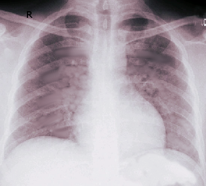
Diffuse bilateral opacities and infiltrates classically present on chest radiograph, but these findings could be variable showing lobar, dependent or unilateral opacities. Computed tomography usually shows widespread patchy airspace opacities more evident in the dependent areas.

In most situations, the diagnosis and etiology will be evident and confirmed after an initial evaluation. In a small number of patients, the diagnosis and/or etiology is unclear, so further evaluation and diagnostic testing are warranted to confirm the diagnosis and etiology or rule out other diagnoses. These tests could include echocardiography to assess for ventricular function and valvular abnormalities, right side heart catheterizations and bronchoscopy with bronchoalveolar lavage and possibly lung tissue biopsy.[6]

Treatment / Management

Management of patients with ARDS requires the collaboration of the medical team with the goal of preventing further complications. Besides the focus on mechanical ventilation, management of these patients requires attention to treatment and recognition of the underlying cause, minimization of unnecessary procedures, prophylaxis for venous thromboembolism, stress ulcers, and aspiration. Additionally, one should ensure adequate nutrition via the enteral route when possible, and minimize the risk of acquired nosocomial infections. 

Safe mechanical ventilation with avoiding further lung injury is the cornerstone of treatment in ARDS. Current guidelines recommend lung protective ventilation consists of low tidal volumes (4 to 8 ml/kg of ideal body weight) with a target of plateau airway pressure less than 30 cm of water.  Usually, tidal volume starts at 6ml/kg of ideal body weight and lowers further for 4 ml/kg if plateau airway pressure is less than 30 and could increase to 8 ml/kg if inspiratory plateau pressure below PEEP or patient is double triggering or in cases of severe hypercapnia and metabolic acidosis. This approach is associated with decreased mortality and improved clinical outcomes at 30 days as shown in different trials.[7][8] Furthermore, in patients with moderate-to-severe ARDS (PaO2/FiO2 less than or equal to 150 mmHg), the American Thoracic Society and European Respiratory Society (ATS/ERS) recommend the prone position for 12 hours per day especially in patients with resistant hypoxemia. Conservative fluid strategy is recommended in patients with ARDS to decrease the risk of fluid accumulation in alveolar space as it associated with a reduction of the duration of mechanical ventilation and ICU stay with an improvement of lung function without causing non-pulmonary organ dysfunction.[9]

Several other measures are used in patients with severe ARDS. Early use of neuromuscular blockade and deep sedation in patients with moderate-to-severe ARDS correlates with 90-day survival without increasing the risk for muscle weakness.[10] Higher rather than lower PEEP has a conditional recommendation in patients with moderate-to-severe ARDS. It might be required to prevent atelectotrauma and to achieve oxygenation, but it has potential risk factors including end-expiratory alveolar distention, barotrauma, increasing intra-pulmonary shunts and higher pulmonary vascular resistance that could lead to cor-pulmonale.[7] Furthermore, recruitment maneuvers involve elevation of applied ventilation pressures transiently to open collapsed alveoli which increases the tidal volume participating in the gas exchange. Several recruitment maneuvers are used including prolonged high continuous PEEP, an incremental increase in PEEP and high driving pressures. However, ATS/ERS has a conditional recommendation for lung recruitment in patients with ARDS, and it should be used cautiously especially in patients with hypovolemia and shock.[7]

Several measures may have promising results in the management of ARDS and are under investigation by many ongoing randomized controlled trials. These measures include extracorporeal membrane oxygenation (ECMO) is reserved for very severe ARDS (PaO2/FiO2 ratio less than or equal to 60). As well, PEEP titration guided by transpulmonary plateau pressure which involves estimating pleural pressures using esophageal manometer results in lower mortality in a small trial and is being investigated by two ongoing studies. Furthermore, other trials are studying ultra-low tidal volume (less than 4ml/kg IBW) with CO2 removal by extracorporeal methods which will allow for low pressure and avoiding hypercapnia and acidosis. 

Differential Diagnosis

  • Acute cardiogenic pulmonary edema
  • Bilateral pneumonia
  • Diffuse alveolar hemorrhage
  • Inflammatory or autoimmune conditions
  • Acute eosinophilic pneumonia
  • Pulmonary vasculitis
  • Cryptogenic organizing pneumonia
  • Acute interstitial pneumonitis
  • Disseminated malignancy

Prognosis

ARDS has high mortality that increases with increasing severity with an incidence of 27%, 32.5% and 45% in mild, moderate and severe classes, respectively.[6] Other risk factors that correlate with increased mortality include advanced age, pre-existing organ dysfunction, and ARDS arising from direct lung injury.  However, the recovery of maximal lung function occurs within 3 to 12 months. Two-third of patients have mild abnormalities in lung function test while only one third has normal pulmonary function tests after 1 year. 

Complications

Complications associated with ARDS divide into the following:

  • Complications related to mechanical ventilation use (barotrauma and nosocomial infections specially ventilator-associated pneumonia)
  • Complications related to underlying condition and complications secondary to prolonged ICU and hospital stay including delirium
  • Critical care neuromyopathies
  • Deep venous thrombosis, and gastrointestinal bleeding

Patients who develop the fibrotic stage are more likely to need tracheostomy because of prolonged intubation and difficult weaning from mechanical ventilation. Oxygen supply may be necessary for a longer duration until recovery of lung injury.

Enhancing Healthcare Team Outcomes

ARDS is a common and serious condition that requires the collaboration of the medical team to diagnose and treat appropriately. Other clinical conditions that mimic ARDS should be treated appropriately. ARDS carries a high mortality and morbidity, and many ongoing randomized clinical trials are investigating new measures to improve mortality and other clinical outcomes in this critical condition. However, safe mechanical ventilation with low-pressure ventilation remains the cornerstone of management.


(Click Image to Enlarge)
Acute Respiratory Distress Syndrome
Image courtesy S Bhimji MD

(Click Image to Enlarge)
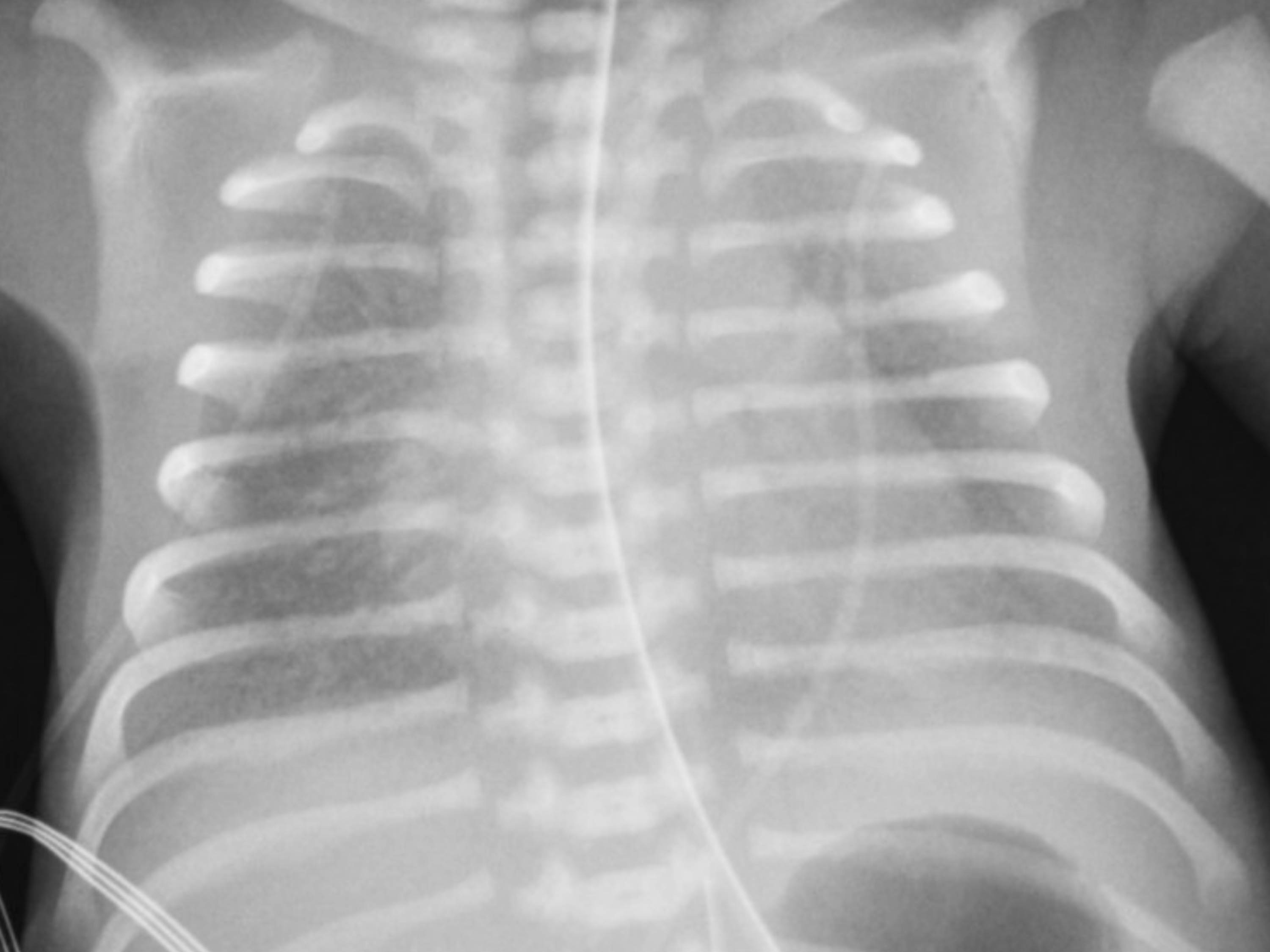
This is a chest radiograph of a preterm neonate with respiratory distress syndrome showing diffuse ground glass haziness bilaterally with air bronchograms
This is a chest radiograph of a preterm neonate with respiratory distress syndrome showing diffuse ground glass haziness bilaterally with air bronchograms.
Contributed by Ranjith Kamity, MD
Details

Author

Yazan Zayed

Editor:

Reza Askari

Updated:

8/8/2023 12:14:43 AM

References


[1]

Thompson BT, Chambers RC, Liu KD. Acute Respiratory Distress Syndrome. The New England journal of medicine. 2017 Aug 10:377(6):562-572. doi: 10.1056/NEJMra1608077. Epub     [PubMed PMID: 28792873]


[2]

Fan E, Brodie D, Slutsky AS. Acute Respiratory Distress Syndrome: Advances in Diagnosis and Treatment. JAMA. 2018 Feb 20:319(7):698-710. doi: 10.1001/jama.2017.21907. Epub     [PubMed PMID: 29466596]

Level 3 (low-level) evidence

[3]

Meyer NJ, Christie JD. Genetic heterogeneity and risk of acute respiratory distress syndrome. Seminars in respiratory and critical care medicine. 2013 Aug:34(4):459-74. doi: 10.1055/s-0033-1351121. Epub 2013 Aug 11     [PubMed PMID: 23934715]


[4]

Bellani G, Laffey JG, Pham T, Fan E, Brochard L, Esteban A, Gattinoni L, van Haren F, Larsson A, McAuley DF, Ranieri M, Rubenfeld G, Thompson BT, Wrigge H, Slutsky AS, Pesenti A, LUNG SAFE Investigators, ESICM Trials Group. Epidemiology, Patterns of Care, and Mortality for Patients With Acute Respiratory Distress Syndrome in Intensive Care Units in 50 Countries. JAMA. 2016 Feb 23:315(8):788-800. doi: 10.1001/jama.2016.0291. Epub     [PubMed PMID: 26903337]


[5]

Rubenfeld GD, Caldwell E, Peabody E, Weaver J, Martin DP, Neff M, Stern EJ, Hudson LD. Incidence and outcomes of acute lung injury. The New England journal of medicine. 2005 Oct 20:353(16):1685-93     [PubMed PMID: 16236739]


[6]

ARDS Definition Task Force, Ranieri VM, Rubenfeld GD, Thompson BT, Ferguson ND, Caldwell E, Fan E, Camporota L, Slutsky AS. Acute respiratory distress syndrome: the Berlin Definition. JAMA. 2012 Jun 20:307(23):2526-33. doi: 10.1001/jama.2012.5669. Epub     [PubMed PMID: 22797452]


[7]

Fan E, Del Sorbo L, Goligher EC, Hodgson CL, Munshi L, Walkey AJ, Adhikari NKJ, Amato MBP, Branson R, Brower RG, Ferguson ND, Gajic O, Gattinoni L, Hess D, Mancebo J, Meade MO, McAuley DF, Pesenti A, Ranieri VM, Rubenfeld GD, Rubin E, Seckel M, Slutsky AS, Talmor D, Thompson BT, Wunsch H, Uleryk E, Brozek J, Brochard LJ, American Thoracic Society, European Society of Intensive Care Medicine, and Society of Critical Care Medicine. An Official American Thoracic Society/European Society of Intensive Care Medicine/Society of Critical Care Medicine Clinical Practice Guideline: Mechanical Ventilation in Adult Patients with Acute Respiratory Distress Syndrome. American journal of respiratory and critical care medicine. 2017 May 1:195(9):1253-1263. doi: 10.1164/rccm.201703-0548ST. Epub     [PubMed PMID: 28459336]

Level 1 (high-level) evidence

[8]

Acute Respiratory Distress Syndrome Network, Brower RG, Matthay MA, Morris A, Schoenfeld D, Thompson BT, Wheeler A. Ventilation with lower tidal volumes as compared with traditional tidal volumes for acute lung injury and the acute respiratory distress syndrome. The New England journal of medicine. 2000 May 4:342(18):1301-8     [PubMed PMID: 10793162]


[9]

National Heart, Lung, and Blood Institute Acute Respiratory Distress Syndrome (ARDS) Clinical Trials Network, Wiedemann HP, Wheeler AP, Bernard GR, Thompson BT, Hayden D, deBoisblanc B, Connors AF Jr, Hite RD, Harabin AL. Comparison of two fluid-management strategies in acute lung injury. The New England journal of medicine. 2006 Jun 15:354(24):2564-75     [PubMed PMID: 16714767]


[10]

Papazian L, Forel JM, Gacouin A, Penot-Ragon C, Perrin G, Loundou A, Jaber S, Arnal JM, Perez D, Seghboyan JM, Constantin JM, Courant P, Lefrant JY, Guérin C, Prat G, Morange S, Roch A, ACURASYS Study Investigators. Neuromuscular blockers in early acute respiratory distress syndrome. The New England journal of medicine. 2010 Sep 16:363(12):1107-16. doi: 10.1056/NEJMoa1005372. Epub     [PubMed PMID: 20843245]