Transjugular Intrahepatic Portosystemic Shunt

Earn CME/CE in your profession:


Continuing Education Activity

Transjugular intrahepatic portosystemic shunt (TIPS) is a well-established percutaneous modality for decreasing portal hypertension. The major clinical indications for transjugular intrahepatic portosystemic shunts include refractory variceal hemorrhage and refractory ascites. The shunt itself is created by placing a stent between the portal vein and the hepatic vein. The resultant shunting of portal venous flow to the systemic circulation helps reduce the portosystemic gradient and alleviate bleeding and ascites without changing the extrahepatic anatomy. This activity reviews the indications, contraindications, and technique involved in performing transjugular intrahepatic portosystemic shunt placement and highlights the role of the interprofessional team in the care of patients undergoing this procedure.

Objectives:

  • Describe the technique involved in performing transjugular intrahepatic portosystemic shunt placement.
  • Review the indications for transjugular intrahepatic portosystemic shunt placement.
  • Summarize the complications associated with transjugular intrahepatic portosystemic shunt placement.
  • Explain a structured interprofessional team approach to provide effective care to and appropriate surveillance of patients undergoing transjugular intrahepatic portosystemic shunt placement.

Introduction

Transjugular intrahepatic portosystemic shunt (TIPS) is now a well established percutaneous means of decreasing portal hypertension. The major clinical indications for transjugular intrahepatic portosystemic shunt are refractory variceal hemorrhage and refractory ascites. The shunt itself is created by placing a stent between the portal vein and the hepatic vein. The resultant shunting of portal venous flow to the systemic circulation helps reduce the portosystemic gradient and alleviate bleeding and ascites without changing the extrahepatic anatomy.

History and Development

The transjugular intrahepatic portosystemic shunt was initially described in 1969, originally by prolonged balloon dilation of a percutaneously created tract between the portal and hepatic veins. Unfortunately, short patency of the track remained a major clinical hurdle to its widespread use. The development of balloon expandable metallic stents and the subsequent introduction of flexible bare-metal stents helped improve patency rates. Intimal hyperplasia within the stent, especially at the hepatic venous end, required frequent secondary interventions such as balloon dilation and re-stenting. Early thrombosis was felt to be secondary to placing the transjugular intrahepatic portosystemic shunt across a major bile duct. Advances in stent technology and the introduction of polytetrafluoroethylene-covered stents over a decade ago has led to further increase in clinical patency and improved long-term results with TIPS.[1][2][3][4]

Anatomy and Physiology

A thorough understanding of the anatomy and patency of vessels as it relates to portal hypertension, including portal venous anatomy, hepatic venous anatomy, and pathways of collateral venous/variceal flow are paramount. These need to be evaluated for before undertaking a transjugular intrahepatic portosystemic shunt procedure as it can have a significant impact on the procedure itself. The presence or absence of shunts such as splenorenal shunts should be assessed before undertaking a transjugular intrahepatic portosystemic shunt.

Indications

Indications for TIPS

  • Secondary prevention of variceal hemorrhage (1A level of evidence)
  • Refractory Ascites (1A level of evidence)
  • Refractory hepatic hydrothorax
  • Budd-Chiari syndrome
  • Hepatorenal syndrome
  • Hepato-pulmonary syndrome 

Contraindications

Absolute Contraindications

  • Primary prevention of variceal hemorrhage
  • Congestive heart failure
  • Pulmonary hypertension
  • Severe sepsis
  • Biliary obstruction
  • Extensive hepatic cysts

Relative Contraindications

  • Liver cancer
  • Portal vein thrombosis or hepatic vein occlusion
  • Severe coagulopathy and/or thrombocytopenia
  • Advanced hepatic encephalopathy

Relative Contraindications for TIPS

  • Primary prevention of variceal bleeding
  • Congestive heart failure/Pulmonary hypertension
  • Severe pulmonary hypertension
  • Unrelieved biliary obstruction
  • Multiple hepatic cysts
  • Portal vein thrombosis
  • Hepatic encephalopathy
  • Central hepatocellular carcinoma

Equipment

The procedure should be undertaken in an interventional suite with adequate fluoroscopy and ultrasound equipment. Basic angiographic supplies such as access needles, guide wires, catheters, and balloons should be on hand. Equipment specifically designed for transjugular intrahepatic portosystemic shunt procedures should be on hand. Covered stents specially designed for TIPS in appropriate sizes need to be on hand. Additionally, embolization materials including coils, plugs and sclerosis agents should be available to embolize varices/shunts. Physiological monitoring equipment and adequate personnel are needed, including nurses and technologists.

Personnel

Experienced Interventionalists should perform a transjugular intrahepatic portosystemic shunt in a dedicated interventional suite with adequate personnel, including experienced technologists and nursing staff. Ideally, the anesthesiology team should be available to provide anesthesia as appropriate. an interprofessional team approach is very useful in the management of these patients including interventional radiology, gastrointestinal (hepatology) medicine, and transplantation surgery as they each add their unique management skills.

Preparation

A thorough clinical evaluation of the patient begins in the interventional radiology clinic, but many times has to be performed emergently in the case of bleeding. A baseline laboratory evaluation, including liver function tests, renal function, and coagulation status, needs to be obtained. Underlying coagulation issues should be corrected. In cases of impaired renal function pre-hydration as well as the use of lower osmolality nonionic contrast agents should be used. Pre-procedure cross-sectional imaging is important to document patency of the portal and hepatic veins and  to exclude the presence of tumor along the parenchymal tract.  Large-volume ascites may be drained immediately before the procedure, especially if advanced techniques need to be utilized such as placement of a percutaneous guide wire into the portal vein. The MELD (model for end-stage liver disease) score should be calculated, as it is used as a predictor of post-TIPS mortality. It takes into consideration creatinine, bilirubin, and INR. A score above 18 predicts significantly higher mortality within 3 months of TIPS. Finally, all the risks and benefits of the procedure need to be carefully discussed with the patient, family, and referring physician.

Technique or Treatment

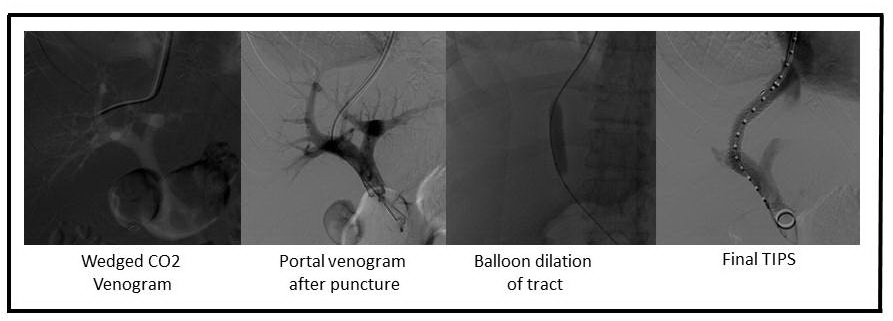
While moderate, conscious sedation may be used for the procedure, general anesthesia is preferred. Pre-procedure antibiotics are necessary. A right internal jugular approach is preferred to cannulate the central venous system. An angled catheter is used to cannulate the right hepatic vein. A balloon occlusion hepatic venogram with CO2 is performed to opacify the portal venous system and provide a fluoroscopic target for portal vein entry with the needle. Several commercial transjugular intrahepatic portosystemic shunt kits are available, and the included needle is used under fluoroscopic/ultrasound guidance to regularly access the right portal vein from the right hepatic vein. Middle and left hepatic veins, as well as the left portal vein, may be used. Real-time ultrasound guidance can help visualize the passage of the needle and portal vein entry. Aspiration of blood, followed by contrast injection, is used to confirm appropriate portal venous access. The ideal entry point in the portal vein is 1 to 2 centimeters from the main bifurcation to avoid an extra-hepatic puncture and possible hemoperitoneum. If the portal vein cannot be accessed in this manner, a percutaneous transhepatic guidewire can be placed into the portal vein and be used as a fluoroscopic target for portal vein entry. An angiographic catheter is advanced into the portal vein for venogram to confirm portal vein access and to confirm suitable location. Pressure measurements in the portal vein and right atrium are obtained to calculate the initial portal to systemic gradient. The initial pressures may be low in patients with a competing spleno-renal shunt, and they may need to be embolized. The tract is then dilated to 8 to 10 millimeters. A partially covered stent is the preferred stent, with the uncovered portion being placed in the portal venous end and the covered end extending to the junction of the hepatic vein and inferior vena cava (IVC). Care should be taken to avoid extending the stent significantly into the IVC or far down into the portal vein as this may impact future liver transplantation. If there is persistent filling of varices after TIPS placement, they should be embolized with coils or plug. They also can be sclerosed at this time to prevent ongoing variceal bleeding. Final TIPS gradients should be recorded to help in future interventions.[5][6][7][8]

The patient should recover in a closely monitored setting, depending on the clinical status. Liver enzymes should be closely followed to monitor for any hepatic dysfunction post-TIPS placement. A TIPS Doppler ultrasound study should be obtained within 48 to 72 hours of TIPS placement. Earlier studies may be inaccurate as residual air bubbles within the wall of the polytetrafluoroethylene material may prevent adequate ultrasound penetration and may simulate decreased or absent flow within the newly created shunt. This study can serve as a baseline for follow-up surveillance scans to assess for shunt dysfunction and the need for follow-up intervention.

Doppler ultrasound velocities of greater than 190 centimeters per second or lower than 90 centimeters per second are associated with shunt dysfunction. Other concerning findings are a change (increase or decrease) in velocity of greater than 50 centimeters per second from baseline. Depending on the clinical scenario and the ultrasound findings a TIPS venogram with direct pressure measurements should be pursued in the angiography suite. Intimal hyperplasia within the stent can be treated with balloon dilation and or re-stenting to improve TIPS flow.[9][10][11][12]

Complications

Complications from a transjugular intrahepatic portosystemic shunt can be broadly categorized into immediate, procedure-related clinical complications and longer-term complications.

Immediate Complications

  • Puncture related bleeds, including hemoperitoneum (may need percutaneous embolization or surgical intervention)
  • Stent malposition/migration
  • Early shunt thrombosis due to technical issues or bile duct injury
  • Shunt-related encephalopathy (30% to 46%)
  • Deterioration of hepatic function (may be treated with shunt narrowing or in severe cases closure of the shunt)
  • Shunt stenosis (leading to secondary intervention with balloon dilation or re-stenting)

Long-term Complications

  • Intimal hyperplasia within the TIPS (may lead to reduced flow and recurrence of portal hypertension in the patient)
  • Similarly, complete TIPS thrombosis (may lead to recurrence of portal hypertension)
  • Both treated by re-establishing TIPS patency with balloon dilation and re-stenting

Clinical Significance

All transjugular intrahepatic portosystemic shunt patients should receive close clinical multi-disciplinary follow-up, both by the interventional radiologist and hepatologist. Regular noninvasive imaging based on local protocols with ultrasound is imperative in this patient population. Patient follow-up may indicate transplant surgery and include being listed for a potential liver transplant, depending on their clinical status.

TIPS has established itself as a percutaneous, minimally invasive means of treating a patient with severe complications of portal hypertension.

Enhancing Healthcare Team Outcomes

The managment of patients with portal hypertension is with an interprofessional team that consists of a pathologist, radiologist, gastroenterologist, general surgeon, dietitian, and an internist. As outpatients the majority of them are followed by the nurse practitioner and primary care provider. Some of these patients may develop variceal bleeding and require TIPS. Only experienced interventionalists should perform a transjugular intrahepatic portosystemic shunt in a dedicated interventional suite with adequate personnel, including experienced technologists and nursing staff. Ideally, the anesthesiology team should be available to provide anesthesia as appropriate. an interprofessional team approach is very useful in the management of these patients including interventional radiology, gastrointestinal (hepatology) medicine, and transplantation surgery as they each add their unique management skills. Because TIPS is not 100% effective, patients need close follow up and the transplant team consulted. Some of these patients may benefit from a liver transplant. The overall outcome of patients with portal hypertension is poor. Liver transplant is not the ideal solution because of expense and shortage of organs.[13](Level V)


(Click Image to Enlarge)
TIPS Figure 1
TIPS Figure 1
Contributed by Scott Jones, MD
Details

Editor:

Paula Novelli

Updated:

7/24/2023 11:04:05 PM

References


[1]

Khoury T, Massarwa M, Daher S, Benson AA, Hazou W, Israeli E, Jacob H, Epstein J, Safadi R. Endoscopic Ultrasound-Guided Angiotherapy for Gastric Varices: A Single Center Experience. Hepatology communications. 2019 Feb:3(2):207-212. doi: 10.1002/hep4.1289. Epub 2018 Dec 10     [PubMed PMID: 30766958]


[2]

Lattanzi B, D'Ambrosio D, Merli M. Hepatic Encephalopathy and Sarcopenia: Two Faces of the Same Metabolic Alteration. Journal of clinical and experimental hepatology. 2019 Jan-Feb:9(1):125-130. doi: 10.1016/j.jceh.2018.04.007. Epub 2018 May 5     [PubMed PMID: 30765945]


[3]

Sonavane AD, Amarapurkar DN, Rathod KR, Punamiya SJ. Long Term Survival of Patients Undergoing TIPS in Budd-Chiari Syndrome. Journal of clinical and experimental hepatology. 2019 Jan-Feb:9(1):56-61. doi: 10.1016/j.jceh.2018.02.008. Epub 2018 Mar 1     [PubMed PMID: 30765940]


[4]

Chen SL, Hu P, Lin ZP, Zhao JB. The Effect of Puncture Sites of Portal Vein in TIPS with ePTFE-Covered Stents on Postoperative Long-Term Clinical Efficacy. Gastroenterology research and practice. 2019:2019():2935498. doi: 10.1155/2019/2935498. Epub 2019 Jan 9     [PubMed PMID: 30728835]


[5]

Darrow AW, Gaba RC, Lokken RP. Transhepatic Revision of Occluded Transjugular Intrahepatic Portosystemic Shunt Complicated by Endotipsitis. Seminars in interventional radiology. 2018 Dec:35(5):492-496. doi: 10.1055/s-0038-1676092. Epub 2019 Feb 5     [PubMed PMID: 30728666]


[6]

Valentin N, Weisberg I. The role of transjugular intrahepatic portosystemic shunt in the management of portal vein thrombosis. European journal of gastroenterology & hepatology. 2019 Mar:31(3):403-404. doi: 10.1097/MEG.0000000000001318. Epub     [PubMed PMID: 30720607]


[7]

Bertino F, Hawkins CM, Shivaram G, Gill AE, Lungren MP, Reposar A, Sze DY, Hwang GL, Koo K, Monroe E. Technical Feasibility and Clinical Effectiveness of Transjugular Intrahepatic Portosystemic Shunt Creation in Pediatric and Adolescent Patients. Journal of vascular and interventional radiology : JVIR. 2019 Feb:30(2):178-186.e5. doi: 10.1016/j.jvir.2018.10.003. Epub     [PubMed PMID: 30717948]

Level 2 (mid-level) evidence

[8]

David A, Liberge R, Meyer J, Morla O, Leaute F, Archambeaud I, Gournay J, Trewick D, Frampas E, Perret C, Douane F. Ultrasonographic guidance for portal vein access during transjugular intrahepatic portosystemic shunt (TIPS) placement. Diagnostic and interventional imaging. 2019 Jul-Aug:100(7-8):445-453. doi: 10.1016/j.diii.2019.01.004. Epub 2019 Jan 31     [PubMed PMID: 30711496]


[9]

Wong K, Ozeki K, Kwong A, Patel BN, Kwo P. The effects of a transjugular intrahepatic portosystemic shunt on the diagnosis of hepatocellular cancer. PloS one. 2018:13(12):e0208233. doi: 10.1371/journal.pone.0208233. Epub 2018 Dec 28     [PubMed PMID: 30592722]


[10]

Nardelli S, Gioia S, Ridola L, Riggio O. Radiological Intervention for Shunt Related Encephalopathy. Journal of clinical and experimental hepatology. 2018 Dec:8(4):452-459. doi: 10.1016/j.jceh.2018.04.008. Epub 2018 May 5     [PubMed PMID: 30564003]


[11]

Downing TM, Khan SN, Zvavanjanja RC, Bhatti Z, Pillai AK, Kee ST. Portal Venous Interventions: How to Recognize, Avoid, or Get Out of Trouble in Transjugular Intrahepatic Portosystemic Shunt (TIPS), Balloon Occlusion Sclerosis (ie, BRTO), and Portal Vein Embolization (PVE). Techniques in vascular and interventional radiology. 2018 Dec:21(4):267-287. doi: 10.1053/j.tvir.2018.07.009. Epub 2018 Jul 29     [PubMed PMID: 30545506]


[12]

Khan F, Mehrzad H, Tripathi D. Timing of Transjugular Intrahepatic Portosystemic Stent-shunt in Budd-Chiari Syndrome: A UK Hepatologist's Perspective. Journal of translational internal medicine. 2018 Sep:6(3):97-104. doi: 10.2478/jtim-2018-0022. Epub 2018 Oct 9     [PubMed PMID: 30425945]

Level 3 (low-level) evidence

[13]

Leng X, Zhang F, Zhang M, Guo H, Yin X, Xiao J, Wang Y, Zou X, Zhuge Y. Comparison of transjugular intrahepatic portosystemic shunt for treatment of variceal bleeding in patients with cirrhosis with or without spontaneous portosystemic shunt. European journal of gastroenterology & hepatology. 2019 Jul:31(7):853-858. doi: 10.1097/MEG.0000000000001349. Epub     [PubMed PMID: 30633039]