Neutropenic Enterocolitis (Typhlitis)

Earn CME/CE in your profession:


Continuing Education Activity

Neutropenic enterocolitis is a severe inflammatory condition of the intestines, occurring mainly in the neutropenic, immune-compromised patients. This activity outlines the evaluation and management of neutropenic enterocolitis and highlights the role of an interprofessional team in managing patients with this condition.

Objectives:

  • Identify the etiology and epidemiology of neutropenic enterocolitis
  • Outline the appropriate history and physical findings, as well as determine the appropriate evaluation of a patient with neutropenic enterocolitis.
  • Describe the treatment and management options available for neutropenic enterocolitis.
  • Review some interprofessional team strategies for improving care coordination and communication to advance the care of patient s with neutropenic enterocolitis and improve outcomes.

Introduction

Neutropenic enterocolitis (NEC) is a severe inflammatory disorder of the intestines that occurs in neutropenic patients. It is a critical condition that is associated with high mortality. Its pathogenesis is not fully understood. The thinking is that NEC occurs due to the translocation of bacteria through weak and friable intestinal mucosa that is damaged by chemotherapy. The diagnosis usually results from findings on abdominal CT in a patient with concerning signs or symptoms of neutropenic enterocolitis. Limited evidence exists regarding the treatment of NEC. Treatment primarily consists of IV antibiotics with bowel rest. Surgery is considered, if needed, for complications.

Etiology

The microorganisms causing NEC are mostly polymicrobial that includes gram-negative bacilli, gram-positive cocci, anaerobes, and fungi. The most commonly isolated organisms include Pseudomonas aeruginosa, Escherichia coli, Klebsiella spp, viridans group streptococci, enterococci, Bacteroides spp, Clostridium spp, and Candida spp.[1]Other reported organisms include Clostridium septicum and Stenotrophomonas maltophilia, which correlate with worse outcomes and severe sepsis.[2]

Epidemiology

The incidence of NEC varies amongst studies. In a systemic review done by Gorschlüter et al., the incidence rate from 21 studies was 5.3% in patients hospitalized for hematological malignancies, high-dose chemotherapy for solid tumors, or aplastic anemia.[3] Another cohort study found it in 3.5% of 317 severely neutropenic patients.[4] The incidence of NEC has been rising with the increased use of intensive chemotherapy, especially those agents that cause mucositis and taxanes.[1]

NEC was first described in patients with hematological malignancies, primarily acute myeloid leukemia. Patients with hematologic malignancies are generally at higher risk for developing NEC due to their underlying malignancy but also due to their chemotherapy protocols. However, NEC also reportedly occurs in patients with myelodysplastic syndrome, solid malignancy, and patients with autoimmune conditions on immune suppressive medications.A study on pediatric patients found that patients who had mucositis, chemotherapy in the previous 14 days, or a hematopoietic stem cell transplant were at a higher risk of developing NEC.[5]

Pathophysiology

The pathology if NEC is not yet entirely clear and is likely secondary to multiple underlying causes [6]. One of the mechanisms is exposure to cytotoxic medications that disrupt the mucosal barrier, which allows bacterial translocation from the gut. This mechanism has support from the histologic findings of intestinal wall edema, engorged blood vessels, and disruption of the mucosal surface.[7] Thick and swollen bowels with areas of ulceration and hemorrhage have been present on surgical evaluation and autopsy. Neutropenia further aggravates the risks, causing decreased immunity with failure to control the transmural translocation of pathogens. There are also concerns that direct invasion of the interstitial wall by malignant cells may contribute to the disease. The cecum is most commonly involved in NEC due to its distensibility and limited blood supply.

History and Physical

Patients usually present with fever, abdominal pain, and diarrhea. Diarrhea can be bloody in severe cases. Patients may also have abdominal distension and paralytic ileus. In patients with severe neutropenia, fever sometimes is absent. The abdominal pain can be diffuse or localized, typically occurring in the right lower quadrant. A rigid abdomen may indicate bowel perforation. Also, mucositis can be visible in oral or anal mucosa, which would support the diagnosis of NEC.[1] Other findings on the exam in patients that have complicated NEC will be signs and symptoms of a systemic inflammatory response. These patients may appear ill, dehydrated, and febrile with unstable vital signs.

Evaluation

Typically patients suspected to have NEC undergo computed tomography (CT) scan of their abdomen. CT findings usually include intestinal wall thickening, mesenteric stranding, intestinal dilatation, and pneumatosis.[8]Ultrasound (US) is an option as well, although the sensitivity was variable in different studies. A study done by Tamburrini et al. showed that the US is comparable to the CT scan. US characteristic findings include circumferential wall thickening with predominant submucosa.[9] Moreover, in a study done by Pugliese N et al., patients were started on treatment for NEC based on US findings.[10]

The diagnosis of NEC is usually based on a combination of clinically and radiologic findings. However, the absence of a disease definition makes it difficult to have a consensus on the criteria for the diagnosis of NEC. Gorschlüter et al. suggested diagnostic criteria in a systematic review include fever, abdominal pain, and any bowel wall thickening more than 4 mm seen on imaging in addition to the exclusion of Clostridium difficile as a cause of the colitis.[3] [Table 1]

Table 1

  1. Presence of fever (axillary temperature over 38.0 C or rectal temperature above 38.5 C)
  2. Abdominal pain (at least degree 3 determined by the patient using a visual analogous scale pain score ranging from degree 1 to 10)
  3. Demonstration of bowel wall thickening by more than 4 mm (transversal scan) over more than 30 mm (longitudinal scan) in any segment by US or CT

Treatment / Management

Management is through medical and surgical treatment. Medical treatment includes supportive therapy plus antimicrobials. Supportive measures are mainly bowel rest with nasogastric suction, intravenous fluids, and parenteral nutrition if indicated. Replacement of platelets should start if severe thrombocytopenia is present or if bleeding occurs. Any coagulopathy should be corrected.There are no trials to date to evaluate different treatment regimens. The guidance for antimicrobial therapy should be according to the patient's antimicrobial exposure, bacteremia, and local resistance pattern. The patient should receive a broad-spectrum antimicrobial that covers for gram-negative and anaerobic microorganisms as they are the most common organisms causing NEC. Monotherapy with piperacillin-tazobactam, carbapenem, or anti-pseudomonal cephalosporin such as cefepime with metronidazole can start empirically.[11] However, if there is a suspicion of mucositis, then concern should be taken for and treatment directed against gram-positive bacteria. Vancomycin is a consideration in this setting.[12]There is one single-center retrospective study done on 100 patients with NEC that found that patients treated with a combination of antimicrobial therapy, including tigecycline, led to improved survival.[10] However, the results might be related to specific microbial epidemiologic characteristics in that center. More trials are necessary to evaluate and determine optimal treatment regimens.The routine use of granulocyte colony-stimulating factor (G-CSF) is still debatable, and the benefit of this therapy is uncertain.[13]Patients should also undergo evaluation for surgical treatment, and early surgical consultation is indicated in all patients with NEC. Surgical intervention is indicated for patients with complicated NEC, such as those who develop bowel perforation, pneumoperitoneum, or persistent gastrointestinal bleeding. In a study done by Shamberger et al. on pediatric patients, the proposed criteria for surgical intervention include[14]: [Table 2] 

Table 2

The persistence of gastrointestinal bleeding despite correction of coagulopathies, thrombocytopenia, and neutropenia can cause:

  1. Free air in the intraperitoneal cavity indicative of bowel perforation;
  2. Clinical deterioration despite optimal medical management;
  3. The development of other indications for surgery, such as appendicitis

Surgical treatment, in general, has typically been avoided due to concerns about bleeding, increased risk of infection, and poor healing. However, a metaanalysis done showed that surgical treatment did not cause excess risk compared to conservative treatment.[15] More studies are necessary to ascertain the optimal situations for surgical versus medical therapy. 

Differential Diagnosis

Differential diagnosis includes Clostridium difficile infection, so this condition requires exclusion. Also, the clinical presentation is similar to acute appendicitis, which can be differentiated from NEC by imaging. Other conditions that can cause colitis and intestinal bleeding include Cytomegalovirus colitis, graft versus host disease in transplant patients, Norovirus infection, and ischemic colitis.

Prognosis

Mortality rates reported from NEC are as high as 50%, especially if the patient has transmural inflammation or bowel perforation. This determination came from pathological studies done on patients who died from NEC.[3][16] However, with the early recognition of NEC and improvement in management, the prognosis has improved, as seen by case-controlled studies.[5]

Complications

The complications of NEC are mainly related to the pathology itself (bowel wall inflammation) such as perforation, peritonitis, sepsis, or abscess formation. Other complications are related to pancytopenia, including severe bleeding due to thrombocytopenia and delayed healing.

Deterrence and Patient Education

Neutropenic enterocolitis is a serious condition that affects the bowel wall in affected patients. It mainly occurs in patients on intensive chemotherapy that causes suppression of the patient’s immune system. The weakened immune system and bowel wall inflammation allow bacteria to translocate to the bowel wall and sometimes enter the bloodstream.

The condition usually starts with abdominal pain that progressively worsens. Associated symptoms include diarrhea that is often bloody and fever. Patients may also have weakness and malaise.

It is important for patients who have received intensive chemotherapy or have undergone a stem cell transplant receive emergent care as early recognition and treatment prevent complications and may improve outcomes.

Enhancing Healthcare Team Outcomes

Abdominal pain and fever in a neutropenic patient is a signal for a serious underlying pathology that requires timely intervention and an interprofessional approach. In the case of NEC, early and appropriate intervention helps to prevent complications and improve outcomes. Early surgical evaluation is critical in the management of this condition, as it can be life-saving for some patients who present with a complicated NEC.

Infectious disease specialists can also help with guiding antibiotic selection because patients also have treatment as febrile neutropenic patients. Pharmacists review prescriptions for antibiotics and granulocyte stimulating factors, verifying dosages, and drug-drug interactions. For recalcitrant infections, a board-certified infectious disease pharmacist can be an invaluable team asset, working with the specialist using the latest antibiogram data to guide antimicrobial therapy. Also, a hematology-oncology evaluation is essential because patients usually have received intensive chemotherapy before or during their symptom onset and presentation. Oncology nurses administer treatments, educate patients, and are invaluable in monitor patients and informing the interprofessional team about changes in the patient's medical condition. This interprofessional team paradigm results in optimal patient outcomes for NEC cases. [Level 5]


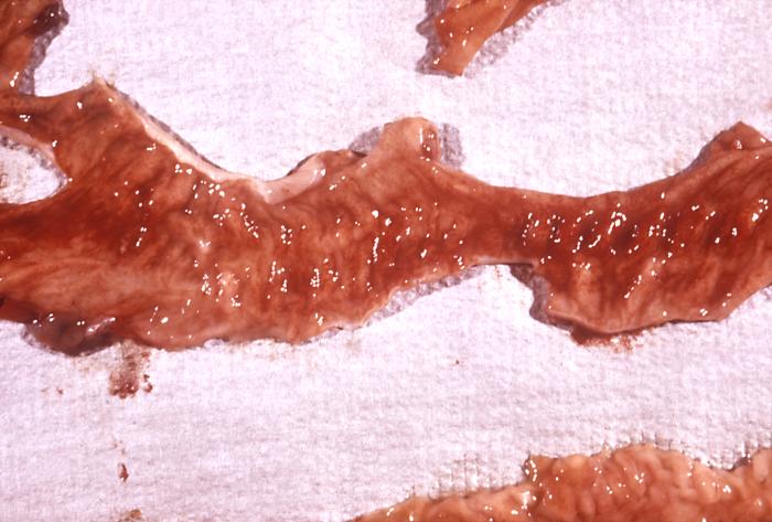
(Click Image to Enlarge)
Pathology, Rhesus monkey, Neutropenic enterocolitis, Typhlitis, Shigella sp
Pathology, Rhesus monkey, Neutropenic enterocolitis, Typhlitis, Shigella sp. infection, Shigellosis, hemorrhagic colonic mucosa
Contributed by The Centers for Disease Control and Prevention (CDC)
Details

Editor:

Joseph Nahas

Updated:

8/7/2023 11:20:59 PM

References


[1]

Nesher L, Rolston KV. Neutropenic enterocolitis, a growing concern in the era of widespread use of aggressive chemotherapy. Clinical infectious diseases : an official publication of the Infectious Diseases Society of America. 2013 Mar:56(5):711-7. doi: 10.1093/cid/cis998. Epub 2012 Nov 29     [PubMed PMID: 23196957]


[2]

Kaito S, Sekiya N, Najima Y, Sano N, Horiguchi S, Kakihana K, Hishima T, Ohashi K. Fatal Neutropenic Enterocolitis Caused by Stenotrophomonas maltophilia: A Rare and Underrecognized Entity. Internal medicine (Tokyo, Japan). 2018 Dec 15:57(24):3667-3671. doi: 10.2169/internalmedicine.1227-18. Epub 2018 Aug 10     [PubMed PMID: 30101922]


[3]

Gorschlüter M, Mey U, Strehl J, Ziske C, Schepke M, Schmidt-Wolf IG, Sauerbruch T, Glasmacher A. Neutropenic enterocolitis in adults: systematic analysis of evidence quality. European journal of haematology. 2005 Jul:75(1):1-13     [PubMed PMID: 15946304]

Level 2 (mid-level) evidence

[4]

Aksoy DY, Tanriover MD, Uzun O, Zarakolu P, Ercis S, Ergüven S, Oto A, Kerimoglu U, Hayran M, Abbasoglu O. Diarrhea in neutropenic patients: a prospective cohort study with emphasis on neutropenic enterocolitis. Annals of oncology : official journal of the European Society for Medical Oncology. 2007 Jan:18(1):183-189. doi: 10.1093/annonc/mdl337. Epub 2006 Oct 5     [PubMed PMID: 17023562]


[5]

Moran H, Yaniv I, Ashkenazi S, Schwartz M, Fisher S, Levy I. Risk factors for typhlitis in pediatric patients with cancer. Journal of pediatric hematology/oncology. 2009 Sep:31(9):630-4. doi: 10.1097/MPH.0b013e3181b1ee28. Epub     [PubMed PMID: 19644402]


[6]

Urbach DR, Rotstein OD. Typhlitis. Canadian journal of surgery. Journal canadien de chirurgie. 1999 Dec:42(6):415-9     [PubMed PMID: 10593241]


[7]

Katz JA, Wagner ML, Gresik MV, Mahoney DH Jr, Fernbach DJ. Typhlitis. An 18-year experience and postmortem review. Cancer. 1990 Feb 15:65(4):1041-7     [PubMed PMID: 2404562]


[8]

Kirkpatrick ID, Greenberg HM. Gastrointestinal complications in the neutropenic patient: characterization and differentiation with abdominal CT. Radiology. 2003 Mar:226(3):668-74     [PubMed PMID: 12601214]


[9]

Tamburrini S, Setola FR, Belfiore MP, Saturnino PP, Della Casa MG, Sarti G, Abete R, Marano I. Ultrasound diagnosis of typhlitis. Journal of ultrasound. 2019 Mar:22(1):103-106. doi: 10.1007/s40477-018-0333-2. Epub 2018 Oct 27     [PubMed PMID: 30367357]


[10]

Pugliese N, Salvatore P, Iula DV, Catania MR, Chiurazzi F, Della Pepa R, Cerchione C, Raimondo M, Giordano C, Simeone L, Caruso S, Pane F, Picardi M. Ultrasonography-driven combination antibiotic therapy with tigecycline significantly increases survival among patients with neutropenic enterocolitis following cytarabine-containing chemotherapy for the remission induction of acute myeloid leukemia. Cancer medicine. 2017 Jul:6(7):1500-1511. doi: 10.1002/cam4.1063. Epub 2017 May 26     [PubMed PMID: 28556623]


[11]

Freifeld AG, Bow EJ, Sepkowitz KA, Boeckh MJ, Ito JI, Mullen CA, Raad II, Rolston KV, Young JA, Wingard JR, Infectious Diseases Society of America. Clinical practice guideline for the use of antimicrobial agents in neutropenic patients with cancer: 2010 update by the infectious diseases society of america. Clinical infectious diseases : an official publication of the Infectious Diseases Society of America. 2011 Feb 15:52(4):e56-93. doi: 10.1093/cid/cir073. Epub     [PubMed PMID: 21258094]

Level 1 (high-level) evidence

[12]

Salazar R, Solá C, Maroto P, Tabernero JM, Brunet J, Verger G, Valentí V, Cancelas JA, Ojeda B, Mendoza L, Rodríguez M, Montesinos J, López-López JJ. Infectious complications in 126 patients treated with high-dose chemotherapy and autologous peripheral blood stem cell transplantation. Bone marrow transplantation. 1999 Jan:23(1):27-33     [PubMed PMID: 10037047]


[13]

Smith TJ, Khatcheressian J, Lyman GH, Ozer H, Armitage JO, Balducci L, Bennett CL, Cantor SB, Crawford J, Cross SJ, Demetri G, Desch CE, Pizzo PA, Schiffer CA, Schwartzberg L, Somerfield MR, Somlo G, Wade JC, Wade JL, Winn RJ, Wozniak AJ, Wolff AC. 2006 update of recommendations for the use of white blood cell growth factors: an evidence-based clinical practice guideline. Journal of clinical oncology : official journal of the American Society of Clinical Oncology. 2006 Jul 1:24(19):3187-205     [PubMed PMID: 16682719]

Level 1 (high-level) evidence

[14]

Shamberger RC, Weinstein HJ, Delorey MJ, Levey RH. The medical and surgical management of typhlitis in children with acute nonlymphocytic (myelogenous) leukemia. Cancer. 1986 Feb 1:57(3):603-9     [PubMed PMID: 3484659]


[15]

Saillard C, Zafrani L, Darmon M, Bisbal M, Chow-Chine L, Sannini A, Brun JP, Ewald J, Turrini O, Faucher M, Azoulay E, Mokart D. The prognostic impact of abdominal surgery in cancer patients with neutropenic enterocolitis: a systematic review and meta-analysis, on behalf the Groupe de Recherche en Réanimation Respiratoire du patient d'Onco-Hématologie (GRRR-OH). Annals of intensive care. 2018 Apr 19:8(1):47. doi: 10.1186/s13613-018-0394-6. Epub 2018 Apr 19     [PubMed PMID: 29675758]

Level 1 (high-level) evidence

[16]

Wade DS, Nava HR, Douglass HO Jr. Neutropenic enterocolitis. Clinical diagnosis and treatment. Cancer. 1992 Jan 1:69(1):17-23     [PubMed PMID: 1727660]