Continuing Education Activity
Xanthelasma palpebrarum is a condition in which soft, yellow papules or plaques filled with cholesterol form over the medial canthus of the upper lid. Several medical conditions are associated with the appearance of xanthelasma palpebrarum, including hyperlipidemia, diabetes, and thyroid dysfunctions. This activity explains when xanthelasma palpebrarum should be considered on differential diagnosis and reviews the proper evaluation and management of this condition. This activity highlights the role of the interprofessional team in caring for patients with this condition.
Objectives:
- Explain the pathophysiology of xanthelasma palpebrarum.
- Outline the factors associated with increased risk of xanthelasma palpebrarum.
- Review management options for patients affected by xanthelasma palpebrarum.
- Explain the interprofessional team strategies for improving care coordination and communication regarding the management of patients with xanthelasma palpebrarum.
Introduction
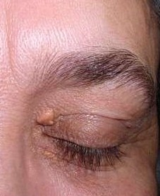
The medical term xanthelasma palpebrarum is composed of two words. Xanthelasma, derived from ancient Greece, where "xanthos" means yellow and "elesma" means plate. Palpebrarum is a Latin word that means "near or related to the eyelid." It is a lipid-rich deposition, mainly cholesterol. It is mostly semisolid yellowish deposits that are commonly found on the medial aspect of the eyes. It is often seen along the corners of the upper and lower eyelids. Xanthelasma palpebrarum is the commonest coetaneous presentation of xanthoma, which occurs over the eyelids, even with the absence of any other cutaneous or mucosal lesions. It is characterized by soft, yellowish papules and plaques that occur more commonly near the inner canthus of the eyelid, more often at the upper eyelid. Lesions are usually symmetrical. There might be one or multiple.[1][2][3]
Xanthelasma palpebrarum is a benign condition that never leads to serious consequences, but it is cosmetically bothersome, and most of the patients are unhappy about their image, and they seek medical advice. The patients visit dermatology offices, reconstructive surgery clinics, and ophthalmology clinics looking for permanent solutions.
Etiology
About 50% of patients who develop xanthelasma have lipid disorder. It is commonly seen in patients with:
- Type II hyperlipidemia that includes type IIa, also known as familial hypercholesterolemia, and type IIb, which is commonly referred to as familial combined hyperlipidemia
- Type IV hyperlipidemia, which is also known as familial hypertriglyceridemia
- Diabetes mellitus
- Hypothyroidism
- Those with low levels of HDL
- Fatty diet
- Excess alcohol intake
- Weight gain
Xanthelasma palpebrarum are observed also in patients who had previous erythroderma, generalized cutaneous inflammatory dermatosis, and in cases who had previous contact dermatitis. It may be a predictor of ischemic heart disease, myocardial infarction, or systemic atherosclerosis.[4]
Epidemiology
Xanthelasma palpebrarum is an uncommon skin lesion in the general population with a crude incidence of 1.2%. It is more common in women than in men. The age of onset ranges from 20 to 70 years, but it is most commonly seen between the age of 35 and 55 years.[5][6]
Pathophysiology
Xanthelasmas are the most widely recognized type of xanthoma. They are yellowish deposits composed of a cholesterol-filled substance that appear mostly over the medial aspects of the eyelids. Several medical conditions are associated with the appearance of xanthelasma, including hyperlipidemia, diabetes, and thyroid dysfunctions. It might appear in patients with normal lipid profile, namely, cholesterol and triglyceride levels.
Histopathology
In xanthelasma palpebrarum, the epidermis is spared and the papillary dermis as well. In the reticular dermis, the inflammatory infiltrate is perivascular, and it is composed of mixed cells, including mono and multinucleated foam cells within the lipid-laden cytoplasmic vacuoles in the superficial reticular dermis. The subcutaneous fatty layer is usually spared.
History and Physical
These lesions are often seen in the fourth and fifth decades of life. Once xanthelasma has developed, it will not spontaneously disappear but will remain the same or increase in size. The majority of people come to attention because of cosmetic concerns. Typically, xanthelasma palpebrarum patient presents with soft, yellowish papules and plaques over the medial canthus of the upper lid. Sometimes it might be solid, semi-solid, or firm. These papules cannot be squeezed. These deposits usually will not go away on their own and typically will grow up with the time.[7][8]
Evaluation
Since a high number of individuals with xanthelasma have a lipid disorder, measurement of serum lipid profile is recommended. Also, liver panel, thyroid function test, fasting blood glucose can be done, or if the patient has diabetes, the glycosylated hemoglobin level measures the blood sugar control over the last 3 months. In most of the cases, cholesterol and triglyceride levels are elevated, and the high-density lipoprotein (HDL) level is reduced. To measure the lipid profile accurately, the patients should fast for at least 12 hours.
Treatment / Management
The treatment of xanthelasma involves changes in lifestyle and taking medications to lower serum lipids. Even though a low-fat diet and statins are often recommended, they have a limited effect on xanthelasmas once they have developed. If the lesions are a cosmetic concern, they can be excised by simple surgical procedures, cauterized, or removed with liquid nitrogen sessions. Surgery around the eyelids is fraught with complications and can result in ectropion, eyelid retraction, and injury to the eye itself. The use of cryotherapy and chemical cauterization can lead to severe scarring and skin discoloration.[1][9][10][11]
Other methods include chemical peels utilizing trichloroacetic acid (TCA) in a high percentage of 50% or above to reach the optimum depth for the cholesterol deposits in the reticular dermis; similar substances like salicylic acid might be used as well. Deep peels may be complicated by hyperpigmentation, especially in dark-skin individuals, so the treating physician should pay attention to this point very well.
Lasers can be used to treat selected cases of xanthelasma palpebrarum using carbon dioxide, erbium, pulsed dye, argon, and Nd:YAG lasers. Most of the patients might accept laser treatments since they are not associated with tissue destructions or loss, and its a better alternative for surgery. Complications of laser therapy include pain, erythema, pigmentations, scars, and eye injuries. Fractionated Er:YAG and/or fractionated CO2 lasers are the most commonly used machines to treat xanthelasmas.
Radiofrequency machines can be used to treat some cases of xanthelasma because it is a very safe method compared to other modalities, but it is less effective, and it might be expensive.
Even after removal, the recurrence of xanthelasma is common, especially due to genetics and high cholesterol levels. Lipid-lowering medications can be used in patients with high cholesterol and triglyceride levels to prevent ischemic attacks and further deposits.
Differential Diagnosis
- Necrobiotic xanthogranuloma
- Orbital lipogranulomas
- Juvenile xanthogranulomata
- Erdheim-Chester disease
- Wegener granulomatosis
- Lipoid proteinosis
- Primary systemic amyloidosis
- Necrobiosis lipoidica
- Sarcoid
- Atypical lymphoid infiltrate
- Syringoma
- Microcystic adnexal carcinoma
- Milia
- Sebaceous hyperplasia
- Steatocystoma multiplex
- Trichoepithelioma
- Apocrine hidrocystoma
Treatment Planning
A treatment plan for any patient with xanthelasma should be interprofessional involving a dietician, internist, dermatologist, and oculoplastic surgeon. Always remember to start with changing the dietary lifestyle of the patient, and if he or she requires lipid-lowering drugs, refer them to an internist before establishing treatment sessions. Simple and safe treatment methods should be tried first. Invasive procedures should be the last option. Liquid nitrogen sessions are the first to be offered for the patients.
Prognosis
Unfortunately, the recurrence rate of xanthelasma is high despite the treatment mode. Furthermore, it is important to treat the underlying medical conditions like hyperlipidemia, liver diseases, diabetes, and thyroid disorders.
Complications
- The cosmetic appearance of the lesion
- Complications of the treatment which include: pain, erythema, scar and pigmentation
Consultations
- Dietician
- Internist
- Endocrinologist
- Dermatologist
- Oculoplastic surgeon
Enhancing Healthcare Team Outcomes
Xanthelasmas have many causes and are often associated with an independent risk factor for ischemic heart disease. These lesions are best managed by an interprofessional team to improve outcomes.
Assessment of any patients with xanthelasma palpebrarum starts with taking a full, detailed history and doing a proper physical examination. The interprofessional team can optimize the treatment of these patients through communication and coordination of care. Primary care physicians, dermatologists, endocrinologists, and nurse practitioners provide diagnoses and care plans. Specialty care nurses should work with the team for coordination of care and are involved in patient education. Pharmacists should evaluate medications prescribed, recognize drug-drug interactions, provide patient education, and monitor compliance. A family history of ischemic heart diseases is important, and it might give a clue for the depth of hyperlipidemia in the family. Dietary lifestyle modifications like reducing carbohydrate and fat intake and replacing these with vegetables and fruits will help in reducing the cholesterol and triglyceride levels. Physical exercise is very important to lower the lipid level. Treatment of the underlying causes such as diabetes, thyroid problems, and obesity will improve the overall result of the treatment sessions. The patient should be educated that there is no good remedy for xanthelasma, and prevention is the best treatment.[12][13]