Anatomy, Shoulder and Upper Limb, Profunda Brachii Artery


Introduction

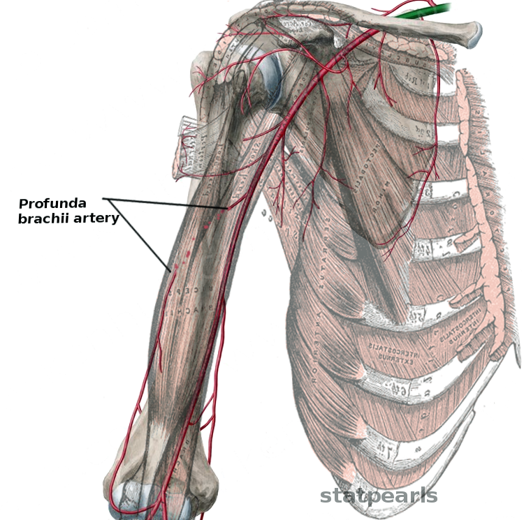
The profunda brachii artery also referred to as the deep brachial artery, is the first and largest branch of the brachial artery. It originates from the posterior portion of the brachial artery, just inferior to the lower border of the teres major muscle.  It then traverses the triangular interval into the posterior compartment of the arm along with the radial nerve.  Once in the posterior compartment, the profunda brachii artery gives off an arterial branch that anastomoses with the posterior circumflex humeral artery before continuing inferiorly along the radial sulcus (radial groove).  Below the inferior border of the latissimus dorsi muscle, it branches into the radial collateral artery and middle collateral artery. The radial collateral artery further divides into the anterior radial collateral artery and the posterior radial collateral artery.[1][2] The anterior radial collateral artery anastomoses with the radial recurrent artery while the middle collateral artery and posterior radial collateral artery both anastomose with the recurrent interosseous artery. 

Structure and Function

The profunda brachii is classified histologically as a muscular artery.  Muscular arteries serve as a bridge between elastic arteries and arterioles.  Defining characteristics of muscular arteries are the multitude of smooth muscle layers in the tunica media and a clearly defined internal elastic lamina. 

Blood supply to the upper extremity is traceable back to the aorta.  Blood initially flows into the brachiocephalic artery and then the subclavian artery on the right and directly into the subclavian artery on the left.  The subclavian artery turns into the axillary artery at the lateral border of the first rib.  The axillary artery turns into the brachial artery at the lower border of the teres major muscle. The brachial artery has five major branches; listed in order of branching are the profunda brachii artery, superior ulnar collateral artery, inferior ulnar collateral artery, ulnar artery, and radial artery. These arteries serve to provide blood flow to the upper extremity musculature. 

The profunda brachii is the first of five major brachial artery branches. It arises from the posterior portion of the brachial artery, just below the inferior border of the teres major muscle.[3] From here, the profunda brachii follows the radial nerve through the triangular interval into the posterior compartment of the arm.  The triangular interval forms from the teres major muscle superiorly, the long head of the triceps brachii muscle medially, and the humerus laterally.  The posterior compartment of the arm is divided from the anterior portion by a medial and lateral intermuscular septum and contains the three heads of triceps brachii muscle.  After traversing the triangular interval into the posterior compartment, the profunda brachii artery gives off its first branch, a small artery that courses superiorly to anastomose with the posterior circumflex humeral artery; this provides additional blood flow to the deltoid muscle. 

The profunda brachii artery continues on an inferior and lateral course in the radial sulcus, lying between the humerus and the lateral head of the triceps brachii muscle. Below the inferior border of the latissimus dorsi muscle, the profunda brachii artery divides into two terminal branches, the middle collateral artery, and radial collateral artery.  The middle collateral artery courses inferiorly in the medial head of the triceps brachii muscle and anastomoses with the inferior ulnar collateral artery and the recurrent interosseous artery superior to the olecranon of the ulna.  The anastomosis between the middle collateral artery and the inferior ulnar collateral artery contributes to the peri-articular anastomoses of the elbow.[1]  The radial collateral artery continues running in the radial sulcus and pierces the lateral intermuscular septum.  At the level of the origin of the brachioradialis muscle, the radial collateral artery further divides into the anterior radial collateral artery and posterior radial collateral artery. The posterior radial collateral artery continues traveling within the lateral intermuscular septum and anastomoses with the recurrent interosseous artery at the level of the olecranon.[4] The anterior radial collateral artery passes anterior to the lateral intermuscular septum and anastomoses with the radial recurrent artery at the level of the lateral epicondyle of the humerus.[1][2]

Embryology

The developing limb buds blood supply comes from intersegmental arteries, which are a group of thirty arteries arising from the embryonic aorta. The lateral branch of the seventh intersegmental artery gives rise to the primary axial artery.   The primary axial artery becomes the brachial artery and its branches, including the profunda brachii artery.

Nerves

The radial nerve, which arises from the C5-T1 nerve roots, provides motor and sensory function to portions of the upper extremity. It initially traverses the triangular interval with the profunda brachii artery and courses with it until the profunda brachii artery splits into the radial collateral artery and middle collateral artery.  At this point, the radial nerve takes a more superficial course, following the anterior radial collateral artery into the anterior compartment of the arm. 

Muscles

While upper extremity muscles receive vascular supply from more than one artery, the profunda brachii artery assists in supplying blood to the deltoid muscle, triceps brachii muscle, and anconeus muscle.  

Physiologic Variants

There is a significant body of literature that discusses the anatomical variations in the origin and termination of the profunda brachii artery.  The most common physiologic variant is the anomalous origin of the profunda brachii artery from the axillary artery rather than the brachial artery.  However, there is a great variation amongst studies in regards to the prevalence of this variant; ranging from 2% to 16.6%.[3]

Surgical Considerations

The posterior radial collateral artery, a branch of the profunda brachii artery, is used as a pedicle in lateral arm flaps.  The pedicle of a skin flap is the narrow base of the flap that is left connected to its original site and provides blood supply during grafting.[5][6][7] 

Surgeons perform the free lateral arm flap procedure when there is a defect in skin coverage of the forearm or hand that necessitates fat and skin coverage.  This procedure allows for a skin flap that can be up to five centimeters wide and span from the lateral epicondyle to the insertion of the deltoid muscle.  The flap is disconnected from its blood supply at the distal portion and elevated proximally, where it is left intact with the posterior radial collateral artery.  At this point, the surgeon can relocate the flap to the primary defect.

Clinical Significance

The brachial artery ranks as the most commonly injured upper extremity artery and accounts for 28% of all vascular injuries.[8] When assessing a brachial artery injury, it is essential to note whether the insult is proximal or distal to the profunda brachii artery as this correlates to the degree of ischemia.[9]

If the brachial artery is injured distal to the origin of the profunda brachii artery at the lower border of the teres major muscle, the profunda brachii artery will serve as the primary source of collateral circulation to the distal extremity. 

Other Issues

The terminal branches of the profunda brachii artery have been a topic of great debate.  Many sources make no mention of the posterior radial collateral artery or anterior radial collateral artery and list only the radial collateral artery and middle collateral artery as the terminal branches.  These sources state that the radial collateral artery anastomoses with the radial recurrent artery and the middle collateral artery anastomoses with the recurrent interosseous artery.[10][11] 


(Click Image to Enlarge)
Profunda brachii a
Profunda brachii a
Image courtesy Dr Chaigasame
Details

Updated:

8/22/2022 7:58:38 PM

References


[1]

Casoli V, Kostopoulos E, Pélissier P, Caix P, Martin D, Baudet J. The middle collateral artery: anatomic basis for the "extreme" lateral arm flap. Surgical and radiologic anatomy : SRA. 2004 Jun:26(3):172-7     [PubMed PMID: 14730394]


[2]

Sun R, Ding Y, Sun C, Li X, Wang J, Li L, Yang J, Ren Y, Zhong Z. Color Doppler Sonographic and Cadaveric Study of the Arterial Vascularity of the Lateral Upper Arm Flap. Journal of ultrasound in medicine : official journal of the American Institute of Ultrasound in Medicine. 2016 Apr:35(4):767-74. doi: 10.7863/ultra.15.01032. Epub 2016 Mar 11     [PubMed PMID: 26969598]


[3]

Chakravarthi KK, Ks S, Venumadhav N, Sharma A, Kumar N. Anatomical variations of brachial artery - its morphology, embryogenesis and clinical implications. Journal of clinical and diagnostic research : JCDR. 2014 Dec:8(12):AC17-20. doi: 10.7860/JCDR/2014/10418.5308. Epub 2014 Dec 5     [PubMed PMID: 25653931]


[4]

Lai CS, Lin SD, Chou CK, Tsai CC. The reverse lateral arm flap, based on the interosseous recurrent artery, for cubital fossa burns. British journal of plastic surgery. 1994 Jul:47(5):341-5     [PubMed PMID: 8087373]


[5]

Meirer R, Schrank C, Putz R. Posterior radial collateral artery as the basis of the lateral forearm flap. Journal of reconstructive microsurgery. 2000 Jan:16(1):21-4; discussion 24-5     [PubMed PMID: 10668750]


[6]

Sauerbier M, Germann G, Giessler GA, Sedigh Salakdeh M, Döll M. The free lateral arm flap-a reliable option for reconstruction of the forearm and hand. Hand (New York, N.Y.). 2012 Jun:7(2):163-71. doi: 10.1007/s11552-012-9395-3. Epub     [PubMed PMID: 23730235]


[7]

Rivet D, Buffet M, Martin D, Waterhouse N, Kleiman L, Delonca D, Baudet J. The lateral arm flap: an anatomic study. Journal of reconstructive microsurgery. 1987 Jan:3(2):121-32     [PubMed PMID: 3560037]


[8]

Ekim H, Tuncer M. Management of traumatic brachial artery injuries: a report on 49 patients. Annals of Saudi medicine. 2009 Mar-Apr:29(2):105-9     [PubMed PMID: 19318753]


[9]

McCready RA. Upper-extremity vascular injuries. The Surgical clinics of North America. 1988 Aug:68(4):725-40     [PubMed PMID: 3046002]


[10]

Devale MM, Munot RP, Bhansali CA, Bhaban ND. Awkward defects around the elbow: The radial recurrent artery flap revisited. Indian journal of plastic surgery : official publication of the Association of Plastic Surgeons of India. 2016 Sep-Dec:49(3):357-361. doi: 10.4103/0970-0358.197235. Epub     [PubMed PMID: 28216816]


[11]

Maruyama Y, Takeuchi S. The radial recurrent fasciocutaneous flap: reverse upper arm flap. British journal of plastic surgery. 1986 Oct:39(4):458-61     [PubMed PMID: 3779192]