Anatomy, Head and Neck, Posterior Auricular Artery


Introduction

The word "auricle" translates to a structure that looks similar to an ear. Cartilage comprises the entire auricular structure of the ear. The function of the ear is to funnel in sound, and then the sound is converted into various signals in the brain. Without the ear, sounds will be less amplified and seem to have lower volume.

The ear receives the majority of its blood from a branch of the external carotid artery. The external carotid artery gives off a branch called the posterior auricular artery. The meaning of the "posterior auricular" is an area posterior to the auricular structure; this is the exact territory of the posterior auricular artery's perfusion. The posterior auricular artery is also the main blood supply to the external ear.

Structure and Function

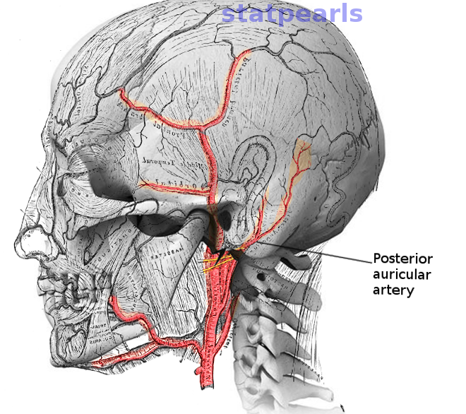
The posterior auricular artery is a smaller muscular artery that branches from the external carotid artery. This artery travels to the region posterior to the ear. The function of the posterior auricular artery is to perfuse the scalp and the ear.[1] The minor function of the posterior auricular artery is to provide collateral blood flow via the anastomoses that form with the superior temporal artery, the parietal artery, and the anterior auricular artery. The anastomoses with the superior temporal artery provide collateral blood supply to the temporal region of the head. The connection between the posterior auricular artery and the anterior auricular artery provides complete perfusion of the auricular part (cartilage part of the ear) of the ear. The connection between the posterior auricular artery and the parietal artery provides additional blood to the parietal region of the skull.

Embryology

During embryology, the arteries in the face, neck, and chest form from the aortic arches. The aortic arches will form around the same time as the formation brachial apparatus. The third aortic arch will develop into the common carotid artery. The common carotid artery will ascend laterally along the face, neck, and head. At the level of cervical vertebra four, the common carotid artery will bifurcate into the external carotid artery and internal carotid artery. The external carotid artery is responsible for perfusing the face and head. One of the branches that come off the external carotid artery is the posterior auricular artery. The external carotid artery branches into the posterior auricular artery at the level of the angle of the mandible. The posterior artery will then ascend towards the region posterior to the ear.

Blood Supply and Lymphatics

The posterior auricular artery will ascend towards the posterior side of the ear. The posterior auricular artery then perfuses the majority of the ear and the posterior region of the scalp. The posterior auricular artery will give off small auricular branches that perfuse the ear. Another branch that comes from the posterior auricular artery is the stylomastoid artery. The stylomastoid artery will perfuse the styloid process of the temporal bone, the mastoid process of the temporal bone, and the facial nerve. The facial nerve receives blood from the stylomastoid artery as it exits the stylomastoid foramen.[2][3] 

The posterior auricular artery forms anastomoses that will provide collateral blood flow to other regions on the face and head. Three main anastomoses form with the superior temporal artery, the parietal artery, and the anterior auricular artery. 

The lymph drainage from the scalp near the ear and the ear itself is towards the posterior auricular lymph nodes. The lymph fluid will eventually drain into the main circulation via the right lymphatic duct and the thoracic duct. The right ear will drain towards the right lymphatic duct. While the left ear drains toward the thoracic duct.[4]

Nerves

The posterior auricular artery perfuses the majority of the ear and the scalp posterior to the ear. This perfusion region will also supply blood to the greater auricular nerve (C2, C3 spinal roots) and the lesser occipital nerve (C2 spinal roots). The facial nerve also receives perfusion from the posterior auricular artery at the stylomastoid foramen opening.

Muscles

The posterior auricular artery's perfusion territory includes four main muscles:

  • The posterior auricular muscles
  • The occipitalis muscle
  • The temporalis muscle
  • The sternocleidomastoid muscle (origin)

These muscles will also derive blood from other arteries, but some of their muscle fibers receive some blood from the perfusion territory of the posterior auricular artery. 

Physiologic Variants

The external carotid artery gives off many branches to perfuse the head and face. The posterior auricular artery is one of the branches that come from the external carotid artery. The order and the location of the posterior auricular artery branch can vary. The posterior auricular artery can sometimes branch from the occipital artery instead of the external carotid artery. But for the majority of individuals, the posterior auricular artery consistently branches from the external carotid artery. The origin, length, and termination of the posterior auricular artery can vary greatly.[5] 

Surgical Considerations

In surgery, the posterior auricular artery is important when it comes to procedures performed on the scalp and posterior region of the ear. The knowledge of the location of the artery and the anastomoses are crucial when it comes to biopsy of the superficial temporal artery. The superficial temporal artery requires biopsy in giant cell temporal arteritis". This condition is a large vessel vasculitis that affects the superficial temporal artery. The definitive diagnosis of this condition is via biopsy. After the biopsy, the posterior auricular artery will provide blood supply to the region that was predominately perfused by the superficial temporal artery.[6] 

In neurosurgery, the posterior auricular artery is grafted in rare instances to restore the blood supply in the circle of Willis. The restoration of blood supply in the circle of Willis is via bypass grafting.[7][8]

In plastic surgery, the tissue and skin that is perfused by the posterior auricular artery has utility in ear reconstruction. The pedicle of tissue posterior to the ear can be dissected and rotated to reconstruct or create the ear. The rotation of the pedicle of tissue will continue to have the majority of its blood supply from the posterior auricular artery.[9][10][11][12][13]

The posterior auricular artery can also be used as a landmark during dissection to find the facial nerve. The facial nerve exits the stylomastoid foramen. Perfusion of this foramen is via the posterior auricular artery.[2][3][14]

Clinical Significance

In the clinical setting, if the ear or the scalp posterior to the ear becomes perforated or lacerated. There will be significant bleeding since there are many anastomoses in that region. With the knowledge of the vasculature of the scalp, hemorrhaging from a scalp laceration can be successfully controlled. The achievement of hemostasis in the scalp is possible with prolonged compression or cauterization of the bleeding arteries. 

Other Issues

The main blood supply of the face, neck, and head derives from the carotid arteries. If there were to be damaged or compromise of the carotid arteries, the territories perfused by the posterior auricular artery will suffer from the lack of blood. The lack of blood supply will lead to ischemia and necrosis.


(Click Image to Enlarge)
Post auricular artery
Image courtesy O.Chaigasame
Details

Editor:

Hieu Duong

Updated:

6/5/2023 9:28:27 PM

References


[1]

Zilinsky I, Erdmann D, Weissman O, Hammer N, Sora MC, Schenck TL, Cotofana S. Reevaluation of the arterial blood supply of the auricle. Journal of anatomy. 2017 Feb:230(2):315-324. doi: 10.1111/joa.12550. Epub 2016 Oct 11     [PubMed PMID: 27726131]


[2]

Liu M, Wang SJ, Benet A, Meybodi AT, Tabani H, Ei-Sayed IH. Posterior auricular artery as a novel anatomic landmark for identification of the facial nerve: A cadaveric study. Head & neck. 2018 Jul:40(7):1461-1465. doi: 10.1002/hed.25127. Epub 2018 Mar 22     [PubMed PMID: 29566447]


[3]

Upile T, Jerjes W, Nouraei SA, Singh SU, Kafas P, Sandison A, Sudhoff H, Hopper C. The stylomastoid artery as an anatomical landmark to the facial nerve during parotid surgery: a clinico-anatomic study. World journal of surgical oncology. 2009 Sep 28:7():71. doi: 10.1186/1477-7819-7-71. Epub 2009 Sep 28     [PubMed PMID: 19785731]


[4]

Pan WR, le Roux CM, Levy SM, Briggs CA. Lymphatic drainage of the external ear. Head & neck. 2011 Jan:33(1):60-4. doi: 10.1002/hed.21395. Epub     [PubMed PMID: 20848416]


[5]

Tokugawa J, Cho N, Suzuki H, Sugiyama N, Akiyama O, Nakao Y, Yamamoto T. Novel classification of the posterior auricular artery based on angiographical appearance. PloS one. 2015:10(6):e0128723. doi: 10.1371/journal.pone.0128723. Epub 2015 Jun 1     [PubMed PMID: 26030595]


[6]

Kobayashi S, Nagase T, Ohmori K. Colour Doppler flow imaging of postauricular arteries and veins. British journal of plastic surgery. 1997 Apr:50(3):172-5     [PubMed PMID: 9176003]


[7]

Torazawa S, Hasegawa H, Kin T, Sato H, Sora S. Long-Term Patency of Posterior Auricular Artery-Middle Cerebral Artery Bypass for Adult-Onset Moyamoya Disease: Case Report and Review of Literature. World neurosurgery. 2017 Dec:108():994.e1-994.e5. doi: 10.1016/j.wneu.2017.09.005. Epub 2017 Sep 20     [PubMed PMID: 28939540]

Level 3 (low-level) evidence

[8]

Tokugawa J, Nakao Y, Kudo K, Iimura K, Esaki T, Yamamoto T, Mori K. Posterior auricular artery-middle cerebral artery bypass: a rare superficial temporal artery variant with well-developed posterior auricular artery-case report. Neurologia medico-chirurgica. 2014:54(10):841-4     [PubMed PMID: 24140773]

Level 3 (low-level) evidence

[9]

Oufkir AA, Kajout M, Chadli R, Alami MN. [Comments on article "Scalp flap pedicled on posterior auricular artery. Anatomical study and clinical application"]. Annales de chirurgie plastique et esthetique. 2015 Jun:60(3):252-3. doi: 10.1016/j.anplas.2015.03.002. Epub 2015 Apr 1     [PubMed PMID: 25841768]

Level 3 (low-level) evidence

[10]

Caughlin BP, Redleaf M. Posterior auricular artery fasciocutaneous island flap: lateral temporal soft tissue reconstruction. The Laryngoscope. 2016 Mar:126(3):722-6. doi: 10.1002/lary.25499. Epub 2015 Jul 30     [PubMed PMID: 26228008]


[11]

Zhang YZ, Li YL, Yang C, Fang S, Fan H, Xing X. Reconstruction of the postauricular defects using retroauricular artery perforator-based island flaps: Anatomical study and clinical report. Medicine. 2016 Sep:95(37):e4853. doi: 10.1097/MD.0000000000004853. Epub     [PubMed PMID: 27631246]


[12]

Hénoux M, Espitalier F, Hamel A, Dréno B, Michel G, Malard O. Vascular Supply of the Auricle: Anatomical Study and Applications to External Ear Reconstruction. Dermatologic surgery : official publication for American Society for Dermatologic Surgery [et al.]. 2017 Jan:43(1):87-97. doi: 10.1097/DSS.0000000000000928. Epub     [PubMed PMID: 28027200]


[13]

Gómez Díaz OJ, Cruz Sánchez MD. Anatomical and Clinical Study of the Posterior Auricular Artery Angiosome: In Search of a Rescue Tool for Ear Reconstruction. Plastic and reconstructive surgery. Global open. 2016 Dec:4(12):e1165. doi: 10.1097/GOX.0000000000001165. Epub 2016 Dec 27     [PubMed PMID: 28293515]


[14]

Elvan Ö, Bobuş A, Erdoğan S, Aktekin M, Olgunus ZK. Fetal anatomy of the facial nerve trunk and its relationship with posterior auricular artery. Surgical and radiologic anatomy : SRA. 2019 Feb:41(2):153-159. doi: 10.1007/s00276-018-2126-x. Epub 2018 Oct 26     [PubMed PMID: 30367188]