Anatomy, Head and Neck, Nasopalatine Nerve


Introduction

Also known as nervus incisivus, the nasopalatine nerve is a division of the maxillary branch of the trigeminal nerve. Its function is to provide sensation to the anterior palate. The sphenopalatine artery supplies the same area it innervates. The pathological development of a cyst in the incisive canal can cause impingement of the nasopalatine nerve and limit its function. Clinically, injecting local anesthesia or performing nasopalatine nerve block facilitates procedures of the palate/maxilla.

Structure and Function

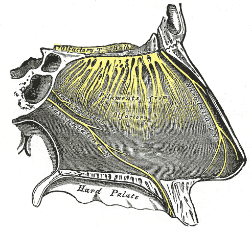
The nasopalatine nerve is a division of the maxillary branch of the trigeminal nerve. As the trigeminal nerve is sensory, the nasopalatine nerve is also a sensory nerve. It courses into the nasal cavity through the pterygopalatine ganglion to enter the sphenopalatine foramen. It passes inferior to the ostium of the sphenoid sinus to reach the septum, lying between the periosteum and the mucous membrane where it provides sensation to the nasal septum. From here, it courses inferiorly to pierce the hard palate anteriorly through the nasopalatine canal, which links the nasal and oral cavities. Other structures in this canal include the terminal branch of the nasopalatine artery, a small amount of fat, and palatine glands. The inferior opening of the nasopalatine canal is called the incisive foramen.[1] Here, the nasopalatine nerve communicates with the anterior palatine nerve. The extent to which it innervates the local mucosa, as well as its relationship with the anterior (greater) palatine nerve, has historically been a topic of debate.[2]

The anterior palatine nerve innervates the gingivae and the structures of the maxillary teeth as far forward as the canines, while the nasopalatine nerve primarily innervates the mucosa surrounding the incisive papilla and the gingival margins of the maxillary incisors.[3] However, there is an overlap in the watershed area between these two nerve distributions.

The territory of the nasopalatine nerve also differs depending on the age of the subject in question. From data of nasopalatine nerve resection during the removal of supernumerary teeth, younger patients have been shown not to have sensory deficits, while the adolescent and adult groups did. This finding led to the conclusion that the nasopalatine nerve assumes the function of the anterior palatine nerve around age fourteen years.[4] The clinical implication here is that different anesthetic plans may be necessary to accommodate these changes with development depending on the age of the patient.

Embryology

As the nasopalatine nerve is a derivative of the maxillary nerve, the V2 branch of the trigeminal nerve (CN V); originates from the first pharyngeal arch. The first pharyngeal arch appears at the beginning of the fourth week of gestation, and cranial nerve V2 spreads to provide sensation to the maxillary region of the face.

The nasopalatine canal forms as part of the primary palate, which itself results from the fusion of the two medial nasal prominences in the midline. It is then the point of union between the primary and secondary palate, which is composed of the palatine shelves- outgrowths of the maxillary prominences.  As development occurs, the nasopalatine nerve passes through a wide tissue space initially, but as ossification of the incisive bone takes place at 12 to 15 weeks, the incisive canal becomes narrow and fills with fibrous tissue.[5]

Blood Supply and Lymphatics

The nasopalatine (or sphenopalatine) artery is a branch of the internal maxillary artery that enters the nasal cavity via the sphenopalatine foramen to supply the frontal, maxillary, ethmoid, and sphenoid sinuses. One branch descends into the incisive canal to anastomose with the descending palatine artery, entering the nasopalatine canal with the nasopalatine nerve.

The veins of the palate are diffuse and variable. The primary vessel draining this region is the facial vein which courses from the medial canthus, behind the facial artery, to join with the retromandibular vein below the mandible; this union is sometimes termed the common facial vein. Venous drainage of the hard palate is via the pterygoid plexus of veins in the infratemporal fossa, while the soft palate drains via the pharyngeal venous plexus.

Lymphatics from the palate generally terminate in the jugulodigastric nodes. The drainage of the palatal gingivae varies, as sometimes it occurs directly via the jugulodigastric nodes and sometimes indirectly via the submandibular nodes. Posterior soft palate lymphatics drain into the pharyngeal lymph nodes.

Surgical Considerations

One entity that can affect the nasopalatine nerve is a nasopalatine duct cyst, wherein remnants of the nasopalatine duct epithelium are stimulated to proliferate, either by trauma, infection, or mucus retention. It is the most common non-odontogenic cyst of the maxilla, occurring in 1% of the population- most commonly arising in middle age. Standard treatment requires surgical excision via a sublabial or palatal approach. As the cyst is not a true neoplasm, it can also be treated by marsupialization to the nasal cavity.[6][7]

Dysesthesia of the anterior palate or maxillary teeth may occur after septoplasty, especially if the procedure addressed low septal spurs, and there is a need for the use of cautery or osteotomes.[8]

Clinical Significance

Nasopalatine nerve blocks are often necessary for oral-maxillofacial procedures, including cleft palate repair. This procedure requires the deposition of anesthetic in the incisive canal to anesthetize the nasopalatine nerves bilaterally, blocking pain fibers in the area surrounding the anterior six maxillary teeth. The initial technique involved a single penetration into the canal, but the prevailing opinion is that this is traumatic and unnecessarily painful to the patient. The current practice involves multiple injections, first injecting on the labial side in the interdental papilla, entering horizontally with a 30-gauge needle. After achieving buccal anesthesia, the solution spreads by diffusion to the palate. However, in patients with complete cleft palate, this block cannot be done as the premaxilla is malformed.[9][10]

Although anterior (greater) palatine nerve and nasopalatine nerve blocks are often both performed before procedures of the palate or anterior maxillary teeth, innervation is variable. Extraction of supernumerary teeth in adults with the related sacrifice of the nasopalatine nerve has shown to correlate with a persistent alteration in sensation in adults (see above), while other data demonstrates that sacrifice of the nasopalatine nerve will not result in the patient reporting of altered sensation. The anterior palatine nerve alone in this study could maintain adequate sensation, while the nasopalatine nerve alone could not.[3]

Nasopalatine canal measurement is also important in the field of implant dentistry. It is useful in preventing intraoperative complications, such as perforation of the nasopalatine canal or buccal bone plate. To date, traditional imaging modalities have proven ineffective, given the bony complexity of this region. For this purpose, cone beam computed tomography has seen increased use with success.[1]


(Click Image to Enlarge)
The Olfactory Nerve, Olfactory Tract and Bulb, Filaments, Nasociliary nerve, Nasopalatine nerve
Contributed by Gray's Anatomy Plates
Details

Editor:

Brian W. Downs

Updated:

7/24/2023 9:58:12 PM

References


[1]

Demiralp KÖ, Kurşun-Çakmak EŞ, Bayrak S, Sahin O, Atakan C, Orhan K. Evaluation of Anatomical and Volumetric Characteristics of the Nasopalatine Canal in Anterior Dentate and Edentulous Individuals: A CBCT Study. Implant dentistry. 2018 Aug:27(4):474-479. doi: 10.1097/ID.0000000000000794. Epub     [PubMed PMID: 30028392]


[2]

Souza SS, Raggio BS. Anatomy, Head and Neck, Sphenopalatine Foramen. StatPearls. 2023 Jan:():     [PubMed PMID: 31751101]


[3]

Langford RJ. The contribution of the nasopalatine nerve to sensation of the hard palate. The British journal of oral & maxillofacial surgery. 1989 Oct:27(5):379-86     [PubMed PMID: 2804040]


[4]

Liu J, Li X, Ma L, Pan J, Tang X, Wu Y, Hua C. A Hypothesis and Pilot Study of Age-Related Sensory Innervation of the Hard Palate: Sensory Disorder After Nasopalatine Nerve Division. Medical science monitor : international medical journal of experimental and clinical research. 2017 Jan 29:23():528-534     [PubMed PMID: 28132066]

Level 3 (low-level) evidence

[5]

Kim JH, Oka K, Jin ZW, Murakami G, Rodríguez-Vázquez JF, Ahn SW, Hwang HP. Fetal Development of the Incisive Canal, Especially of the Delayed Closure Due to the Nasopalatine Duct: A Study Using Serial Sections of Human Fetuses. Anatomical record (Hoboken, N.J. : 2007). 2017 Jun:300(6):1093-1103. doi: 10.1002/ar.23521. Epub 2017 Jan 27     [PubMed PMID: 27860365]


[6]

Honkura Y, Nomura K, Oshima H, Takata Y, Hidaka H, Katori Y. Bilateral endoscopic endonasal marsupialization of nasopalatine duct cyst. Clinics and practice. 2015 Jan 28:5(1):748. doi: 10.4081/cp.2015.748. Epub 2015 Feb 5     [PubMed PMID: 25918636]


[7]

Wu YH, Wang YP, Kok SH, Chang JY. Unilateral nasopalatine duct cyst. Journal of the Formosan Medical Association = Taiwan yi zhi. 2015 Nov:114(11):1142-4. doi: 10.1016/j.jfma.2015.08.005. Epub 2015 Aug 29     [PubMed PMID: 26329385]


[8]

Chandra RK, Rohman GT, Walsh WE. Anterior palate sensory impairment after septal surgery. American journal of rhinology. 2008 Jan-Feb:22(1):86-8. doi: 10.2500/ajr.2008.22.3114. Epub     [PubMed PMID: 18284865]


[9]

Reena, Bandyopadhyay KH, Paul A. Postoperative analgesia for cleft lip and palate repair in children. Journal of anaesthesiology, clinical pharmacology. 2016 Jan-Mar:32(1):5-11. doi: 10.4103/0970-9185.175649. Epub     [PubMed PMID: 27006533]


[10]

Echaniz G, De Miguel M, Merritt G, Sierra P, Bora P, Borah N, Ciarallo C, de Nadal M, Ing RJ, Bosenberg A. Bilateral suprazygomatic maxillary nerve blocks vs. infraorbital and palatine nerve blocks in cleft lip and palate repair: A double-blind, randomised study. European journal of anaesthesiology. 2019 Jan:36(1):40-47. doi: 10.1097/EJA.0000000000000900. Epub     [PubMed PMID: 30308523]

Level 1 (high-level) evidence