Minimally Invasive Aortic Valve Surgery

Earn CME/CE in your profession:


Continuing Education Activity

Aortic valve replacement is the standard treatment for patients with severe or symptomatic aortic insufficiency or aortic stenosis. In the past, the standard method of replacing the aortic valve has been via a median sternotomy approach. However, in some patients, this midline anterior chest incision may not heal properly, cause pain, and be associated with a prolonged recovery. In patients with osteoporosis or diabetes, the thinned out sternum may take a long time to heal and be associated with a significant amount of pain. This activity describes the technique of minimally aortic valve surgery, its indications, contraindications and highlights the role of the interprofessional team in the management of patients with aortic valve disease.

Objectives:

  • Describe the technique of minimally aortic valve surgery.
  • Review the indications for minimally invasive aortic valve surgery.
  • Summarize the complications of minimally invasive aortic valve surgery.
  • Outline the importance of an interprofessional team to improve outcomes for patients undergoing minimally invasive aortic valve surgery.

Introduction

Aortic valve replacement is the standard treatment for patients with severe or symptomatic aortic insufficiency or aortic stenosis. In the past, the standard method of replacing the aortic valve has been via a median sternotomy approach. However, in some patients, this midline anterior chest incision may not heal properly, cause pain, and be associated with a prolonged recovery. In patients with osteoporosis or diabetes, the thinned out sternum may take a long time to heal and be associated with a significant amount of pain.[1][2][3][4]

Over the past two decades, surgeons have developed alternative access routes to the aortic valve. The two main approaches to the aortic valve include a mini-thoracotomy and the minimally thoracoscopic approach. A third way of implanting the aortic valve is via a percutaneous approach combined with a mini-anterior thoracotomy. All these surgical approaches to the heart require general anesthesia, use of transesophageal echocardiogram, and full cardiac monitoring. In addition, the perfusionist must have the open heart-lung machine ready in case a complication occurs and there is a need to convert to a median sternotomy. The operating room is fully prepared in the same way as when a conventional aortic valve surgery is going to be performed.

The minimally invasive aortic valve surgery is performed by making a 4 cm to 6 cm incision on the anterior chest wall (to the left of the sternum) which gives direct access to the left ventricle. Another variation is to make a 4 cm to 6 cm horizontal incision in the right third intercostal space to access the root of the aorta. In both these scenarios, the femoral vessels are used to place the patient on the heart-lung machine. The most difficult part of the surgery is getting proper access to the aortic root. The rest of the procedure is then done the same as a conventional aortic valve replacement.

Percutaneous aortic valve surgery is performed by making a 6 cm incision at the left costal margin and accessing the heart. A transesophageal probe is placed in the esophagus, and special instruments are used to make a hole in the left ventricle apex, and a valve-containing device is guided to the aortic annulus. The stenotic valve is dilated first. Once the device is in place, the valve is released at the aortic annulus, and the instruments are removed. The hole in the ventricle is closed with sutures.There also is a robotic-assisted valve surgery procedure where three to five small incisions are made, and the surgeon uses robotic arms to conduct the surgery. Again, the most difficult part of this technique is to perform the actual aortic valve anastomosis. Most experts believe that the robotic surgery should be combined with an open incision so that the surgeon can manually perform the aortic anastomosis. The minimally aortic valve surgeries are also more expensive than the routine method of replacing the aortic valve as they require expensive disposable equipment.

Anatomy and Physiology

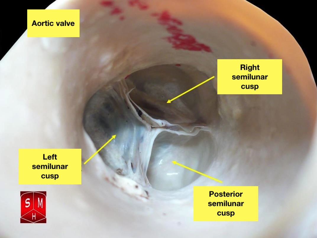
The human heart has four different anatomical valves that play a role in the regulation of blood flow into and out of the myocardium. The two semilunar valves include the pulmonic and aortic valves, and the two atrioventricular valves include the mitral and tricuspid valves.

Except for the mitral valve, all the cardiac valves are tri-leaflet. Each of the heart valves is surrounded by a complete or partial fibrous ring (annulus), which helps supports the valves. The aortic valve is located between the ascending aorta and the left ventricular outflow tract. It is a very important valve and closely approximates other myocardial structures like the mitral valve posterolaterally, the pulmonic valve anteriorly, and the tricuspid valve posteromedially.

Indications

Percutaneous aortic valve replacement is indicated for the following patients: 

  • High-risk
  • Predicted EuroSCORE greater than 30%
  • 30-day actual mortality lower than 10%

The procedure is contraindicated in patients with severe aortic valve calcification and severe peripheral vascular disease.

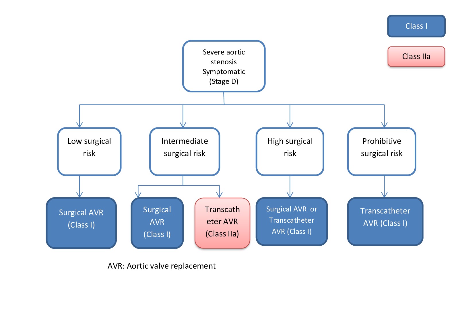
In general, aortic valve surgery is done for all symptomatic patients and those with severe aortic stenosis.

Contraindications

The median sternotomy approach to the aortic valve is associated with an increased risk of perioperative complications; thus an alternative surgical approach to the aortic valve, such as a mini-thoracotomy or a left lateral thoracotomy, may be safer sometimes. Other relative contraindications to median sternotomy include COPD, obesity, or prior radiation exposure to the anterior chest.

Equipment

The new tri-leaflet aortic valve can either be sutured or attached to the annulus by first expanding the annulus and then deploying a stent mounted catheter system. The initial percutaneously implanted aortic stent valves failed because they were too large, but over the years, the newer generation of aortic valves have a better profile and hemodynamic performance. The smaller catheter-based valves do not obstruct the coronary arteries or interfere with the function of the mitral valve. However, they still need to be accurately placed at the annulus.

 The following is a summary of the different types of aortic valves that are currently available:

  • Cribier-Edwards Valve (Edwards Lifesciences, Irvine, CA): Stainless steel stent; equine pericardial valve; currently available in 23 and 26 mm diameters
  • CoreValve (CoreValve, Inc, Irvine, CA): Self-expanding nitinol stent; porcine pericardial tissue valve; repositionable and retrievable
  • Direct Flow percutaneous aortic valve (Direct Flow Medical, Inc, Santa Rosa, CA): Stentless, inflatable, fabric-cuff; equine pericardial tissue valve
  • Lotus Valve System (Sadra Medical, Campbell, CA): Self-expanding nitinol frame; bovine pericardial tissue valve; repositionable and retrievable
  • AorTx percutaneous aortic valve (Hansen Medical, Mountain View, CA): Low-profile, folded, metallic frame; pericardial tissue valve; repositionable and retrievable

To deploy the stent valve, either expand the balloon on a balloon-expandable stent or withdraw the sheath that releases a self-expanding stent. 

Once the valve is deployed it must remain secure around the diseased aortic valve and be able to resume hemodynamic function right away

Once the valve is functioning, its hemodynamic performance should be arranged in the same way as the valves that are implanted via a median sternotomy. The left ventricular outflow gradient must be 10 mmHg or less and the aortic valve area should be between 1.5 and 2.0 cm2. Once the prosthetic valve has been deployed, it is vital that it does not occlude the native coronary vessels or affect the function of the mitral valve which is located just underneath the aortic valve. In addition, the newly seated valve should be firmly seated on the aortic annulus and prevent the development of a paravalvular leak or aortic insufficiency.[5][6][7]

Preparation

General anesthesia for sternotomy is dependent upon the type of operation to be performed. If single lung ventilation is required, patients should be intubated with a double lumen endotracheal tube.

The patient must be sufficiently sedated and paralyzed before opening the chest to avoid tachyarrhythmias and hypertension during anesthesia. In patients with severe aortic stenosis, the surgeon must be scrubbed and ready to open the chest in case an arrest occurs.

The lungs are usually deflated before sternal division.

Technique or Treatment

Today, several minimally invasive approaches to the aortic valve aim to avoid a full median sternotomy. Of these incisions, the right parasternal approach appears to be the most widely used. However, it is important to remember that the median sternotomy is still the gold standard for replacing the aortic valve.

Complications

Complications may include:

  • Heart failure
  • Sudden cardiac death
  • Calcific embolization
  • Sudden death

Clinical Significance

In general, the minimally invasive procedures are not the standard of care for aortic valve replacement. These procedures are done in patients who are too ill to undergo a formal median sternotomy and use of a heart-lung machine. Short-term results of these minimally aortic valve replacement procedures are fraught with complications. Complications of minimally valve surgery include bleeding, failure of the procedure to work, immediate opening of the chest due to a complication, thrombosis of the valve, perforation of the esophagus, and damage to the femoral vessels (usually dissection). The recovery of the patient is done in the same way as a normal open-heart surgery patient.

Several large clinical trials have been performed on the use of the percutaneous method of aortic valve implantation, and early results indicate that the morbidity and mortality are the same as the conventional technique. However, more long-term data are needed to determine if the results are durable and sustained for at least a decade.[8][9][10]

Enhancing Healthcare Team Outcomes

an interprofessional approach to minimally invasive aortic surgery

Even though there are several ways for cardiac surgeons to operate on the aortic valve, the gold standard is the open procedure. The minimally invasive procedures do offer some advantages but they also require expensive equipment, extensive exposure to radiation and one must have an operating room on standby in case something goes wrong. Healthcare workers who see patients with valvular disorders must be aware that these minimally invasive procedures are only done for very ill patients who are not fit for the open procedure. In the end, a an interprofessional team must fully evaluate the patient and decide which is the ideal procedure for him/her.[11][12]


(Click Image to Enlarge)
Aortic Valve
Aortic Valve
By Anatomist90 - Own work, CC BY-SA 3.0, https://commons.wikimedia.org/w/index.php?curid=17215093

Valveguru [CC BY-SA 3.0 (https://creativecommons.org/licenses/by-sa/3.0)]

(Click Image to Enlarge)
Indication for aortic valve replacement
Contributed by Dr. Sai Harika Pujari
Details

Author

Akarsh Parekh

Author

Poonam Bhyan

Editor:

Nauman Khalid

Updated:

5/22/2023 7:31:10 AM

References


[1]

Ramchandani MK, Wyler von Ballmoos MC, Reardon MJ. Minimally Invasive Surgical Aortic Valve Replacement Through a Right Anterior Thoracotomy: How I Teach It. The Annals of thoracic surgery. 2019 Jan:107(1):19-23. doi: 10.1016/j.athoracsur.2018.11.006. Epub 2018 Nov 20     [PubMed PMID: 30468728]


[2]

Raptis DA, Beal MA, Kraft DC, Maniar HS, Bierhals AJ. Transcatheter Aortic Valve Replacement: Alternative Access beyond the Femoral Arterial Approach. Radiographics : a review publication of the Radiological Society of North America, Inc. 2019 Jan-Feb:39(1):30-43. doi: 10.1148/rg.2019180059. Epub 2018 Nov 23     [PubMed PMID: 30468629]


[3]

Tamadon I, Soldani G, Dario P, Menciassi A. Novel Robotic Approach for Minimally Invasive Aortic Heart Valve Surgery. Annual International Conference of the IEEE Engineering in Medicine and Biology Society. IEEE Engineering in Medicine and Biology Society. Annual International Conference. 2018 Jul:2018():3656-3659. doi: 10.1109/EMBC.2018.8513309. Epub     [PubMed PMID: 30441166]


[4]

Senst B, Kumar A, Diaz RR. Cardiac Surgery. StatPearls. 2023 Jan:():     [PubMed PMID: 30422530]


[5]

Nguyen DH, Vo AT, Le KM, Vu TT, Nguyen TT, Vu TT, Pham CVT, Truong BQ. Minimally Invasive Ozaki Procedure in Aortic Valve Disease: The Preliminary Results. Innovations (Philadelphia, Pa.). 2018 Sep/Oct:13(5):332-337. doi: 10.1097/IMI.0000000000000556. Epub     [PubMed PMID: 30394956]


[6]

El-Essawi A, Breitenbach I, Haupt B, Brouwer R, Morjan M, Harringer W. Aortic valve replacement with or without myocardial revascularization in octogenarians. Can minimally invasive extracorporeal circuits improve the outcome? Perfusion. 2019 Apr:34(3):217-224. doi: 10.1177/0267659118811048. Epub 2018 Nov 3     [PubMed PMID: 30394847]


[7]

Vasanthan V, Kent W, Gregory A, Maitland A, Cutrara C, Bouchard D, Asch F, Adams C. Perceval Valve Implantation: Technical Details and Echocardiographic Assessment. The Annals of thoracic surgery. 2019 Mar:107(3):e223-e225. doi: 10.1016/j.athoracsur.2018.08.091. Epub 2018 Oct 24     [PubMed PMID: 30367839]


[8]

Rodríguez-Caulo EA, Guijarro-Contreras A, Otero-Forero J, Mataró MJ, Sánchez-Espín G, Guzón A, Porras C, Such M, Ordóñez A, Melero-Tejedor JM, Jiménez-Navarro M. Quality of life, satisfaction and outcomes after ministernotomy versus full sternotomy isolated aortic valve replacement (QUALITY-AVR): study protocol for a randomised controlled trial. Trials. 2018 Feb 17:19(1):114. doi: 10.1186/s13063-018-2486-x. Epub 2018 Feb 17     [PubMed PMID: 29454380]

Level 2 (mid-level) evidence

[9]

Herold J, Herold-Vlanti V, Sherif M, Luani B, Breyer C, Bonaventura K, Braun-Dullaeus R. Analysis of cardiovascular mortality, bleeding, vascular and cerebrovascular events in patients with atrial fibrillation vs. sinus rhythm undergoing transfemoral Transcatheter Aortic Valve Implantation (TAVR). BMC cardiovascular disorders. 2017 Dec 20:17(1):298. doi: 10.1186/s12872-017-0736-6. Epub 2017 Dec 20     [PubMed PMID: 29262768]


[10]

Oakley L, Pritchard W, Colletta J, Penny W, Romero S, Cox J, Boswell G, Kindelan J, Gramins D, Nayak K. Development and Early Experience of the First Joint Military Health System-Veterans Affairs Transcatheter Aortic Valve Replacement Program. Military medicine. 2017 Nov:182(11):e2036-e2040. doi: 10.7205/MILMED-D-16-00398. Epub     [PubMed PMID: 29087877]


[11]

Kolte D, Abbott JD, Aronow HD. Interventional Therapies for Heart Failure in Older Adults. Heart failure clinics. 2017 Jul:13(3):535-570. doi: 10.1016/j.hfc.2017.02.009. Epub     [PubMed PMID: 28602371]


[12]

Yücel S, Ince H, Kische S, Sherif MA, Bushnaq H, Bärisch A, Öner A. [Interdisciplinary differential treatment of structural heart disease : When operation and when catheter-based intervention?]. Herz. 2016 Aug:41(5):443-58. doi: 10.1007/s00059-016-4444-2. Epub     [PubMed PMID: 27460051]


[13]

Khoshbin E, Prayaga S, Kinsella J, Sutherland FW. Mini-sternotomy for aortic valve replacement reduces the length of stay in the cardiac intensive care unit: meta-analysis of randomised controlled trials. BMJ open. 2011:1(2):e000266. doi: 10.1136/bmjopen-2011-000266. Epub 2011 Nov 24     [PubMed PMID: 22116090]

Level 1 (high-level) evidence