Oral Mucosa Cancer

Earn CME/CE in your profession:


Continuing Education Activity

Oral mucosal cancer is cancer that arises from the lining (mucosa) of the oral cavity. The main risk factors are smoking and alcohol consumption. The mainstay of treatment is surgery, often with adjuvant radiotherapy. This activity outlines the evaluation and management of oral mucosal cancer and highlights the role of the interprofessional healthcare team in managing patients with this condition.

Objectives:

  • Outline the etiology of oral mucosal cancer.
  • Review the evaluation of oral mucosal cancer.
  • Explain the management options available for oral mucosal cancer.

Introduction

Oral mucosal cancer is cancer that arises from the lining (mucosa) of the oral cavity. The oral cavity is compromised of the mucosa lining of the lips and cheeks, the teeth, gingiva (gums), anterior two-thirds of the tongue, the floor of the mouth, hard palate, and the retromolar trigone posterior to the wisdom teeth. It has a close anatomical relationship with the oropharynx, the boundary of which is the border between the hard and soft palate, the border between the anterior 2/3rds and posterior 1/3rd of the tongue, and the anterior pillars of the tonsils. The main risk factors are smoking and alcohol consumption. The mainstay of treatment is surgery, often with adjuvant radiotherapy.

Etiology

Tobacco 

Smoking tobacco is the greatest risk factor and cause of oral cancer due to its carcinogenic chemicals, including nitrosamines, benzopyrenes, and aromatic amines.[1] The risk of developing oral cancer is 3 times higher in smokers compared with non-smokers.[2] Individuals are also at risk from secondary passive smoking environments.[3] Studies have shown a synergistic relationship with alcohol consumption, resulting in a higher risk of malignancy. 

In various parts of the world, tobacco is chewed or held in the mouth rather than smoked.  Nicotine is absorbed via the mucous membranes to provide the desired effect. It is most closely linked to oral cavity cancers due to direct contact with tissues affected but is also associated with oropharyngeal malignancies. 

  • Chewing betel quid: also known as ‘pan’ or ‘paan,’ uses a combination of betel leaf, areca nut, slaked lime, and tobacco, which is then chewed. It is a widespread practice in Asia and is associated with an even higher risk of malignancy compared with smoking tobacco alone due to the prolonged exposure of the carcinogens to cells in the mouth.[2]
  • Snuff/Snus: a moist form of smokeless tobacco commonly placed under the upper lip for prolonged periods of time. This practice is most common in Scandinavia and North America.[4] 

Alcohol 

Consuming alcohol, particularly in conjunction with smoking, increases the risk of oral cancer. Despite no clear carcinogenic properties of ethanol itself, alcohol has been shown to increase the permeability of oral mucosa, thus enabling damage by other carcinogens.[5] 

HPV 

Human Papilloma Virus (HPV), mainly types 16 and 18, have been shown to be associated with malignancy. Although it is most closely associated with cervical cancer and oropharyngeal cancer (particularly tonsillar and base of tongue tumors), there is some evidence to suggest that there is an association with oral cancers. In the oral cavity, HPV infection is 4 times more likely in those with squamous cell carcinomas by comparison to healthy mucous membranes.[6] The spread of infection is mostly through oral sexual contact. 

Stem Cell Transplants 

Patients who have undergone hemopoietic stem cell transplants are 4 to 7 times more likely to develop oral cancer compared with the average population. Evidence of graft versus host disease in the oral cavity often precedes this. Symptoms include mucositis, xerostomia, and lichenoid changes. The tongue and salivary gland are the most common cancer sites developing 5 to 9 years after transplant.[7]

Epidemiology

It can be challenging to differentiate the incidence and prevalence of oral cavity cancers independently of oropharyngeal cancers as there is a huge variation in the categorization of these cancers. Many institutions will group data despite being distinct diseases, which can be confusing. According to the American Cancer Society's Global Cancer Facts and Figures 4th Edition Report in 2018, the global incidence is reported as approximately 2% of all cancers. In males in medium human development (HDI) index countries, lip and oral cavity cancer (together with lung cancer) are the most commonly diagnosed cancers.

Although oropharyngeal cancer incidence is increasing (particularly HPV positive cancers), the incidence of oral cavity cancers is, in fact, decreasing. Countries in Southern Asia, such as India and Sri Lanka, and the Pacific Islands, have the highest incidence of oral cavity and lip cancers worldwide.[8] The incidence is approximately double in males versus females, likely due to the higher rate of carcinogenic activities such as smoking and alcohol consumption.

Histopathology

Squamous cell carcinomas account for over 90% of cancers of the oral cavity.[9] Premalignant lesions that display dysplasia such as erythroplakia and leukoplakia are associated with the development of squamous cell carcinomas. 

Other malignant types of tumors are listed below.  

  • Verrucous carcinoma 
  • Mucosal melanoma 
  • Kaposi sarcoma
  • Primary intraosseous squamous cell carcinoma
  • Osteosarcoma
  • Rare malignant tumors: e.g. fibrosarcoma, liposarcoma, lymphoma, chondrosarcoma, plasmasarcoma

History and Physical

Oral mucosal cancer presents clinically in various ways depending on its location. Early disease may manifest as irregular white, red, or mixed patches on the mucosa. More established cancers appear as an indurated raised nodule, often with an ulcerated surface that may cause little pain.  If cancer has spread locally or systemically, patients may present with dysphagia, odynophagia, hoarse voice, otalgia, weight loss, and lymphadenopathy.

A thorough clinical examination of the entire oral cavity is key in identifying potential tumors and spotting concurrent tumors and/or spread. Examination with two tongue depressors and a good light source should be carried out in addition to neck examination to assess for any lymphadenopathy. Regional neck lymphadenopathy is recorded according to the anatomical levels I to VI. A flexible nasendoscopy should be performed to check for any concurrent oropharyngeal or laryngeal tumors.

Evaluation

Biopsy 

Initial investigations include a tissue biopsy of the oral lesion. If tolerated and easily accessible many lesions can be biopsied in an outpatient clinic. Ultrasound-guided fine-needle aspiration (FNA) may be carried out if there is associated lymphadenopathy. For tongue base or more posterior lesions, an examination under general anesthetic (EUA) should be performed in order to obtain a tissue sample for histology.[9]

Endoscopy 

In addition to simple nasendoscopy in the clinic, panendoscopy should be performed under general anesthetic to look for concurrent tumors of the pharynx and larynx. 

Imaging 

Magnetic Resonance Imaging (MRI) is the modality of choice in evaluating the tumor itself, soft tissue involvement, and local perineural invasion. Computed tomography (CT) scans should be requested to assess the involvement of the bone, lymph nodes, and chest. Positron emission tomography (PET) scans may be used for assessment in cases where the location of the primary cancer is unclear.

Treatment / Management

Surgical

The mainstay of curative treatment usually involves surgical excision. The extent of surgery will depend on the size and staging of cancer but will usually involve a wide local excision. If there is a local invasion or lymph node spread, additional lymph node excision or neck dissection may be indicated or undertaken electively. Histological examination of excised tumors will be undertaken to ensure that there are clear margins. A temporary tracheostomy may be required in order to provide a safe airway following upper airway swelling from the operation. For more extensive resections, a free flap reconstruction may be required. 

Chemoradiotherapy 

Chemotherapy, radiotherapy, or both may be used in conjunction with surgery in order to eliminate any further malignant cells; this is usually done post-operatively. Its use is associated with multiple short and long-term side effects including xerostomia, nausea, dysphagia, mucositis, hair loss. 

Monoclonal Antibodies 

Cetuximab, an epidermal growth factor receptor (EGFR) inhibitor can also be used in conjunction with radiotherapy. This is usually used for locally advanced, recurrent, or metastatic disease.[10]

Palliation

For aggressive or advanced tumors or when a patient has other significant co-morbidities that precludes them from curative treatment, it may be felt that a palliative approach is in the patient’s best interest. This most often involves palliative radiotherapy, as well as anticipatory medications for symptom control and end of life care.

Differential Diagnosis

  • Pre-cancerous lesions: erythroplakia, leucoplakia 
  • Benign oral mucosal lesions: geographic tongue, median rhomboid glossitis, necrotizing sialometaplasia, hairy tongue, oral hairy leukoplakia, oral candidiasis, herpetic gingivostomatitis, aphthous ulcers, traumatic ulcers, herpes labialis
  • Benign tumors: papilloma, lipoma, lingual thyroid, mucocele, ranula, neurofibroma, haemangioma, oral keratoacanthoma 
  • Odontogenic tumors 

Staging

Staging 

Tumor, Node, Metastasis (TNM) staging is used to categorize tumors and aid prognostication as well as plan treatment. This is carried out using both clinical, histological, and radiological evaluation with attention to the primary tumor, lymph nodes in the neck, and distant metastases. In 2017, The American Joint Committee on Cancer published changes in the 8th edition Cancer Staging Manual on TNM classification for oral cavity and oropharyngeal cancers in order to better distinguish between the two cancer types.[11] 

T Classification

  • T1: size ≤2 cm and DOI ≤5 mm 
  • T2: size 2-4 cm and DOI ≤10 mm or size ≤2 cm and DOI 5-10 mm
  • T3: size >4 cm or any tumor>10 mm DOI
  • T4a: tumor invades through the cortical bone of the mandible or maxillary sinus, or invades the skin of the face
  • T4b: tumor invades the masticator space, pterygoid plates, or skull base, or encases internal carotid artery

N Classification 

Clinical 

  •  NX: regional node involvement cannot be assessed
  •  N0: no LN involved
  •  N1: single ipsilateral LN ≤3 cm 
  •  N2a: single ipsilateral LN, 3-≤6 cm 
  •  N2b: multiple ipsilateral LNs, all ≤6 cm
  •  N2c: any bilateral or contralateral LNs, all ≤6 cm

(all above no extra-nodal involvement ENE(-))

  •  N3a: size >6 cm and ENE(-)
  •  N3b: any ENE(+), either clinical or radiographic

Pathologic 

  • N1–N2: same as above and ENE(-) with exception of:
  • N2a includes lymph node ≤3 cm, ENE(+)
  • N3a size >6 cm and ENE(-)
  • N3b: ≥3 cm and ENE(+) LN or >1 ENE(+) LNs
  • M Classification 
  • M0 No distant metastases
  • M1 Distant metastases 
  • LN = lymph node 

DOI = depth of invasion 

ENE(+) = extra-nodal extension present 

ENE(-) = extra-nodal extension absent

Prognosis

Prognosis and survival rates depend on the staging of cancer at diagnosis, adequate and prompt treatment, and local expertise to deliver this. 5-year survival rates drop significantly in those whose disease has locally spread and even further if there are distant metastases, highlighting the importance of early diagnosis. The American Cancer Society estimates survival rates of oral and oropharyngeal cancers together. They state that for those with local disease, 5-year survival is around 84%. This drops to 66% and 39% with regional and distant spread of disease respectively. It has been shown that HPV positive disease results in higher survival rates – given many statistics are grouped with oropharyngeal cancer, with a higher incidence of HPV positive cancers, drawing conclusions on survival for oral cavity cancer alone can be difficult. Tumor recurrence is common with oral squamous cell carcinoma, either at the primary site, in the lymph nodes, or as distant metastases in the lungs, liver, or bone.[12] Recurrence is associated with very high mortality and early recurrence is linked with the worst prognosis.

Complications

Complications occur either due to untreated disease and subsequent spread of the tumor or commonly due to side-effects of treatment. Surgery involving tumor excision, neck dissection, and free flap reconstruction carries the risk of flap failure, wound dehiscence, damage to local motor and sensory nerves, vocal cord palsy, trismus, dysarthria, and the potential long-term requirement of tracheostomy and/or feeding tubes. Patients may require an extended stay in intensive care. 

Chemo or radiotherapy can result in a wide range of debilitating, chronic symptoms. Specifically, in the oral cavity, patients may experience mucositis (inflammation of the mucosa) pain, bleeding, trismus, and dry mouth. Together with dysphagia, this can lead to reduced oral intake and malnutrition. Speech can commonly be affected, requiring therapy from Speech and Language teams. Systemic effects of therapy may also result in neutropenia and subsequent associated infections due to immunocompromise. 

The psychological impact of a cancer diagnosis, together with the above complications and side-effects of treatment, can have a devastating and life-long impact on mental wellbeing and quality of life.

Deterrence and Patient Education

The mainstay of patient education and public health measures to prevent oral cancer centers around lifestyle modification and reducing key risk factors. Smoking cessation advice is imperative in addition to information about safe levels of alcohol consumption and balanced nutrition. This is beneficial as a wider public health message to prevent new incidences of oral cancer but also for patients with a diagnosis in order to prevent the recurrence of the disease.

Enhancing Healthcare Team Outcomes

A multidisciplinary team (MDT) approach is vital in the diagnosis, investigation, and treatment of patients with an oral mucosal cancer diagnosis. Primary care and dental professionals may spot early pre-cancerous changes or tumors themselves and refer to secondary care. Radiologists are key in interpreting imaging and performing ultrasound-guided biopsies for diagnosis. A combination of otolaryngologists, maxillofacial, and plastic surgeons may be involved in the surgical evaluation, planning, and surgical procedure itself. Histopathologists will be involved in diagnosis and staging. Oncologists provide specialist input with regards to chemoradiotherapy.

Cancer nurse specialists are vital in co-ordinating care and being the first point of call for support and advice for patients. After treatment, surveillance for disease recurrence must be carried out and will involve a range of teams for some years to come. MDT meetings where inter-professional discussions on individual patient cases take place are key to establishing integrated decision making and good outcomes.


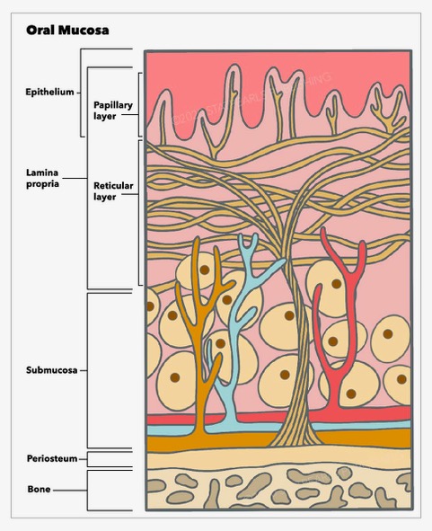
(Click Image to Enlarge)
Oral Mucosa, epithelium, lamina propria, submucosa, periosteum, bone, papillary layer, reticular layer
Contributed From StatPearls Publishing Illustration by Emma Gregory
Details

Author

Sabrina Brar

Editor:

Tom Pepper

Updated:

6/10/2022 6:50:55 PM

References


[1]

Rivera C. Essentials of oral cancer. International journal of clinical and experimental pathology. 2015:8(9):11884-94     [PubMed PMID: 26617944]


[2]

Gandini S,Botteri E,Iodice S,Boniol M,Lowenfels AB,Maisonneuve P,Boyle P, Tobacco smoking and cancer: a meta-analysis. International journal of cancer. 2008 Jan 1;     [PubMed PMID: 17893872]

Level 1 (high-level) evidence

[3]

Lee YC, Marron M, Benhamou S, Bouchardy C, Ahrens W, Pohlabeln H, Lagiou P, Trichopoulos D, Agudo A, Castellsague X, Bencko V, Holcatova I, Kjaerheim K, Merletti F, Richiardi L, Macfarlane GJ, Macfarlane TV, Talamini R, Barzan L, Canova C, Simonato L, Conway DI, McKinney PA, Lowry RJ, Sneddon L, Znaor A, Healy CM, McCartan BE, Brennan P, Hashibe M. Active and involuntary tobacco smoking and upper aerodigestive tract cancer risks in a multicenter case-control study. Cancer epidemiology, biomarkers & prevention : a publication of the American Association for Cancer Research, cosponsored by the American Society of Preventive Oncology. 2009 Dec:18(12):3353-61. doi: 10.1158/1055-9965.EPI-09-0910. Epub     [PubMed PMID: 19959682]

Level 2 (mid-level) evidence

[4]

Kumar M, Nanavati R, Modi TG, Dobariya C. Oral cancer: Etiology and risk factors: A review. Journal of cancer research and therapeutics. 2016 Apr-Jun:12(2):458-63. doi: 10.4103/0973-1482.186696. Epub     [PubMed PMID: 27461593]


[5]

Jafarey NA, Mahmood Z, Zaidi SH. Habits and dietary pattern of cases of carcinoma of the oral cavity and oropharynx. JPMA. The Journal of the Pakistan Medical Association. 1977 Jun:27(6):340-3     [PubMed PMID: 413946]

Level 3 (low-level) evidence

[6]

Candotto V, Lauritano D, Nardone M, Baggi L, Arcuri C, Gatto R, Gaudio RM, Spadari F, Carinci F. HPV infection in the oral cavity: epidemiology, clinical manifestations and relationship with oral cancer. ORAL & implantology. 2017 Jul-Sep:10(3):209-220. doi: 10.11138/orl/2017.10.3.209. Epub 2017 Nov 30     [PubMed PMID: 29285322]


[7]

Kruse AL, Grätz KW. Oral carcinoma after hematopoietic stem cell transplantation--a new classification based on a literature review over 30 years. Head & neck oncology. 2009 Jul 22:1():29. doi: 10.1186/1758-3284-1-29. Epub 2009 Jul 22     [PubMed PMID: 19624855]


[8]

Bray F, Ferlay J, Soerjomataram I, Siegel RL, Torre LA, Jemal A. Global cancer statistics 2018: GLOBOCAN estimates of incidence and mortality worldwide for 36 cancers in 185 countries. CA: a cancer journal for clinicians. 2018 Nov:68(6):394-424. doi: 10.3322/caac.21492. Epub 2018 Sep 12     [PubMed PMID: 30207593]


[9]

Montero PH, Patel SG. Cancer of the oral cavity. Surgical oncology clinics of North America. 2015 Jul:24(3):491-508. doi: 10.1016/j.soc.2015.03.006. Epub 2015 Apr 15     [PubMed PMID: 25979396]


[10]

Naruse T, Yanamoto S, Matsushita Y, Sakamoto Y, Morishita K, Ohba S, Shiraishi T, Yamada SI, Asahina I, Umeda M. Cetuximab for the treatment of locally advanced and recurrent/metastatic oral cancer: An investigation of distant metastasis. Molecular and clinical oncology. 2016 Aug:5(2):246-252     [PubMed PMID: 27446558]


[11]

Kato MG, Baek CH, Chaturvedi P, Gallagher R, Kowalski LP, Leemans CR, Warnakulasuriya S, Nguyen SA, Day TA. Update on oral and oropharyngeal cancer staging - International perspectives. World journal of otorhinolaryngology - head and neck surgery. 2020 Mar:6(1):66-75. doi: 10.1016/j.wjorl.2019.06.001. Epub 2020 Mar 6     [PubMed PMID: 32426706]

Level 3 (low-level) evidence

[12]

Wang B,Zhang S,Yue K,Wang XD, The recurrence and survival of oral squamous cell carcinoma: a report of 275 cases. Chinese journal of cancer. 2013 Nov;     [PubMed PMID: 23601241]

Level 3 (low-level) evidence

[13]

Renou A, Guizard AV, Chabrillac E, Defossez G, Grosclaude P, Deneuve S, Vergez S, Lapotre-Ledoux B, Plouvier SD, Dupret-Bories A, Francim Network. Evolution of the Incidence of Oral Cavity Cancers in the Elderly from 1990 to 2018. Journal of clinical medicine. 2023 Jan 30:12(3):. doi: 10.3390/jcm12031071. Epub 2023 Jan 30     [PubMed PMID: 36769722]


[14]

Uguru CC, Chukwubuzor O, Otakhoigbogie U, Ogu UU, Uguru NP. Awareness and knowledge of risk factors associated with oral cancer among military personnel in Nigeria. Nigerian journal of clinical practice. 2023 Jan:26(1):73-80. doi: 10.4103/njcp.njcp_322_22. Epub     [PubMed PMID: 36751827]


[15]

Rochefort J, Karagiannidis I, Baillou C, Belin L, Guillot-Delost M, Macedo R, Le Moignic A, Mateo V, Soussan P, Brocheriou I, Teillaud JL, Dieu-Nosjean MC, Bertolus C, Lemoine FM, Lescaille G. Defining biomarkers in oral cancer according to smoking and drinking status. Frontiers in oncology. 2022:12():1068979. doi: 10.3389/fonc.2022.1068979. Epub 2023 Jan 11     [PubMed PMID: 36713516]


[16]

Ferrándiz-Pulido C, Leiter U, Harwood C, Proby CM, Guthoff M, Scheel CH, Westhoff TH, Bouwes Bavinck JN, Meyer T, Nägeli MC, Del Marmol V, Lebbé C, Geusau A. Immune Checkpoint Inhibitors in Solid Organ Transplant Recipients With Advanced Skin Cancers-Emerging Strategies for Clinical Management. Transplantation. 2023 Jul 1:107(7):1452-1462. doi: 10.1097/TP.0000000000004459. Epub 2023 Jun 20     [PubMed PMID: 36706163]


[17]

Tsai CY, Wen YW, Lee SR, Ng SH, Kang CJ, Lee LY, Hsueh C, Lin CY, Fan KH, Wang HM, Hsieh CH, Yeh CH, Lin CH, Tsao CK, Fang TJ, Huang SF, Lee LA, Fang KH, Wang YC, Lin WN, Hsin LJ, Yen TC, Cheng NM, Liao CT. Early relapse is an adverse prognostic factor for survival outcomes in patients with oral cavity squamous cell carcinoma: results from a nationwide registry study. BMC cancer. 2023 Feb 7:23(1):126. doi: 10.1186/s12885-023-10602-1. Epub 2023 Feb 7     [PubMed PMID: 36750965]


[18]

Noel CW, Sutradhar R, Chan WC, Fu R, Philteos J, Forner D, Irish JC, Vigod S, Isenberg-Grzeda E, Coburn NG, Hallet J, Eskander A. Gaps in Depression Symptom Management for Patients With Head and Neck Cancer. The Laryngoscope. 2023 Feb 7:():. doi: 10.1002/lary.30595. Epub 2023 Feb 7     [PubMed PMID: 36748910]